/
SPINAL CORD, DISEASES AND DIFFERENTIAL DIAGNOSIS SPINAL CORD, DISEASES AND DIFFERENTIAL DIAGNOSIS

SPINAL CORD, DISEASES AND DIFFERENTIAL DIAGNOSIS - PowerPoint Presentation

cheryl-pisano
cheryl-pisano . @cheryl-pisano
Follow
523 views
Uploaded On 2015-11-15

SPINAL CORD, DISEASES AND DIFFERENTIAL DIAGNOSIS - PPT Presentation

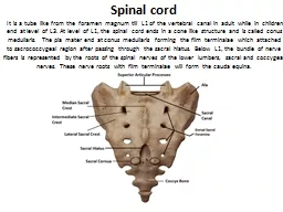
ProfDrAyse ALTINTAS IUCERRAHPASA MEDICAL SCHOOL NEUROLOGY DEPT 3RD GRADE 2011 OCTOBER MEDULLA SPINALIS The spinal cord the grayishwhite oblong cylindrical continuation of the medulla oblongata of the brain ID: 194052

cord spinal myelopathy syndromes spinal cord syndromes myelopathy description myelopathies syndrome posterior anterior involvement compression vertebral dysfunction acute column vascular sensory disorders

Share:

Link:

Embed:

Download Presentation from below link

Download Presentation The PPT/PDF document "SPINAL CORD, DISEASES AND DIFFERENTIAL D..." is the property of its rightful owner. Permission is granted to download and print the materials on this web site for personal, non-commercial use only, and to display it on your personal computer provided you do not modify the materials and that you retain all copyright notices contained in the materials. By downloading content from our website, you accept the terms of this agreement.


Presentation Transcript

Slide1

SPINAL CORD, DISEASES AND DIFFERENTIAL DIAGNOSIS

Prof.Dr.Ayse ALTINTASI.U.CERRAHPASA MEDICAL SCHOOL, NEUROLOGY DEPT.

3RD GRADE, 2011 OCTOBERSlide2

MEDULLA SPINALIS

The spinal cord, the grayish-white oblong cylindrical continuation of the medulla oblongata of the brain,

***begins at the foramen magnum of the skull and

***extends within the vertebral canal to terminate as the cone-shaped conus

medullarisSlide3

MEDULLA SPINALIS

In the adult the caudal tip of the conus

medullaris is located between vertebral levels L1 and L2.Thus, the adult spinal cord is approximately 45 cm in length with an average diameter of 1–1.5 cmSlide4

MEDULLA SPINALIS

The spinal cord is a two-way conduit to and from the brain. It functions as a

“central relay station”, receiving incoming information from

the body and the brain, and as a “central processing station”, conveying

outgoing information to the body and the brain.Slide5
Slide6
Slide7

Each spinal nerve leaves the

vertebral column via its i

ntervertebral foramen and is named accordingly. The first eight spinal nerves, C1–C8, exit the vertebral canal

above the correspondingly numbered cervical vertebrae, whereas all subsequent spinal nerves

(T1–T12, L1–L5, S1–S5, and Co1) exit

below the correspondingly named vertebrae Hence there are 31 pairs of spinal nerves emerging from the spinal

cord.

The region of the spinal cord associated with a particular

pair of spinal nerves is called a

spinal segment.Slide8

Lumbar puncture (LP)Slide9
Slide10
Slide11

VASCULAR SUPPLY OF THE SPINAL CORD

***The

anterior

spinal arteries

,

direct branches of the vertebral

arteries, join

with

each

other to form a single

median

vessel

,

the

anterior

spinal

artery

,

which

provides

small

branches

that penetrate and supply the white and gray matter of the

spinal

cord

.Slide12

VASCULAR SUPPLY OF THE SPINAL CORD

***The

posterior spinal arteries also arise from the vertebral arteries directly or frequently indirectly by way of the inferior

cerebellar branch of the vertebral artery. **

penetrate and serve the white and gray matter of the

spinal cord.Slide13

VASCULAR SUPPLY OF THE SPINAL CORDSlide14
Slide15

SPINAL CORD LESION-LOCATIONSSlide16

SPINAL CORD

Acute spinal cord dysfunction

Can

develop over seconds to wee

k

a common reason for neurology referral Slide17

SPINAL CORD-STRUCTURE

The

neurologist’s tasks are to determine

the etiology,

institute treatment to reverse the acute deficit,

subsequently

determine whether there is a risk of recurrence (especially for inflammatory demyelinating myelopathies)

and

where necessary, institute prophylactic treatment to prevent recurrence.Slide18

SPINAL CORD

Acute and subacute myelopathy occurs for numerous reasons and should always be considered a medical

emergency.

Unfortunately, the diagnosis of myelopathy is often missed at first, leading to missed opportunities for

treatment.Slide19

SPINAL CORD

Any patient who reports, or is found to have a sensory level on exam should be evaluated as a potential

myelopathy patient. Also, patients who present with urinary retention should be considered a spinal cord

dysfunction patient until proven otherwise.

Patients with either a new sensory level or urinary retention should not

be discharged from an office or ER to home without complete imaging of the spinal cord with MRI.Slide20

SPINAL CORD DISEASES- DIAGNOSTIC ALGORITMSlide21

SPINAL CORD AND ROOT COMPRESSION

CAUSES:-TUMOURS (PRIMARY, SECONDARY)

EXTRADURAL, INTRADURAL, INTRAMEDULLARYINFECTIONS (ACUTE, CHRONIC)DISC DISEASE AND SPONDYLOSISHAEMATOMA (AVM, SPONTANEOUS, TRAUMA)

- CYSTIC LESIONS (EXTRADURAL, INTRADURAL, INTRAMEDULLARY)Slide22

SPINAL CORD AND ROOT COMPRESSION

LEVEL OF THE LESION: A lesion above the L1 vertebral body may damage both the cord and its roots.

Below this, only roots are damaged.Slide23

Categories of non-compressive acute myelopathies

Disorders that present as acute myelopathy can be grouped as follows:

1. Vascular

2. Demyelinating

3. Infectious

4. Non-infectious inflammatory

5. Cancer relatedSlide24
Slide25

LOCALIZING A PROCESS AT A SPECIFIC LEVEL IN THE SPINAL CORD

Foramen magnum and upper cervical cord:

suboccipital pain and neck stiffness,

Lhermitte’s symptom,

occipital and

finger tip paresthesias, syringomyelic-type sensory dissociation,

spastic tetraparesis,

long tract sensory signs,

bladder

disturbance,

Slide26

LOCALIZING A PROCESS AT A SPECIFIC LEVEL IN THE SPINAL CORDSlide27

LOCALIZING A PROCESS AT A SPECIFIC LEVEL IN THE SPINAL CORDSlide28

APPROACH TO MYELOPATHIES WITH A NORMAL MRI

Has a compression been missed?

(Epidural lipomatosis, Dynamic compression (flexion-extension studies)

Is it really a myelopathy?

(CNS: parasagittal meningioma, venous sinus thrombosis, ACA thrombosis,

hydrocephalus, vascular parkinsonism, other EP disorders/ Ganglionopathy: Sjogren’s , B6, paraneoplastic/

Plexopathy:

paraneoplastic/ PNS: AIDP, NMJn: MG or LEMS, Muscle: periodic paralysis/ Motor

neuronopathy: ALS/ PLSSlide29

APPROACH TO MYELOPATHIES WITH A NORMAL MRI

• Is it a

metabolic/ toxic/ degenerative/ infective myelopathy?

Are the images of adequate

quality? (Include contrast?)

Were the images taken

too early or too late?

• Is the lesion

too small to be seen on MRI?

• Is there subtle focal or generalized

cord atrophy?Slide30

APPROACH TO MYELOPATHIES WITH A NORMAL MRI

Is there a history of

radiation

?, Is there evidence of CSF hypovolemia?,

Is there evidence of superficial

siderosis?

• Is it

functional

?Slide31

SPINAL CORD-SYNDROMES

Clinical presentation of spinal cord disorders:

The differential diagnosis of an acute myelopathy is dictated by the clinical setting in which the myelopathy

occurs, by the

specific spinal syndrome

,

the occurrence of systemic/non-neurological symptoms,

Radiographic

appearance,

C

linical and radiological response to

c

ortico

-

steroid therapySlide32

SPINAL CORD-SYNDROMES

Spinal cord disorders are

conventionally classified as

syndromes’

due to the typical signs and symptoms produced as a result of the

location of lesions and tract involvement.Slide33

SPINAL CORD-SYNDROMES

For example, Brown-Séquard hemi cord syndrome indicates a

structural myelopathy, either compressive or inflammatory and less often neoplastic disorders; it would be rarely

seen in vascular, paraneoplastic and metabolic disorders. In contrast, myelopathies with

selective tract

involvement are characteristic of metabolic, paraneoplastic and degenerative myelopathies, rather than

inflammatory or infectious; for example, corticospinal and posterior columns involvement is typical of B12 andSlide34

SPINAL CORD-SYNDROMES

In contrast, myelopathies with

selective tract

i

nvolvement are characteristic of metabolic, paraneoplastic and degenerative myelopathies, rather than

inflammatory or infectious; for example, corticospinal and posterior columns involvement is typical of B12 and copper deficiency, adrenomyeloneuropathy, Friedreich’s ataxia.Slide35

SPINAL CORD-SYNDROMES

Complete spinal cord syndrome:

Description: Bilateral involvement of all tracts

Causes: Myelitis (parainfectious/ postvaccinal/ “transverse”), NMO, compression (trauma, tumor, hematoma,

abscess), vascular, (rare with MS)Slide36

SPINAL CORD-SYNDROMES

Brown-Sequard syndrome (often incomplete):

Description: Pain and temperature loss contralateral to weakness, ipsilateral spastic weakness and loss of

proprioceptive function

Causes: compression or intrinsic cord lesion like demyelinationSlide37

BROWN-SEQUARD SYNDROMESlide38
Slide39
Slide40

SPINAL CORD-SYNDROMES

Central cord syndrome:

Description: suspended analgesia, sacral sparing, dissociation of sensory loss, ipsilateral Horner, anterior

horn dysfunction

Causes: syrinx, intramedullary tumors, hyperextension neck injuries (“man-in-a-barrel”)Slide41

CENTRAL CORD LESIONSlide42

SPINAL CORD-SYNDROMES

Posterolateral column disease:

Description: foot > hand paresthesias, dorsal column dysfunction, intact pain and temperature, corticospinal

dysfunction, associated PN

Causes: Cbl/ copper deficiency, HTLV or HIV, extrinsic compression as in spondylotic myelopathySlide43

SPINAL CORD-SYNDROMES

Posterior column disease:

Description: sensory ataxia, absent reflexes, hypotonic but not weak, hyperextensible joints, trophic changes,crises, decreased deep pain, Lhermitte’s

Causes: TabesSlide44

SPINAL CORD-SYNDROMES

Anterior horn cell syndrome:

Description: weakness, atrophy, fasciculations, reduced tone and reflexes, intact sensations, possible

involvement of cranial motor nuclei

Causes: spinal muscular atrophy (infantile/ Werdnig-Hoffman, intermediate, juvenile-Kugelberg-Welander, motor neuron disease)Slide45

SPINAL CORD-SYNDROMES

Combined anterior horn cell and pyramidal tract disease:

Description: anterior horn cells and pyramidal tract, sphincteric sparing, asymmetric, cramping, bulbar or

pseudobulbar involvement, sensory sparing, preserved superficial abdominals, reflexes variable, rare for

extraocular muscles to be involved

Causes: ALSSlide46

SPINAL CORD-SYNDROMES

Anterior spinal artery syndrome:

Description: Bilateral corticospinal and spinothalamic involvement without dorsal column, bladder

dysfunctionCauses: Anterior spinal artery infarct or MSSlide47

SPINAL CORD-SYNDROMES

Posterior spinal artery syndrome;

Description: posterior column dysfunction with loss of segmental reflexes, infarction of unilateral posterior

horn and lateral column with sparing of posterior columns may also occurCauses: posterior spinal artery infarctSlide48
Slide49
Slide50

SPINAL CORD-SYNDROMESSlide51

THANK YOU VERY MUCH