/
Care of Patients with Problems of the Central Nervous System: The Spinal Cord Care of Patients with Problems of the Central Nervous System: The Spinal Cord

Care of Patients with Problems of the Central Nervous System: The Spinal Cord - PowerPoint Presentation

luanne-stotts
luanne-stotts . @luanne-stotts
Follow
385 views
Uploaded On 2018-07-12

Care of Patients with Problems of the Central Nervous System: The Spinal Cord - PPT Presentation

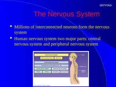
Chapter 45 Spinal Cord Lumbosacral Back Pain Low Back Pain Herniated nucleus pulposus Health Promotion and Maintenance Good posture Proper lifting Exercise Ergonomics PatientCentered Collaborative Care ID: 663715

spinal assessment care cord assessment spinal cord care management patient injuries injury patients cont

Share:

Link:

Embed:

Download Presentation from below link

Download Presentation The PPT/PDF document "Care of Patients with Problems of the Ce..." is the property of its rightful owner. Permission is granted to download and print the materials on this web site for personal, non-commercial use only, and to display it on your personal computer provided you do not modify the materials and that you retain all copyright notices contained in the materials. By downloading content from our website, you accept the terms of this agreement.


Presentation Transcript

Slide1

Care of Patients with Problems of the Central Nervous System: The Spinal Cord

Chapter 45Slide2

Spinal CordSlide3

Lumbosacral Back Pain (Low Back Pain)

Herniated nucleus pulposusSlide4

Health Promotion and Maintenance

Good posture

Proper lifting

Exercise

Ergonomics Slide5

Patient-Centered Collaborative Care

Assessment

Diagnostic assessmentSlide6

Nonsurgical Management

Positioning

Drug therapy

Heat therapy

Physical therapy

Weight control

Complementary and alternative therapiesSlide7

Surgical Management

Minimally invasive surgery:

Percutaneous lumbar diskectomy

Thermodiskectomy

Laser-assisted laparoscopic lumbar diskectomy

Conventional open surgical procedures:

Diskectomy

Laminectomy

Spinal fusionSlide8

Postoperative Care

Prevention and assessment of complications

Neurologic assessment; vital signs

Patient’s ability to void

Pain control

Wound care

CSF check

Patient positioning and mobilitySlide9

Community-Based Care

Home care management

Health teaching

Health care resourcesSlide10

Cervical Neck Pain

Conservative treatment is the same as described for back pain except that the exercises focus on shoulder and neck.

If these treatments do not work, soft collar may be used at night for a period of no longer than 10 days.

If conservative treatment is ineffective, surgery such as an anterior cervical diskectomy and fusion is commonly performed.Slide11

Spinal Cord Injuries

Hyperflexion injury

Hyperextension injury

Axial loading injury or vertical compression such as those that occur in jumping

Excessive rotation of the head beyond its range

Penetration injury, such as those wounds caused by a bullet or a knife Slide12

Spinal Cord Injuries (Cont’d)Slide13

Spinal Cord Injuries (Cont’d)Slide14

Common Spinal Cord Syndromes

Complete lesion

Anterior cord syndrome

Posterior cord lesion

Brown-S

é

quard syndrome

Central cord syndromeSlide15

Common Spinal Cord Syndromes (Cont’d)Slide16

Anterior Cord Syndrome

Damage to the anterior portion of both gray and white matter of the spinal cord

Usually a result of decreased blood supply

Motor function and pain and temperature lost below the level of the injury

Sensations of touch, position, and vibration remain intactSlide17

Posterior Cord Lesion

Damage to the posterior gray and white matter of the spinal cord

Motor function remains intact

Patient experiences loss of vibratory sense, touch, and position sensationSlide18

Brown-Séquard Syndrome

Results from penetrating injuries that cause hemisection of the spinal cord, or injuries that affect half of the spinal cord.

Motor function, proprioception, vibration, deep touch sensations are lost on the same side (ipsilateral) of the body as the lesion.

Opposite side (contralateral) of the body sensations of pain, temperature, light touch are affected.Slide19

Central Cord Syndrome

Lesions of the central portion of the spinal cord.

Loss of motor function is more pronounced in the upper extremities than in the lower extremities.

Varying degrees and patterns of sensation remain intact.Slide20

SCI: Etiology

Trauma is the leading cause

Incidence/prevalenceSlide21

Patient with SCI: Initial Assessment

First priority is assessment of the patient’s airway, breathing pattern, and circulation status

Assessment for indications of intra-abdominal hemorrhage or hemorrhage or bleeding around fracture sites

Assessment of level of consciousness using Glasgow Coma ScaleSlide22

Initial Assessment (Cont’d)

Establishment of level of injury: tetraplegia, quadriplegia, quadriparesis, paraplegia, and paraparesisSlide23

Spinal Shock/Spinal Shock Syndrome

This condition occurs immediately as a concussion response to the injury. The patient has:

Flaccid paralysis

Loss of reflex activity below the level of the lesion

Usually resolves within 24 hours

Muscle spasticity begins in patients with cervical or high thoracic injuriesSlide24

Assessment of Sensory and Motor Ability

Hypoesthesia

Hyperesthesia Slide25

Cardiovascular and Respiratory Assessment

Cardiovascular dysfunction is usually the result of disruption of the autonomic nervous system especially if the injury is above the 6th thoracic vertebra.

Cardiac dysrhythmias may result.

Systolic BP below 90 requires treatment because lack of perfusion to the spinal cord could worsen the patient’s condition.

Hypothermia.

Slide26

Cardiovascular and Respiratory Assessment (Cont’d)

Patients with cervical SCI are at risk for respiratory problems resulting from immobility or from an interruption of spinal innervations to the respiratory muscles.

Continued respiratory assessment including vital capacity and minute volume.Slide27

Gastrointestinal and Genitourinary Assessment

Assess abdomen for indications of hemorrhage, distention, or paralytic ileus.

Assess for reflex or hypotonic bowel.

Assess for areflexic bladder, which later leads to urinary retention.

Assess for neurogenic bladder.Slide28

Other Assessments

Lower motor neuron assessment

Upper motor neuron assessment

Skin assessment

Heterotrophic ossification assessment

Psychosocial assessment

Laboratory assessment

Imaging assessmentSlide29

Nonsurgical Management

Constant assessment

Assess for neurogenic shock. Neurogenic shock is spinal shock with:

Bradycardia

Decreased or absent bowel sounds

Warm, dry skin

Hypothermia

HypotensionSlide30

Immobilization for Cervical Injuries

Fixed skeletal traction to realign the vertebrae, facilitate bone healing, and prevent further injury

Halo fixation and cervical tongs

Stryker frame, rotational bed, kinetic treatment table

Pin site care and monitoring of traction ropesSlide31

Immobilization of Thoracic and Lumbosacral Injuries

For patients with thoracic injuries

bedrest and possible immobilization with a fiberglass or plastic body cast

For patients with lumbar and sacral injuries

immobilization of the spine with a brace or corset worn when the patient is out of bed; custom-fit thoracic lumbar sacral orthoses preferredSlide32

Drug Therapy

Methylprednisolone (controversial)

Dextran

Atropine sulfate

Dopamine hydrochloride

Tizanidine

Intrathecal baclofenSlide33

Surgical Management

Emergency surgery necessary for spinal cord decompression

Decompressive laminectomy

Spinal fusion

Harrington rods to stabilize thoracic spinal injuriesSlide34

Ineffective Airway Clearance and Breathing Pattern

Interventions for the patient with spinal cord injury:

Airway management is the priority.

Patients with injuries at or above the 6th thoracic vertebra are especially at risk for respiratory complications.

Provide measures to maintain airway.Slide35

Ineffective Airway Clearance and Breathing Pattern (Cont’d)

Assisted coughing, quad cough, cough assist

Use of incentive Spiro meterSlide36

Impaired Physical Mobility; Self-Care Deficit

Interventions include:

In patients with spinal cord injury, monitor for risk of pressure ulcers, contractures, and deep vein thrombosis or pulmonary emboli.

Proper positioning, skin inspection, ROM exercises, heparin, and graduated compression stockings.

Slide37

Impaired Physical Mobility; Self-Care Deficit (Cont’d)

Prevent orthostatic hypotension.

Promote self-care.Slide38

Impaired Urinary Elimination; Constipation

Interventions include:

A bladder retraining program

Spastic bladder

manipulating external area

Flaccid bladder

Valsalva maneuver

Encouraging consumption of 2000 to 2500 mL of fluid daily to prevent urinary tract infectionSlide39

Impaired Urinary Elimination; Constipation (Cont’d)

Long-term renal complication

Signs and symptoms of urinary tract infection not perceived by the patientSlide40

Autonomic Dysreflexia

Commonly seen in patients with upper spinal cord injury

Severe hypertension

Bradycardia

Severe headache

Nasal stuffiness

Flushing

Treatment Slide41

Establishing a Bowel Retraining Program

Consistent time for bowel elimination

High fluid intake

High-fiber diet

Rectal stimulation (with or without suppositories)

Stool softener medications, as neededSlide42

Impaired Adjustment

Interventions include:

Invite patients to ask questions about significant life changes; reply openly and honestly.

Encourage patients to discuss their perceptions of their situation and coping strategies that can be used.

Begin a patient education program to clarify misconceptions.Slide43

Community-Based Care

Home care management

Health teaching

Health care resourcesSlide44

Spinal Cord Tumors

Primary spinal cord tumors

Intramedullary tumors

Extramedullary tumorsSlide45

Patient-Centered Collaborative Management

Assessment

Diagnostic assessment

Surgical management

need for emergency surgery

Nonsurgical management

radiation, chemotherapySlide46

Community-Based Care

Home care management

Health teaching

Health care resources Slide47

Multiple Sclerosis

Chronic autoimmune disease affecting the myelin sheath and conduction pathway of the CNS

Characterized by periods of remission and exacerbation

Inflammatory response resulting in random or patchy areas of plaque in the white matter of the CNSSlide48

Multiple Sclerosis (Cont’d)

Etiology

Genetic risk

Incidence

Prevalence Slide49

Major Types of Multiple Sclerosis

Relapsing-remitting

Primary progressive

Secondary progressive

Progressive-relapsing Slide50

Patient-Centered Collaborative Care

Patient history

Physical assessment/clinical manifestations

FatigueSlide51

Common Physical Assessment

Findings include:

Flexor spasms at night

Intention tremor

Dysmetria

Blurred vision, diplopia, decreased visual acuity, scotomas, nystagmus

Hypalgesia, numbness, tingling, or burning

Bowel and bladder dysfunctionSlide52

Assessment

Psychosocial assessment

Laboratory assessment

Other diagnostic testsSlide53

Drug Therapy

Therapies include:

Interferon beta

Monoclonal antibodies

Copaxone

Novantrone

Immunosuppressive therapy

MethylprednisoloneSlide54

Drug Therapy (Cont’d)

Muscle relaxants

Treatment of paresthesia

Treatment of bladder dysfunctionSlide55

Other Interventions

Promoting mobility

Managing symptoms

Complementary and alternative therapiesSlide56

Community-Based Care

Home care management

Health teaching

Health care resourcesSlide57

Amyotrophic Lateral Sclerosis

Known as

Lou Gehrig’s disease,

an adult onset upper and lower motor neuron disease characterized by progressive weakness, muscle wasting, and spasticity eventually leading to paralysis

Early symptoms

fatigue while talking, tongue atrophy, dysphagia, weakness of the hands and arms, fasciculations, nasal quality of speech, dysarthriaSlide58

Interventions

No known cure, no treatment, no preventive measures

Riluzole, only drug approved by FDA to extend survival time

Exercise and mobility program

Management of swallowing difficulties

Respiratory support