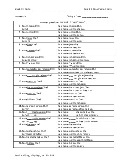
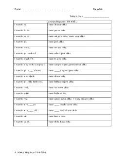
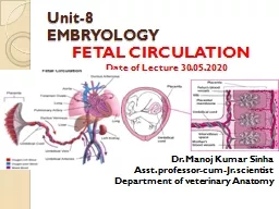
/
Fetal Circulation Ms.Ashwini.S.Mane Fetal Circulation Ms.Ashwini.S.Mane

Fetal Circulation Ms.Ashwini.S.Mane - PowerPoint Presentation

kylie
kylie . @kylie
Follow
66 views
Uploaded On 2023-05-20

Fetal Circulation Ms.Ashwini.S.Mane - PPT Presentation

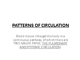
Anatomy and Physiology Fetal Circulation Umbilical cord 2 umbilical arteries return nonoxygenated blood fecal waste CO2 to placenta 1umbilical vein brings oxygenated blood and nutrients to the fetus ID: 998508

ductus blood left fetal blood ductus fetal left fetus atrium umbilical oxygenated arteriosus venous liver vein inferior ovale pulmonary

Share:

Link:

Embed:

Download Presentation from below link

Download Presentation The PPT/PDF document "Fetal Circulation Ms.Ashwini.S.Mane" is the property of its rightful owner. Permission is granted to download and print the materials on this web site for personal, non-commercial use only, and to display it on your personal computer provided you do not modify the materials and that you retain all copyright notices contained in the materials. By downloading content from our website, you accept the terms of this agreement.


Presentation Transcript

1. Fetal CirculationMs.Ashwini.S.Mane

2. Anatomy and PhysiologyFetal CirculationUmbilical cord2 umbilical arteries: return non-oxygenated blood, fecal waste, CO2 to placenta1umbilical vein: brings oxygenated blood and nutrients to the fetus

3. Fetus depends on placenta to meet O2 needs while organs continue formationOxygenated blood flows from the placentaTo the fetus via the umbilical veinAfter reaching fetus the blood flows through the inferior vena cava

4. The differences between fetal and newborn circulationThe fetus received oxygen from the placenta and through the lungs after birth.The fetal liver doesn’t have the metabolic function that it will have after birth because the mother performs these functions.Three shunts are present in fetal life:Ductus venosus: connects the umbilical vein to the inferior vena cavaDuctus arteriosus: connects the main pulmonary artery to the aortaForamen ovale: anatomic opening between the right and left atrium.

5. Fetal Circulation

6. The umbilical vein carrying the oxygenated blood (80% saturated) from the placenta , enters the fetus at the umbilicus and run along the free margins of the falciform ligaments of the liver , in the liver , it gives off branches to the lobe of the liver and receives the deoxygenated blood from the portal vein The greater portion of the oxygenated blood , mixed with some portal venous blood , short circuits the liver through the ductus venous to enter the inferior vena cava and then to right atrium of the heart

7. The O2 content of this mixed blood is thus reduced , although both the ductus venous and hepatic portal/fetal trunk blood enters the right atrium through the inferior vana cava, there is a little mixing The terminal part of the IVC receives blood from the right hepatic vein

8. In the right atrium , most of the well oxygenated (75%) ductus venous blood is preferentially directed into the foramen ovale by the valve of the inferior vana cava and crista dividens and passes in to the left atrium , here it is mixed with small amount of venous blood returning from the lungs through the pulmonary veins The left atrial blood is passed on through the mitral opening into the left ventricle

9. Remaining the lesser amount of the blood (25%), after reaching the right atrium via the superior and inferior vena cava (carrying the venous blood from the cephalic and caudal parts of the fetus respectively) passes through the tricuspid opening into the right ventricle

10. During the ventricular systole , the left ventricular blood is pumped into the ascending and arch of aorta and distributed by their branches to heart , neck , brain and arms The right ventricular blood with low oxygen content is discharged into the pulmonary trunk , sinces the resistance in the pulmonary arteries during fetal life is very high , the main portion of the blood passes directly through the ductus arteriosus into the descending aorta bypassing the lungs where it mixes with the blood from the proximal aorta

11. 70% of cardiac output is carried out by ductus arteriosus to the descending aorta About 40% of the combined output goes to the placenta through the umbilical arteries

12. The deoxygenated blood leaves the body by of two umbilical arterie s to reach the placenta where it is oxygenated and get ready for recirculation The mean cardiac output is comparatively high in fetus and is estimated to be 350ml per kg per minute

13.

14. Conversion of Fetal to Infant CirculationAt birthClamping the cord shuts down low-pressure systemIncreased atmospheric pressure(increased systemic vascular resistance) causes lungs to inflate with oxygenLungs now become a low-pressure system

15. Conversion: Fetal to Infant CirculationPressure from increased blood flow in the left side of the heart causes the foramen ovale to closeMore heavily oxygenated blood passing by the ductus arteriosus causes it constrictFunctional closure of the foramen ovale and ductus arteriosus occurs soon after birthOverall anatomic changes are not complete for weeks

16. What happens to these special structures after birth?Umbilical arteries atrophyUmbilical vein becomes part of the fibrous support ligament for the liverThe foramen ovale, ductus arteriosus, ductus venosus atrophy and become fibrous ligaments

17. Overview of ConversionUmbilical cord is clampedLoose placentaClosure of ductus venosusBlood is transported to liver and portal systemIncreased systemic resistancePressure in right atrium decreasedChange from right to left shunting to left to right blood flowIncreased O2 levels in pulmonary circulationClosure of the ductus arteriosus

18. Thank You….