/
Unit-8 EMBRYOLOGY      FETAL CIRCULATION Unit-8 EMBRYOLOGY      FETAL CIRCULATION

Unit-8 EMBRYOLOGY FETAL CIRCULATION - PowerPoint Presentation

roxanne
roxanne . @roxanne
Follow
342 views
Uploaded On 2022-05-14

Unit-8 EMBRYOLOGY FETAL CIRCULATION - PPT Presentation

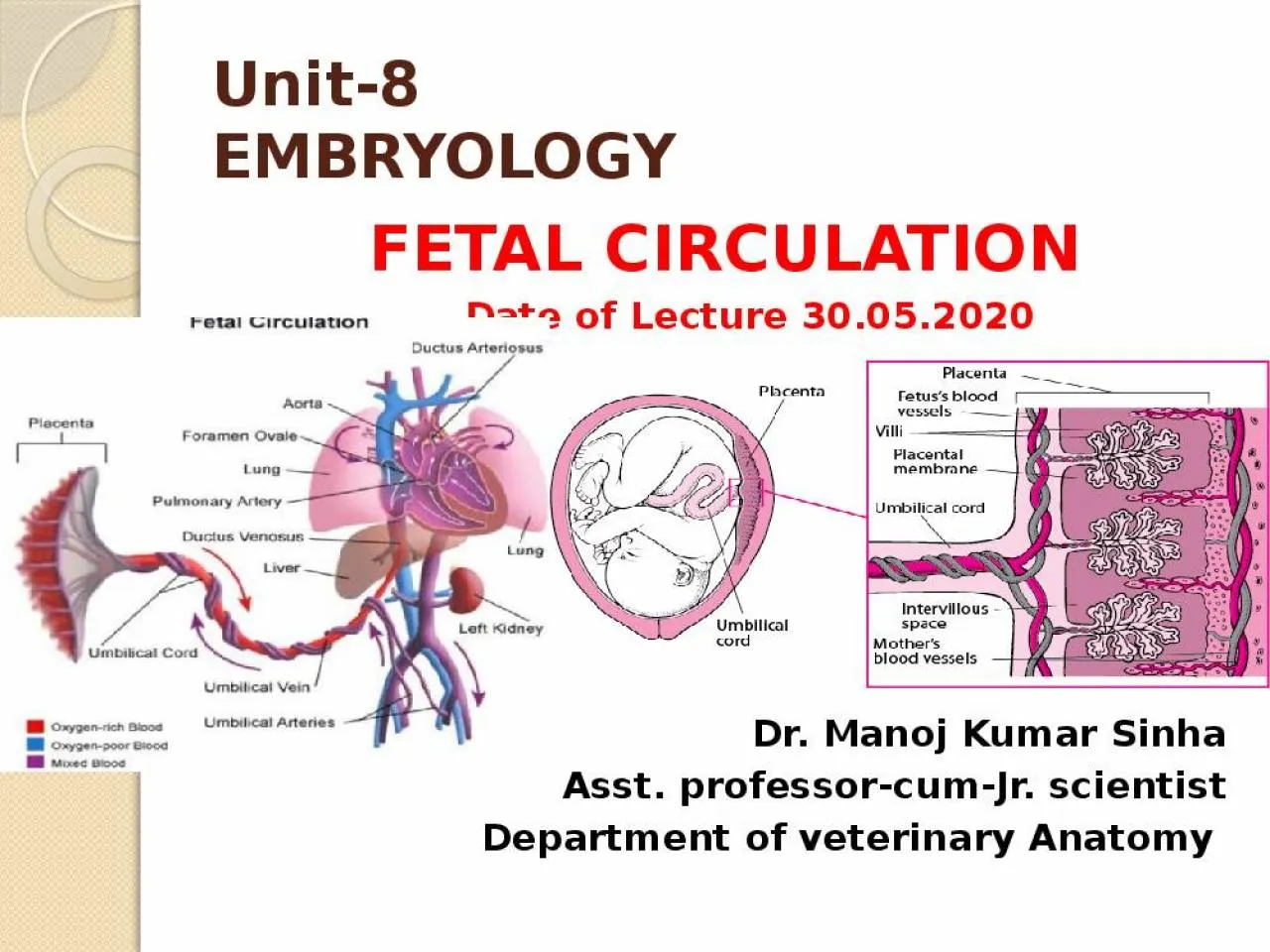
Date of Lecture 30052020 Dr Manoj Kumar Sinha Asst professorcumJr scientist Department of veterinary Anatomy FETAL CIRCULATION The circulation of oxygenated blood deoxygenated blood and nutritive materials in the fetus is termed as ID: 910938

umbilical blood vein fetus blood umbilical fetus vein fetal cord placenta circulation aorta ductus arteriosus arteries oxygenated birth pulmonary

Share:

Link:

Embed:

Download Presentation from below link

Download Presentation The PPT/PDF document "Unit-8 EMBRYOLOGY FETAL CIRCULATION" is the property of its rightful owner. Permission is granted to download and print the materials on this web site for personal, non-commercial use only, and to display it on your personal computer provided you do not modify the materials and that you retain all copyright notices contained in the materials. By downloading content from our website, you accept the terms of this agreement.


Presentation Transcript

Slide1

Unit-8EMBRYOLOGY

FETAL CIRCULATION Date of Lecture 30.05.2020 Dr. Manoj Kumar SinhaAsst. professor-cum-Jr. scientist Department of veterinary Anatomy

Slide2

FETAL

CIRCULATION

The circulation of oxygenated blood, deoxygenated blood and nutritive materials in the fetus is termed as Fetal CirculationThe circulatory system of the mother is not directly connected to that of the fetus thus the placenta functions as the respiratory center for the fetus as well as a site of filtration of nutrients and wastages water glucose, amino acids, vitamins etc. freely diffuse through the placenta along with oxygenThe uterine arteries carry oxygenated blood to the placenta.

Slide3

Three shunts are present in fetal life which play important role in fetal circulation

Ductus

Venosus: connects the umbilical vein to the inferior venacavaDuctus Arteriosus: connects the main pulmonary artery to the AortaForamen Ovale: Opening between the right and left atrium

Slide4

Slide5

Continue..

The placenta accepts deoxygenated blood from the fetus through blood vessels that leave the fetus through the

Umbilical Cord (Umbilical Arteries )The oxygenated nutrient rich blood then returns to the fetus via the Umbilical cord (Umbilical Vein)

Slide6

UMBILICAL

CORD

Thick cord like structure which extends from center of the abdominal wall of the fetus to the center of the fetal surface of placenta (The structure connecting the fetus with the placenta)The umbilical cord develops from and contains remnants of the yolk sac and allantois

Slide7

Continue…

Umbilical cord is made up of :

Two umbilical arteries one umbilical vein duct of yolk sac and distal part of allantoic diverticulumThus umbilical vein carries blood towards the fetus’s heart while the umbilical arteries carry blood awayAll these structures are surrounded by Wharton’s jelly( a gelatinous substance made from mucopolysaccharides which protects the blood vessels inside) and finally enveloped by amniotic membraneThe Wharton's jelly is formed by the

mucoid degeneration of the

mesodermal

cells of the connecting stalk

Slide8

CROSS SECTION OF UMBILICAL CORD

Slide9

Slide10

Continue…

Early part of embryonic life

two umbilical veins are present , but later only the left vein persistsThe umbilical vein carrying placental (oxygenated) blood, enters the abdomen of the fetus at the umbilicus and then enters into the liverLeft vein then joins the portal vein, hepatic vein and finally opens into the abdominal venacava through Ductus VenosusThen blood conveyed to the right atriumThrough Foramen Ovalae

blood passes into the left atrium and then to the left ventricle The foramen ovale closes at the time of birth

Through the aorta blood goes to the head, fore limbs and small amount of blood passes into the abdominal aorta

Slide11

Slide12

Continue…

From cranial end blood returns through

anterior venacava to the right atriumThis blood along with blood from abdominal venacava passes into the right ventricle The right ventricular blood goes via pulmonary trunk going to the descending aorta through Ductus Arteriosus because fetal lungs become inactiveDuctus Arteriosus is a arterial duct which connects the pulmonary artery with the aorta ( In the fetus there is special connection between the pulmonary artery and the aorta, called Ductus Arteriosus)At birth it becomes occluded but in adult it persists as a ligament

(Ligamentum Arteriosum)

Some of the blood moves from the aorta through the internal iliac arteries and re-enters the placenta, the maternal circulation

Slide13

AT THE TIME OF BIRTH

Closure of Foramen OvaleClosure of Ductus Arteriosus

Closure of Ductus VenosusAFTER BIRTHAfter birth of the baby the lung, renal, digestive and liver functions are working the fetal circulation undergoes some changes since they are no longer needed

Slide14

Slide15