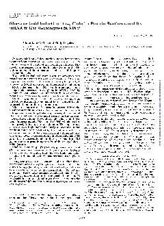
/
Page 317 of 317  KRCFischer rat hepatocytes Aneupoidy 12000 mgkg die Page 317 of 317  KRCFischer rat hepatocytes Aneupoidy 12000 mgkg die

Page 317 of 317 KRCFischer rat hepatocytes Aneupoidy 12000 mgkg die - PDF document

kylie
kylie . @kylie
Follow
342 views
Uploaded On 2022-09-02

Page 317 of 317 KRCFischer rat hepatocytes Aneupoidy 12000 mgkg die - PPT Presentation

Negative Hasmall and Roberts 1997 IARC 2000 Rat kidney Tumor promotion NA Positive Kurokawa al1988 ATSDR 2002 S typhimuriumTA100 Rat hostmediated assay Gene mutation NA Negative Kozum ID: 947012

rats day loael dehp day rats dehp loael group weeks noael weight days liver feeding 2008 dose mice 2000

Share:

Link:

Embed:

Download Presentation from below link

Download Pdf The PPT/PDF document "Page 317 of 317 KRCFischer rat hepatocy..." is the property of its rightful owner. Permission is granted to download and print the materials on this web site for personal, non-commercial use only, and to display it on your personal computer provided you do not modify the materials and that you retain all copyright notices contained in the materials. By downloading content from our website, you accept the terms of this agreement.


Presentation Transcript

Page 317 of 317 KRCFischer rat hepatocytes Aneupoidy 12,000 mg/kg diet 7 days Negative Hasmall and Roberts, 1997; IARC, 2000 Rat kidney Tumor promotion N/A Positive Kurokawa al.,1988; ATSDR, 2002 S. typhimurium(TA100): Rat host-mediated assay Gene mutation N/A Negative Kozumbo al.,1982; ATSDR, 2002 C57BL/6f lacl transgenic mouse liver Transgenic mouse mutation assay 3000, 6000 mg/kg (600, 1200 mg/kg-day) 6000 mg/kg diet 120 days Negative, no inc. in mutant frequency in the lacl gene of liver DNA Gunz 1993; IARC, 2000; ECB, 2008 Cynomolgous monkey liver cells Inhibition of Gap junction intercellular communication 500 x 14 po Negative IARC, 2000 B6C3F mice Sperm morphology 6000 x 5 ip Negative Douglas al.,1986; IARC, 2000 Sprague-Dawley rats Sperm morphology 5200 x 5 ip Negative Douglas al.,1986; IARC, 2000 Insect Systems D. melanogaster (Canton-S)Sex-linked recessive lethal mutation 20 mg/kg single dose injection Negative – percentage of lethal was 0.03 compared to 0.05 in controlsYoon 1985; IARC, 2000; ATSDR, 2002; ECB, 2008 D. melanogaster (Canton-S)Sex-linked recessive lethal mutation 18,600 µg/g in feed, no exposure period Negative – percentage of lethal was 0.07 compared to 0.11 in controlsZimmering al.,1989; IARC, 2000; ECB, 2008 D. melanogasterDNA double strand breakage 7540 µg/g food NegativeKawai, 1998; IARC, 2000 D. melanogasterDNA repair test 7540 µg/g food NegativeKawai, 1998; IARC, 2000 D. melanogasterWing spot test, mutation 7540µg/g food NegativeKawai, 1998; IARC, 2000 GL – Guideline study GLP – Good Laboratory Practice IP – Intraperitoneal N/A – not applicable/ specified NT – not tested PO – er os, oral administration UDS – Unscheduled DNA synthesis Yellow highlight denotes positive tests. Tan highlights denote marginally positive or equivocal tests. Test results in {parentheses} denote occasions where source materials differed in result interpretation. Page 316 of 317 KRCFischer 344 rats (5-10 M per group) Replicative DNA synthesis 1.2% in diet (500 mg/kg-day) for 1, 2, 4, 8, 18, 77, 151, 365 days Pulse labeling index of hepatocyte nuclei increased at only 2 days; Pump infusion tech. in

crease in hepatic nuclear labeling at 8 days Marsman al.,1988; ECB, 2008 Fischer 344 rats (6 M per group) Replicative DNA synthesis 1.2% (600 mg/kg-day) in diet for 8 weeks Urinary bladder epithelium labeling index unaffected Hagiwara al.,1990; ECB, 2008 Fischer 344 rats (M) Replicative DNA synthesis 1000, 2000 mg/kg once via gavage or subcutaneous injection Synthesis increased both doses at 24 hours but not 39 or 48 hours after administration Uno 1994; ECB, 2008 Alderly Park rats (10 M and 10F) Replicative DNA synthesis 2000 mg/kg-day via gavage for 14 days; GLP Significant decrease in H-thymidine uptake in hepatocytes (M) and proximal tubule cells (M&F) ICI, 1982b; ECB, 2008 Marmosets (5M & 5F) Replicative DNA synthesis 2ml/kg-day (1960 mg/kg-day) via gavage for 14 days; GLP No difference in thymidine uptake ICI, 1982b; ECB, 2008 B6C3F mice (48 M per group) Replicative DNA synthesis 0, 6000, 12000 mg/kg (1000, 2000 mg/kg-day) in diet for 2, 8, 24, 40 weeks Hepatic labeling index increased at 2000 mg/kg- day (24, 40 wk), thymidine kinase activity increased at 2 wk and decreased at 8 wk (2000 mg/kg-day), increased at 2 and 40 wk (1000 mg/kg-day) Ward 1988; ECB, 2008 Rat liverStrand breaks N/A NegativeButterworth al.,1984; ATSDR, 2002 Wistar rat liverDNA strand breaks2000 x 28 po NegativeElliott and Elcombe, 1985; IARC, 2000; ATSDR, 2002 Wistar rat liverDNA strand breaks500 x 14 po (MEHP) NegativeElliott and Elcombe, 1985; IARC, 2000 Fischer 344 rat liver (4 M per group)DNA single-strand breaks2, 40, 78 weeks, diet 2% (900 mg/kg-day) 20,000 mg/kg diet 78 wk Negative (positive – 5- fold increase in single strand breaks in tumor bearing rats, ECB)Tamura 1991; IARC, 2000; ATSDR, 2002; ECB, 2008 Fischer 344 rat liverDNA base modification – DNA oxidative damage 12,000 mg/kg diet 22 wk NegativeCattley and Glover, 1993; IARC, 2000; ATSDR, 2002 Fischer 344 rat liver (3 M per group)DNA base modification – DNA oxidative damage 1, 2, 3, 6, 9, 12 months – diet 1.2% (600 mg/kg-day) 12,000 mg/kg diet 1 yr 12,000 mg/kg diet over 1-2 wk Positive (oxidative DNA damage 2-fold increased after 1 month, but no

oxidative damage in kidney DNA)Takagi al.,1990a, 1990b; IARC, 2000; ATSDR, 2002; ECB, 2008 Rat liverTetraploid nuclei N/A PositiveAhmed 1989; ATSDR, 2002 Page 315 of 317 KRCB6C3F Mouse erythrocytesMicronucleus formation6000 x 5 ip NegativeDouglas al.,1986; IARC, 2000; ATSDR, 2002 Mouse bone marrowMicronucleus formationN/A NegativePutman 1983; ATSDR, 2002 Fischer 344 rat hepatocyte DNADNA binding - covalent390 mg/kg-day NegativeGupta 1985; IARC, 2000 Fischer 344 rats (F) DNA binding Radiolabeled 500 mg/kg Radiolabeled carbonyl-or alcohol-C, ± 4 week prefeeding period with 1% in diet Carbonyl-C – negative Alcohol-C - positive BASF, 1982; ECB, 2008 Fischer 344 rat liver DNA DNA binding - covalent 10,000 mg/kg diet 11 days Positive(ATSDR)/Negati ve (IARC)Albro 1982a; IARC, 2000; ATSDR, 2002 Fischer 344 rat liver DNA (3 M per group)DNA binding – covalent – DNA adduct formation2000 mg/kg-day - Once via gavage daily for 3 days Negative for adduct formationGupta 1985; IARC, 2000; ATSDR, 2002; ECB, 2008 Fischer 344 rat liver DNADNA binding - covalent500 x 1 po NegativeLutz, 1986; IARC, 2000; ATSDR, 2002 Fischer 344 rat liver DNADNA binding - covalent10,000 mg/kg diet 4 wk NegativeVon Däniken al.,1984; IARC, 2000; ATSDR, 2002 Fischer 344 rat hepatocytes (M)DNA repair- UDS – alkaline elution assay 150 mg/kg-day, One gavage daily for 14 days; 500 mg/kg, one gavage dose at 2, 12, 24, 48 hours prior to sacrifice, 12,000 mg/kg diet (600 mg/kg-day) for 30 days followed by 500 mg/kg gavage Negative Butterworth al.,1984; IARC, 2000; ATSDR, 2002; ECB, 2008 Fischer 344 rat hepatocytes (liver)DNA repair - UDS12,000 mg/kg diet 28d Negative1988; IARC, 2000; ATSDR, 2002 Sprague-Dawley rat hepatocytes (M; liver)DNA repair - UDS5000 mg/kg gavage for 4-8 weeks; 2% feed (1000 mg/kg-day) for 4-8 weeks followed by 5000 mg/kg gavage NegativeKornbrust al.,1984; IARC, 2000; ATSDR, 2002; ECB, 2008 Rat liverDNA repairN/A PositiveHayashi al.,1998; ATSDR, 2002 B6C3F mouse hepatocytes (M; liver) DNA repair - UDS10, 100, 500 mg/kg for 7, 14, 28 days; 6000 mg/kg feed (1200 mg/kg-day) for 7, 14, 28 days NegativeSmith Oliver and Butterworth, 1987;

IARC, 2000; ATSDR, 2002; ECB, 2008 Primary rat hepatocytes Stimulation of DNA synthesis 200µM (78µg/mL) Positive Reddy 1992; ECB, 2008 Fischer 344 rats (4 M per group) Stimulation of DNA synthesis 1.73 mmol/kg (676 mg/kg) single gavage dose Increased ratio of thymidine incorporation Büsser and Lutz, 1987; ECB, 2008 Page 314 of 317 KRC Genotoxic Effects (retrieved from ECB, 2008; Species/Test System (Strain) End Point Doses (IARC, 2000; ECB, 2008; NICNAS 2008) Conclusion Citation Mammalian Systems Human leucocytes Chromosomal aberrations 0.01-0.16 mg/m NegativeThiess and Fleig, 1978; ATSDR, 2002 Syrian Hamster (SH) embryo cells (F) Chromosomal aberrations Single gavage dose (0, 3750, 7500, 15,000 mg/kg) at Gd 11{7500 x 1 po, PositiveTomita al.,1982b; IARC, 2000; ATSDR, 2002; ECB, 2008 Fischer 344 rat bone marrow (5 M per group) Chromosomal aberrations 0.5, 1.7, 5.0 ml/kg-day (500, 1700, 5000 mg/kg-day) gavage daily for 5 days {4900 x 5 po, IARC} Negative Putman 1983; Nuodex, 1981g; IARC, 2000; ECB, 2008 Syrian Hamster (SH) embryo cells (F)Cell transformation Single gavage dose (0, 3750, 7500, 15,000 mg/kg-day at Gd 11) {7500 po} PositiveTomita al.,1982b; IARC, 2000; ATSDR, 2002; ECB, 2008 Syrian Hamster (SH) embryo cells8AG/6TG-resistant mutation N/A Positive (?)Tomita al.,1982b; ATSDR, 2002 Rat bone marrow Micronucleus formation N/A NegativePutman 1983; ATSDR, 2002 Rat bone marrow Mitotic Index N/A NegativePutman 1983; ATSDR, 2002 Mouse Dominant lethal test N/A NegativeRushbrook al.,1982; ATSDR, 2002 Mouse Dominant lethal test980x3sc PositiveAutian, 1982; IARC, 2000; ATSDR, 2002 ICR Swiss mouse (10 M per group) Dominant lethal test12,530, 18,790, 25,060 mg/kg single intraperitoneal dose {12,780 x 1 ip} PositiveSingh 1974; IARC, 2000; ATSDR, 2002; ECB, 2008 ICR Swiss mouse Dominant lethal test 980 x 3sc Positive Agarwal al.,1985; IARC, 2000 CD-1 mice (10 M per group) Dominant lethal test 12,500, 25,000 mg/kg single gavage Negative Hamano al.,1979; ECB, 2008 ICR/SIM mice (25 M per group) Dominant lethal test 2465, 4930, 9860 mg/kg gavage dose for 5 days; GLP Negative Nuodex, 1981b; ECB, 2008 Mouse b

one marrow Micronucleus formation5000 x 1 po Negative1986; IARC, 2000; ATSDR, 2002 Page 313 of 317 KRCChinese hamster liver cells, Chinese Hamster 1-L primary liver cells Cytogenetic assay - Mitotic aberrations 5, 12.5, 25, 50 mg/mL 50 NT Positive {positive} – spindle effects Parry, 1985; IARC, 2000; ECB, 2008 Chinese hamster liver cells, Chinese Hamster 1-L primary liver cells Cytogenetic assay - Mitotic aberrations Up to 50 µL/mL (49 mg/mL) N/A Positive {positive} – increase in hyperploidy (chromosom e no� 22) Parry 1984; ECB, 2008 Syrian hamster embryo (SHE) cells Ornithine superinduction 39 µL/mL NT Negative Dhalluin al.,1998; IARC, 2000 Human blood - leucocytes Comet assay; DNA damage 156 µL/mL Negative Positive Anderson al.,1999; IARC, 2000; ATSDR, 2002 Sprague-Dawley rat, body fluids Urine, mutagenicity 2000 x 15 po Negative Negative DiVencenzo et al.,1985; IARC, 2000 CA – Chromosome aberrations CT – Cell transformation GL – Guideline study GLP – Good Laboratory Practice IP – Intraperitoneal N/A – not applicable/ specified NT – not tested PO – er os, oral administration UDS – Unscheduled DNA synthesis Yellow highlight denotes positive tests. Tan highlights denote marginally positive or equivocal tests. Test results in {parentheses} denote occasions where source materials differed in interpretation. Page 312 of 317 KRCChinese hamster V79 Inhibition of Gap junction intercellular communication – metabolic cooperation Up to 300 nM (0.12 µg/mL) 0.1 {negative, ATSDR} Negative ATSDR) Kornburst al.,1984; IARC, 2000; ATSDR, 2002; ECB, 2008 Chinese hamster V79 fibroblastsInhibition of Gap junction intercellular communication – metabolic cooperation 0.5-200 µg/mL PositiveElmoore et al.,1985; IARC, 2000; ECB, 2008 Chinese hamster V79 fibroblastsInhibition of Gap junction intercellular communication – metabolic cooperation 0, 0.01, 0.02, 0.05, 0.1, 0.2, 0.5 mM (3.9-195 µg/mL) NegativeUmeda 1985; ECB, 2008 Chinese hamster V79 fibroblastsInhibition of Gap junction intercellular communication10 µg/mL NT PositiveMalcolm and Mills, 1989; IARC, 2000 Chinese hamster V79 fibroblastsInhibition of Gap

junction intercellular communication78 µg/mL NT PositiveVang 1993; IARC, 2000 Syrian hamster embryo cellsInhibition of Gap junction intercellular communication30 µg/mL NT PositiveMikalsen and Sanner, 1993; IARC, 2000 Chinese hamster V79 fibroblasts and Syrian hamster embryo cellsInhibition of Gap junction intercellular communication10 µg/mL NT PositiveCruciani et al.,1997; IARC, 2000 Chinese hamster V79 fibroblasts and Syrian hamster embryo cellsInhibition of Gap junction intercellular communication28 µg/mL (MEHP) PositiveCruciani et al.,1997; IARC, 2000 Rat hepatocytes DNA binding N/A Negative N/A Gupta 1985; ATSDR, 2002 Human fetal lung cells Aneupoidy 6 µg/mL NT {negative, ATSDR}Negative ATSDR} Stenchever al.,1976; IARC, 2000; ATSDR, 2002 Chinese Hamster liver cells Aneupoidy 50 µg/mL NT Positive Danford, 1985; IARC, 2000 Rat liver cells (RL-4) Polyploidy, aneuploidy 1000 µg/mL NT {negative, ATSDR}Negative ATSDR} Priston and Dean, 1985; IARC, 2000; ATSDR, 2002 Page 311 of 317 KRCSyrian hamster embryo (SHE) cellstransformation0, 3, 10, 30, 100 µM (1.2-39 µg/mL) 1.2 Positive (S9 mix) Positive {positive, Tsutsui 1993; IARC, 2000; ECB, 2008 Syrian hamster embryo (SHE) cellstransformation56 µg/mL (MEHP) Positive NegativeTsutsui 1993; IARC, 2000 Mouse C3H/10T½ transformation0-100µM (39µg/mL) 3.9 {negative, ATSDR}Negative ATSDR}Sanchez 1987; IARC, 2000; ATSDR, 2002; ECB, 2008 Mouse C3H/10T½ transformation417 µg/mL (MEHP) NegativeSanchez 1987; IARC, 2000 Primary rat tracheal epithelial cells transformation 37.5 µg/mL N/A Positive Steele 1989; ECB, 2008 Mouse C3H/10T½ transformation10, 20, 40 µg/mL (-S9); 250, 500, 1000 µg/mL (+S9) 40 Positive {negative, Positive {negative, Lawrence and McGregor, 1985; IARC, 2000; ECB, 2008 BALB/3T3 mouse embryo transformation0, 0.9, 3.5, 7.0, 12.0, 21.0 µg/mL (-RLC); 0, 10,000, 25,000, 50,000, nl/mL (9800-49,000 µg/mL; +RLC) 25,000 NegativeNegativeMatthews al.,1985; IARC, 2000; ECB, 2008 BALB/3T3 mouse cells Cell transformation20 µg/mL NegativeNegative1986; IARC, 2000 RLV/Fischer rat Cell transformation 1000 µg/mL NT Positive Suk and Humphreys, 1985;

IARC, 2000 SA7/Syrian hamster embryo transformation 500 µg/mL NT Positive Hatch and Anderson, 1985; IARC, 2000 Syrian hamster embryo (SHE) cellstransformation 39 µg/mL NT Positive Dhalluin al.,1998; IARC, 2000 Syrian hamster embryo (SHE) cellstransformation 13-4000 µg/mL N/A Positive Jones 1988; ECB, 2008 Syrian hamster embryo (SHE) cellstransformation 0-75 µM (0-29 µg/mL) N/A Positive Sanner 1991; ECB, 2008 Mouse Balb/c-3T3 clone I13 C14 cells transformation 0.498, 4.98, 12.5, 24.9, 49.8 µg/mL N/A Negative Nuodex, 1981c; ECB, 2008 Mouse Balb/c-3T3 clone I13 C14 cells transformation 1.64-32.8 µg/mL N/A Negative Nuodex, 1981d; ECB, 2008 Mouse Balb/c-3T3 clone A31 cells transformation 0.1, 0.3, 1.0 µL/mL (98-980 µg/mL; +S9); 0.01, 0.1, 1.0 µL/mL (9.8-980 µg/mL; -S9) Negative (rat S9 mix) Positive Nuodex, 1981f; ECB, 2008 Chinese hamster V79 Inhibition of Gap junction intercellular communication 3 µg/mL NT {positive, ATSDR} Positive ATSDR} Malcolm and Mills, 1989; ; IARC, 2000; ATSDR, 2002 Page 310 of 317 KRCChinese Hamster Ovary Cytogenetic Chromosomal 50, 160, 500, 1600, 2000, 3000, 4000, 5000 µg/mL 5000 Negative (rat S9 mix)NegativeGulati et al.,1985; Gulati et al.,1989; IARC, 2000; ECB, 2008 Chinese hamster lung fibroblasts (CHL) Cytogenetic Chromosomal 1375, 2750, 4130 µg/mL 4130 Negative for CA Negative for Ishidate and Sofuni, 1985; IARC, 2000; ECB, 2008 Syrian hamster embryo (SHE) cells Cytogenetic Chromosomal aberrations 0, 1, 3, 10, 30, 100 µM (0.39-39 µg/mL) 39 Positive (rat S9 mix) Negative (Positive, Tsutsui 1993; IARC, 2000; ECB, 2008 Syrian hamster embryo (SHE) cells Chromosomal aberrations 2.8 µg/mL (MEHP) Positive Negative Tsutsui 1993; IARC, 2000 Human lymphocytes Chromosomal aberrations 160 µg/mL NT Negative Tsuchiya and Hattori, 1976; IARC, 2000 Chinese Hamster Ovary Micronucleus formation 0.1, 1, 10, 100 mM (3.9-3900 µg/mL) 3900 Negative (rat S9 mix) NegativeDouglas 1985; IARC, 2000; ECB, 2008 Rat hepatocytes Micronucleus formation 3900 µg/mL NT NegativeMüller-Tegethoff al.,1995; IARC, 2000 Syrian hamster embryo (SHE) cells Micronucleus formation N/A NT Posi

tive Fritzenschaf al.,1993; IARC, 2000 Chinese Hamster SV40-transformed liver cells Selective DNA amplification NegativeSchmezer al.,1988; ATSDR, 2002 Chinese Hamster Ovary transformation PositiveSanner and Rivedal, 1985; ATSDR, 2002 Mouse JB6 epidermal cells Cell transformation PositiveDiwan 1985; ATSDR, 2002 Syrian hamster embryo (SHE) cells transformation Negative1986; ATSDR, 2002 Syrian hamster embryo (SHE) cells transformation0.01, 0.1, 1.0, 10, 100 µg/mL Positive – 0.2-0.9% transf. �colonies in 0.1 µg/mLBarret and Lamb, 1985; IARC, 2000; ECB, 2008 Syrian hamster embryo (SHE) cells transformation0.8-300 µg/mL Positive – transformati on freq. up to 6%Sanner and Rivedal, 1985; IARC, 2000; ECB, 2008 Syrian hamster embryo (SHE) cellstransformation10 µg/mL NT {positive, ATSDR) Positive ATSDR)Mikalsen al.,1990; IARC, 2000; ATSDR, 2002 Syrian hamster embryo (SHE) cellstransformation23 µg/mL (MEHP) PositiveMikalsen al.,1990; IARC, 2000 Syrian hamster embryo (SHE) cellstransformation77 µM (30 µg/mL) 30 Positive – transf. in 12/1, 197 Mikalsen and Sanner, 1993; IARC, 2000; ECB, 2008 Page 309 of 317 KRCChinese Hamster Ovary chromatid exchangeN/A Positive 1987; ATSDR, 2002 Chinese Hamster Ovary chromatid exchange3.9, 19.5, 39, 195, 390, 1170, 2340, 3900 µg/mL Negative (rat S9 mix) NegativeDouglas 1985, 1986; IARC, 2000; ECB, 2008 Chinese Hamster Ovary chromatid exchange5, 16, 50, 160, 500, 1600, 3000, 4000, 5000 µg/mL 5000 Negative (rat S9 mix) Weak Positive (Negative, Gulati et al.,1985; Gulati et al.,1989; IARC, 2000; ECB, 2008 Chinese Hamster Ovary chromatid exchange N/A Negative 1987; ATSDR, 2002 Rat liver (RL-4) Sister chromatid exchange0, 125, 250, 500, 1000 µg/mL {negative, ATSDR}Negative ATSDR}Priston and Dean, 1985; IARC, 2000; ATSDR, 2002; ECB, 2008 Human peripheral lymphocytes chromatid exchange10, 100, 1000 µg/mL 1000 Negative (rat S9 mix)NegativeObe at al., 1985; IARC, 2000; ECB, 2008 Human lymphocytes (co-culture with rat liver cells) chromatid exchange39 µg/mL Weak Positive Negative Lindahl-Kiessling 1989; IARC, 2000 Chinese hamster V79 cells Sister chromatid exchang

e 25 µg/mL (MEHP) NT Positive Tomita 1982; IARC, 2000 Human lymphocytes {human hepatocytes, ATSDR} Cytogenetic Chromosomal aberrations 3.2, 15.7, 30.6, 45.0, 61.3, 75.4 µg/mL 75 {negative, ATSDR} Negative ATSDR}Turner 1974; IARC, 2000; ATSDR, 2002; ECB, 2008 Human leucocytes {lymphocytes} Cytogenetic Chromosomal 0.06, 0.6, 6, 60 µg/mL 60 {negative, ATSDR} Negative for breaks, gaps, abnormal forms {N/A, ATSDR} Stenchever al.,1976; IARC, 2000; ATSDR, 2002; ECB, 2008 Human fetal lung cells Cytogenetic Chromosomal 6 µg/mL N/A Negative for breaks, gaps, abnormal forms, aneuploidy Stenchever al.,1976; ECB, 2008 Chinese Hamster Ovary Cytogenetic Chromosomal 0.5, 1.0, 2.0 mM (195-780 µg/mL) 781 {negative, ATSDR} Negative for CA {N/A, ATSDR} Phillips 1982; IARC, 2000; ATSDR, 2002; ECB, 2008 Rat liver cells (RL4) Cytogenetic Chromosomal aberrations 0, 125, 250, 500, 1000 µg/mL 1000 {negative, ATSDR} Negative for CA {N/A, ATSDR} Priston and Dean, 1985; Shell, 1983; IARC, 2000; ATSDR, 2002; ECB, 2008 Chinese Hamster Don cells Chromosomal aberrations 3900 µg/mL NegativeAbe and Sasaki, 1977; IARC, 2000 Chinese Hamster lung cells Chromosomal aberrations 160 µg/mL NegativeIshidate and Odashima, 1977; IARC, 2000 Chinese Hamster liver cells Chromosomal 50 µg/mL NegativeDanford, 1985; IARC, 2000 Page 308 of 317 KRCChinese Hamster Ovary DNA damage and repair - single strand breaks391, 1170, 1950, 2730, 3910 µg/mL {39,000, {negative, NegativeDouglas 1985, 1986; IARC, 2000; ECB, 2008 Syrian hamster embryo (SHE) cells DNA damage and repair - DNA (SA7) transformation 0, 0.2, 0.3, 0.6, 1.3, 2.6 mM (78-1016 µg/mL) N/A Equivocal Hatch and Anderson, 1985; ECB, 2008 Human primary hepatocytes DNA repair - UDS 0.1, 1, 10 mM (39-3900 µg/mL); GL NT {negative, ATSDR} Negative ATSDR}Butterworth al.,1984; IARC, 2000; ATSDR, 2002; ECB, 2008 Human primary hepatocytes DNA repair - UDS 139 µg/mL (MEHP) NegativeButterworth al.,1984; IARC, 2000 Rat primary hepatocytes DNA repair- UDS0.1, 1, 10 mM (39-3900 µg/mL; GL) 3900 {negative, ATSDR}Negative ATSDR}Butterworth al.,1984; IARC, 2000; ATSDR, 2002; ECB, 2008 Rat primary h

epatocytes DNA repair- UDS0.01, 0.1, 1, 10 mM (3.9-3900 µg/mL) 3900 {negative, ATSDR}Negative ATSDR}Kornbrust al.,1984; IARC, 2000; ATSDR, 2002; ECB, 2008 Rat primary hepatocytes DNA repair- UDS0.19, 0.39, 1.95, 3.9, 19.5, 39, 195, 390, 1950, 3900 µg/mL 3900 {negative, ATSDR}Negative ATSDR}Probst and Hill, 1985; IARC, 2000; ATSDR, 2002; ECB, 2008 Rat primary hepatocytes DNA repair- URP, UDS0.1, 1, 10, 100, 1000, 10,000 µg/mL 10,000 NegativeWilliams al.,1985; IARC, 2000; ECB, 2008 Rat primary hepatocytes DNA repair- URP, UDS1000 µg/mL NTNegative1986; IARC, 2000 Rat primary hepatocytes DNA repair- URP, UDS0.078, 0.156, 0.313, 0.625, 1.25, 2.5, 5.0, 10.0 µL/mL (76-9800 µg/mL; GLP) NegativeNuodex, 1981e; ECB, 2008 B6C3F Mouse primary hepatocytes DNA repair- UIA, UDS0.01, 0.1, 1 mM (3.9-390 µg/mL) 390 {negative, ATSDR}Negative ATSDR}Smith-Oliver and Butterworth 1987; IARC, 2000; ATSDR, 2002; ECB, 2008 B6C3F Mouse primary hepatocytes DNA repair- UIA, UDS139 µg/mL (MEHP) NegativeSmith-Oliver and Butterworth 1987; IARC, 2000 V79 cells DNA repair N/A Negative Kornbrust al.,1984; ATSDR, 2002 Chinese Hamster Don cells Sister chromatid exchange 3900 µg/mL NT {negative, ATSDR} Negative ATSDR}Abe and Sasaki, 1977; IARC, 2000; ATSDR, 2002 Chinese Hamster Ovary chromatid exchangeN/A Negative Phillips 1982; ATSDR, 2002 Page 307 of 317 KRCMouse lymphoma L5178Y locus Mutagenicity – gene mutation 184-2468 µg/mL(+S9); 22-301 µg/mL (-S9) {2500, IARC} Negative for trifluorothymidine mix) Equivocal Amacher and Turner, 1985; IARC, 2000; ECB, 2008 Mouse lymphoma L5178Y locus Mutagenicity – gene mutation 250, 500, 1000, 2000, 3000, 5000 nL/mL (245-4900 µg/mL) 4900Negative (rat S9 mix) Negative Myhr 1985; IARC, 2000; ECB, 2008 Mouse lymphoma L5178Y locus Mutagenicity – gene mutation 10, 20, 30, 40, 50, 100, 200, 400, 620 µg/mL (-S9), 1.0, 2.5, 5.0, 7.5, 10, 20, 40, 80 µg/mL (+S9) 7.5 Positive (Positive, ECB; S9 mix) Weak Positive (Positive, Oberly 1985; IARC, 2000; ECB, 2008 Mouse lymphoma L5178Y locus, clone 372 Mutagenicity – gene mutation 0, 78, 392, 1962, 9810 µg/mL 9800 Negative (rat S9 mix) Negative Styles 1985; IARC

, 2000; ECB, 2008 Mouse lymphoma cells Mutagenicity N/A Negative Negative Tennant 1987; ATSDR, 2002 CHO-K1-BH4 Chinese hamster ovary (CHO) cells Forward mutation – mutant frequency 5, 10, 20, 40, 80 nl/ml (4.9-78 µg/mL; GLP) Negative (rat S9 mix) Negative CMA, 1985; ECB, 2008 Mouse lymphoma L5178Y cells, ouabain and 6-thioguanine resistance Mutagenicity – gene mutation –fluctuation 12.5-200 µg/mL 200 Negative (rat S9 mix) Negative Garner and Campbell, 1985; IARC, 2000; ECB, 2008 Mouse lymphoma L5178Y cells, ouabain resistance Mutagenicity – gene mutation 9800 µg/mL Negative Negative Styles 1985; IARC, 2000 BALB/c-3T3 mouse embryo cells, ouabain resistance Mutagenicity – gene mutation 0, 79, 250, 791, 2000, 7910 nl/mL (77-7752 µg/mL) {1960, IARC} Negative (rat S9 mix) NT Matthews al.,1985; IARC, 2000; ECB, 2008 Human lymphoblasts (TK6, AHH-1) Gene mutation 0, 200, 250, 400, 600, 750, 800, 1000 µg/mL (TK-6 +S9); (AHH-1 – 1000 Negative (rat S9 mix) Negative Crespi 1985; IARC, 2000; ECB, 2008 Rat hepatocytes DNA damage – DNA single strand breaks 1.25, 2.5, 3.125, 5.0, 12.5, 25.0 µmol/tube (488-9765 µg/tube) 9750 {negative, ATSDR}Negative ATSDR} Schmezer al.,1988; ATSDR, 2002; ECB, 2008 Syrian hamster hepatocytes DNA damage – DNA single strand breaks 1.25, 2.5, 3.125, 5.0, 12.5, 25.0 µmol/tube (488-9765 µg/tube) 9750NT{negative, ATSDR}Negative ATSDR}Schmezer al.,1988; IARC, 2000; ATSDR, 2002; ECB, 2008 Rat hepatocytes DNA repairN/A Negative1986; ATSDR, 2002 Rat hepatocytesDNA repairN/A NegativeHodgson et al.,1982; ATSDR, 2002 Rat hepatocytesDNA damage and repair - single strand breaks391, 1172, 3907 µg/mL NTNegativeBradley, 1985; IARC, 2000; ECB, 2008 Page 306 of 317 KRCAspergillus nidulans (strain P1)Haploid, mutation 0, 2465, 4930, 9860 µg/mL 9900NT Negative1985; IARC, 2000 Aspergillus nidulans (strain P1)Non-disjunction 0, 2465, 4930, 9860 µg/mL 9900NT Negative1985; IARC, 2000 Aspergillus nidulans (strain P1)Mitotic crossing-over 0, 2465, 4930, 9860 µg/mL 9900 NT Negative1985; IARC, 2000; ECB, 2008 Drosphila melanogaster Crossing-over/recombination 39,000 µg/g food NTNegative Würgler 1985; IARC, 2000 D. melanogas

terSomatic mutation mosaic test) 96 hours, addition to culture medium of 10, 20, 40, 80, 160, 320 mM (3.9-125 mg/mL) (6930 µg/cm food Positive (positive at 20 mM, negative at other concentratio ns, ECB) Fujikawa al.,1985; IARC, 2000; ECB, 2008 D. melanogasterSomatic mutation 4 days, feed 2 mM (0.78 mg/mL) 780 µg/g food Positive (questionabl e results) Vogel, 1985; IARC, 2000; ECB, 2008 D. melanogasterSomatic mutation (wing spot tests) 48, 72, 96 hours, feed 200 mM (78 mg/mL) 39,000 µg/g food Negative (negative except for induction of twin spots after 48 hours, ECB) Würgler 1985; IARC, 2000; ECB, 2008 D. melanogasterSomatic mutation (wing spot tests) 48 hours, feed 200 mM (78 mg/mL) Positive for large single spots; ambiguous for twin spots; negative for small single spots Graf 1989; ECB, 2008 Mammalian Systems Mouse lymphoma L5178Y locus Mutagenicity – gene mutation 250 µL/mL Negative Negative Astill 1986; IARC, 2000; ATSDR, 2002 Mouse lymphoma L5178Y locus Mutagenicity – gene mutation 0.016-1.0 µL/mL (15-206 µg/mL; -S9); 0.067-5.0 µL/mL (66-4900 µg/mL; +S9) {980, IARC} Negative (S9 mix) Positive (Negative, ATSDR, Kirby 1983; Nuodex, 1981d; IARC, 2000; ATSDR, 2002; ECB, 2008 Mouse lymphoma L5178Y locus Mutagenicity – gene mutation 0.3 µL/mL (MEHP) Negative Negative Kirby 1983; IARC, 2000 Mouse lymphoma L5178Y locus Mutagenicity – gene mutation 0.3 µL/mL (2-ethylhexanol) Negative Negative Kirby 1983; IARC, 2000 Page 305 of 317 KRCS. cerevisae (D61M, D6)Mitotic aneuploidy 5000 µg/mL Positive Positive Parry and Eckardt, 1985; IARC, 2000; ATSDR, 2002 S. cerevisae (D7) Mitotic aneuploidy Up to 50 µL/mL (49 µL/mL) N/A Positive – increase in hyperploidy (chromosom e no� 22) Parry, 1985; ECB, 2008 S. cerevisae (D61.M) Mitotic aneuploidy Saturated solution N/A Negative Zimmerman al.,1985; ECB, 2008 S. cerevisae (D7)Homozygosis 0, 40, 200, 1000, 5000 µg/mL 5000 Negative(S9 mix) Negative Arni, 1985; IARC, 2000; ECB, 2008 S. cerevisae (PV-2, PV-3, PV-4a,b)Homozygosis 1-1000 µg/mL 1000 Negative Negative Inge-Vechtomov al.,1985; IARC, 2000; ECB, 2008 S. cerevisae (D61M, D6){D7}

Mitotic segregation Up to 50 µL/mL (49µg/mL) {negative – ATSDR} Negative Parry, 1985; ATSDR, 2002; ECB, 2008 S. cerevisae (D7) Gene mutation– point mutation Up to 50 µL/mL (49µg/mL) {mg/mL, N/A Negative Parry, 1985; ECB, 2008 S. cerevisae (PV-1, PV-2, Forward gene mutation 1-1000 µg/mL 1000 Negative (rat S9 mix) Negative Inge-Vechtomov al.,1985; IARC, 2000; ECB, 2008 S. cerevisae (XV185-14C, D7, RM52, D6, D5, D6-1) Reverse gene mutation 200, 500, 1000, 2000, 3000, 5000 µg/mL 5000 Negative (rat S9 mix) Negative Parry and Eckardt, 1985; IARC, 2000; ATSDR, 2002; ECB, 2008 S. cerevisae (D7) Reverse gene mutation – point mutation0, 40, 200, 1000, 5000 µg/mL 5000 Negative (S9 mix) Negative Arni, 1985; IARC, 2000; ECB, 2008 S. cerevisae (PV-1, PV-2, Reverse gene mutation1-1000 µg/mL 1000 Negative (rat S9 mix) Negative Inge-Vechtomov al.,1985; IARC, 2000 S. cerevisae Reverse gene mutation 1541, 3081, 6163, 12325 nl/mL (1510-12080 µg/mL) 1500 Positive ECB; rat S9 mix) Positive Mehta and von Borstel, 1985; IARC, 2000; ECB, 2008 Schizosaccharomyces pombe (P1) Gene mutation N/A Negative Negative Parry 1985; ATSDR, 2002 S. pombe (P1) Forward gene mutation 369, 738, 1467, 2935, 5870 µg/mL 5900 (ECB; rat S9 mix) {negative, Negative al.,1985; IARC, 2000; ECB, 2008 S. cerevisae (RS112)Deletion assay, Interchromosorecombination 0, 3000, 10,000, 30,000, 100,000, 200,000 µg/mL 200,000 Negative (rat S9 mix) Negative Carls and Schiestl, 1994; IARC, 2000; ECB, 2008 Aspergillus niger (P1) Mitotic segregation N/A Negative N/A Parry 1985; ATSDR, 2002 Page 304 of 317 KRCS. typhimurium (TA100, TA102, TA97, Reverse gene mutation 1000µg/plate (5cx-MEPP; V) Negative Negative Dirven 1991; IARC, 2000 S. typhimurium (TA100, TA1535, TA1537, TA1538, TA2637, TA98) Spot test 500 µg/plate N/A Negative Agarwal al.,1985; ECB, 2008 Escherichia coli (PQ37)Gene mutation N/A Negative Negative Sato 1994; ATSDR, 2002 E. coli (WP2uvrA)Reverse gene mutation 50, 100, 200, 500, 1000, 2000 µg/plate Negative (rat S9 mix) Negative Yoshikawa al.,1983; IARC, 2000; ATSDR, 2002; ECB, 2008 E. coli (WP2uvrA) Reverse gene mutation 50, 100, 200, 500, 1000,

2000 µg/plate 2000Negative (rat S9 mix) Negative Yoshikawa al.,1983; ATSDR, 2002; ECB, 2008 S. typhimurium Azaguanine resistance N/A Negative Negative Seed, 1982; ATSDR, 2002 Bacillus subtilis (rec assay)DNA damage – differential toxicity 500 µg/disc (plate) NT {negative, ATSDR} Negative Tomita al.,1982b; IARC, 2000; ATSDR, 2002; ECB, 2008 Bacillus subtilis (rec assay)DNA damage – differential toxicity 50-300 µg/disc (plate) 400-500 µg/disc (plate) (MEHP) NT Positive (400-500); Negative (50-300) Tomita al.,1982b; IARC, 2000; ECB, 2008 Bacillus subtilis (rec assay)DNA damage – differential toxicity 500 µg/disc (plate) (2-ethylhexanol) NT Negative {slightly positive, Tomita al.,1982b; IARC, 2000; ECB, 2008 Bacillus subtilis (rec assay)DNA damage – differential toxicity 500 µg/disc (plate) (phthalic acid) NT Negative Tomita al.,1982b; IARC, 2000 Eukaryotic Systems Saccharomyces cerevisae (JD1, D7-144, D7)Gene conversion 200, 500, 1000, 2000, 3000, 5000 µg/mL 5000 Negative (ATSDR) {eqiovocal for mitotic segregation, ECB, rat S9 mix} Negative Parry and Eckardt, 1985; IARC, 2000; ATSDR, 2002; ECB, 2008 S. cerevisae (D7) Gene conversion 0, 40, 200, 1000, 5000 µg/mL 5000 Weak Positive (positive; S9 mix at 5000, Positive (positive at 5000, ECB) Arni, 1985; IARC, 2000; ECB, 2008 S. cerevisae Gene conversion 2000 µg/mL NegativeNegativeBrooks 1985; IARC, 2000 S. cerevisae (PV-2, PV-3, PV-4a,b)Gene conversion 1-1000 µg/mL 1000 Negative NegativeInge-Vechtomov al.,1985; IARC, 2000; ECB, 2008 S. cerevisae (D7-144)Gene conversion 1541, 3081, 6163, 12,325 nL/mL (1510-12,080 µg/mL) 1500 Positive (negative/posi tive, ECB; S9 mix) Positive (negative/po sitive, ECB) Mehta and von Borstel, 1985; IARC, 2000; ECB, 2008 Page 303 of 317 KRCS. typhimurium (TA100, TA1535, TA1537, Reverse gene mutation 100-2000 µg/plate Negative (rat S9 mix) Negative Agarwal al.,1985; ECB, 2008 S. typhimurium (TM677) Forward gene mutation 50, 200, 500 µg/mL 500 Negative (rat S9 mix) Negative Liber, 1985; IARC, 2000; ECB, 2008 S. typhimurium (TA100, TA98)Reverse gene mutation 4000 µg/mL Negative Negative Robertson al.,1983

; IARC, 2000 S. typhimurium (TA 1537, TA98, TA7001, TA7002, TA7003, TA7004, TA7005, TA7006) Reverse gene mutation 1000 µg/mL Negative Negative Gee 1998; IARC, 2000 S. typhimurium Gene mutation N/A Negative Negative Tennant 1987; ATSDR, 2002 S. typhimurium (TA100)Reverse gene mutation 5 mg/plate Marginally Positive (rat S9 mix, ECB) {positive, ATSDR} N/A Tomita al.,1982b; ATSDR, 2002; ECB, 2008 S. typhimurium (TA100)Reverse gene mutation 1250µg/plate (MEHP) Negative Negative Tomita al.,1982b; IARC, 2000 S. typhimurium (TA98, TA100)Reverse gene mutation 50, 100, 200, 500, 1000, 2000 µg/plate 2000 Negative (rat S9 mix) Negative Yoshikawa al.,1983; IARC, 2000; ATSDR, 2002; ECB, 2008 S. typhimurium (TA100, TA102, TA97, Reverse gene mutation 0, 320, 1000, 3200, 10,000 µg/plate 10,000 Negative (rat S9 mix) Negative Baker and Bonin, 1985; IARC, 2000; ECB, 2008 S. typhimurium (TA100, TA102, TA97, Reverse gene mutation 0, 100, 200, 500, 1000, 2000, 5000 µg/plate 5000 Negative (rat S9 mix) Negative Matsushima al.,1985; IARC, 2000; ECB, 2008 S. typhimurium (TA100, TA1535, TA1537, TA1538, TA98)Reverse gene mutation 50, 100, 500, 1000, 2000 µg/plate 5000 Negative (rat S9 mix) Negative Rexroat and Probst, 1985; IARC, 2000; ECB, 2008 S. typhimurium (TA100, TA1535, TA97, Reverse gene mutation 10,000 µg/mL Negative Negative Zeiger and Haworth, 1985; IARC, 2000 S. typhimurium (TA100, TA1535, TA1537, Reverse gene mutation 100, 333, 1000, 3333, 10,000 µg/plate; GL 10,000 Negative (rat hamster S9 mix) Negative Zeiger et al.,1985a,b {1982}; IARC, 2000; ECB, 2008 S. typhimurium (TA100, TA102, TA97, Reverse gene mutation 10,000 µg/mL Negative Negative Nohmi 1985; IARC, 2000 S. typhimurium (TA100, TA102, TA97, Reverse gene mutation 1000 µg/plate (MEHP) Negative Negative Dirven 1991; IARC, 2000 S. typhimurium (TA100, TA102, TA97, Reverse gene mutation 1000µg/plate (5OH-MEHP; IX) Negative Negative Dirven 1991; IARC, 2000 S. typhimurium (TA100, TA102, TA97, Reverse gene mutation 1000µg/plate (5oxo-MEHP; VI) Negative Negative Dirven 1991; IARC, 2000 Page 302 of 317 KRC Genotoxic Effects(retrieved from ECB, 2008; ATSDR, 2002; and IARC

, 2000) Species/Test System (Strain) End Point Doses (ECB, 2008) – Doses (IARC, 2000) Conclusion - WITH activationConclusion - WITHOUT activationCitation Bacterial Systems Salmonella typhimurium Gene mutation N/A Negative Negative Astill 1986 ; ATSDR, 2002 S. typhimurium Gene mutation N/A Negative Negative Barber 1987; ATSDR, 2002 S. typhimurium (TA100, TA1535, TA1537, TA1538, TA98)Reverse gene mutation 0.1, 0.5, 2.5, 5.0, 10.0 µl/plate (98-9800 µg/plate; GLP) 9860 Negative (arochlor-induced rat liver S9) Negative Kirby 1983; Nuodex, 1980; IARC, 2000; ATSDR, 2002; ECB, 2008 S. typhimurium (TA100, TA1535, TA1537, TA1538, TA98)Reverse gene mutation 0.2 µl/plate (MEHP)Negative Negative Kirby 1983; IARC, 2000 S. typhimurium (TA100, TA1535, TA1537, TA1538, TA98)Reverse gene mutation 1 µl/plate (2-ethylhexanol)Negative Negative Kirby 1983; IARC, 2000 S. typhimurium (TA98, TA100)Reverse gene mutation Up to 1000 µg/plate Negative (rat S9 mix) Negative – (ECB) {Positive, ATSDR} Kozumbo al.,1982; ATSDR, 2002; ECB, 2008 S. typhimurium (TA98)Gene mutation N/A Negative Negative Sato 1994; ATSDR, 2002 S. typhimurium (TA102)Reverse gene mutation 0, 1.0, 2.5, 5.0, 10.0, 20.0 µmol/plate (391-7812 µg/plate) Negative (various enzymes) Negative Schmezer al.,1988; ATSDR, 2002; ECB, 2008 S. typhimurium (TA102) Reverse gene mutation Up to 5000 µg/plate; GL Negative (rat S9 mix) Negative Jung 1992; ECB, 2008 S. typhimurium (TA100)Gene mutation N/A Negative Negative Seed 1982; ATSDR, 2002 S. typhimurium (TA98, TA100)Reverse gene mutation 0, 30, 39, 3900 µg/mL Negative (rat S9 mix) Negative Warren et al.,1982; ECB, 2008 S. typhimurium (TA100, TA1535, TA1537, TA1538, TA98) Reverse gene mutation 0.5, 5.0, 50, 500, 5000 µg/plate; GLP Negative (rat S9 mix) Negative Eastman Kodak, 1984; DiVincenzo 1985; ECB, 2008 S. typhimurium (TA100, TA1535, TA1537, TA1538, TA98) Reverse gene mutation 0.02, 0.06, 0.20, 0.66, 2.00 ml urine from rats treated 15 days with 2000 mg/kg-day Negative (rat S9 mix) Negative Eastman Kodak, 1984; DiVincenzo 1985; ECB, 2008 S. typhimurium (TA100, TA1535, TA1537, TA1538, TA98) Reverse gene mutation 0.15, 0.47, 1.50

, 4.74, 15.0, 47.43, 150.0 µl/plate (147-14,700 µg/plate; GLP) Negative (arochlor-induced rat liver S9) Negative CMA, 1982d; ECB, 2008 Page 301 of 317 KRCGmbHChemicalhttp://www.chemos 68515-43-5 Phthalate1,2-benzoldicarbonsäure di-9-11-verzweigte und lineare Alkylester 68123-46-6 tetracesium 4,4'-carbonylbisphthalate 131-15-7 dicapryl phthalate 67846-42-8 tetraethyl 4,4'-[2,2,2-trifluoro-1-(trifluoromethyl)ethylidene]bis(phthalate) 84-61-7 phthalate 35395-64-3 tetrahydrofurfuryl hydrogen 68443-43-6 didecyl 2-ethyl-2-[[(2-methyl-1-oxoallyl)oxy]methyl]propane-1,3-diylphthalate 68226-87-9 tetralithium 4,4'-carbonylbis 84-77-5 didecyl phthalate; di-n-decyl phthalate 68516-73-4 tetramethyl 2,2'-[1,4-phenylenebis[imino(1-acetyl-2-oxoethane-1,2- diyl)azo]]bisterephthalate 2432-90-8 didoceyl phthalate98% 85050-00-6 tetramethyl 5,5'-[(1,4-dioxo-1,4-butanediyl)diimino]bisisophthalate 65701-07-7 nylbis(hydrogen ); compound with benzene-m-diamine 79723-02-7 tetramethylammonium hydrogen phthalate 65701-06-6 nylbis(hydrogen ); compound with p,p'- methylenedianiline (1:1) 56585-48-9 tetrapotassium 4,4'-carbonylbisphthalate 64139-21-5 diethyl 4-hydroxyphthalate 68123-47-7 tetrarubidium 4,4'-carbonylbis 636-53-3 diethyl isophthalateDEIP 67892-57-3 tetrasodium 4,4'-[(1-methylethylidene)bis(1,4-phenyleneoxy)]bisphthalate 3648-21-3 diheptyl phthalatedi-n-heptyl phthalate 68123-48-8 tetrasodium 4,4'-carbonylbis 13372-18-4 dihexadecyl phthalate 68226-88-0 tricesium hydrogephthalate 84-75-3 dihexyl phthalate 61886-60-0 tridecyl isodecyl phthalate 605-50-5 diisoamyl phthalateDIAP 67907-14-6 triisooctyl (methylstannylidyne)tris(thioethylene) triphthalate 70969-58-3 diisobutyl hexahydrophthalate 71686-04-9 trilithium 5-sulphonatoiso 18699-48-4 diisobutyl terephthalate 68226-92-6 trilithium hydrogen 4,4'carbonylbis 90937-19-2 diisoheptyl phthalate 68226-90-4 tripotassium hydrphthalate 71850-09-4 phthalate 68226-89-1 trirubidium hydrogephthalate 68515-50-4 phthalate 68226-91-5 trisodium hydrogephthalate 68515-48-0 diison

onyl phthalateDINP 52642-40-7 trisodium sulphonatophthalate 28553-12-0 di-''isononyl'' phthalatediisononyl phthalate 51622-03-8 undecyl hydrogen phthalate 67907-15-7 diisooctyl (dimethylstannylene)bis(thioethylene)phthalate 60580-61-2 zinc 5-nitroisophthalate 71850-11-8 diisooctyl isophthalate 2880-85-5 zinc Page 300 of 317 KRC 34262-88-9 cobalt terephthalate 84-76-4 phthalic acid di-n-nonyl ester; di-n-nonyl phthalate 68123-45-5 phthalate 117-84-0 phthalic acid di-n-octyl ester; di-n-octylphthalate 5423-38-1 copper dibutyl diphthalate 25053-15-0 poly(diallyl phthalate); average MW 65000 (gpc) 10027-30-2 copper phthalate 25038-59-9 poly(ethylene terephthalate); polyethylene terephthalate = PET 6190-36-9 cotarnine phthalate 29382-68-1 polyvinyl hydrogen phthalate 1169-98-8 phthalate 10433-41-7 potassium dimethyl 5-sulphonatoisophthalate 5334-09-8 phthalate 877-24-7 potassium hydrogen phthalatephthalic acid monopotassium salt~potassium phthalate 71486-48-1 phthalate potassium monomethyl terephthalate 85391-46-4 decyl 2-ethylhexyl phthalate 29801-94-3 potassium (2:1) 25724-58-7 phthalate 97552-48-2 phthalate 24539-60-4 decyl hydrogen phthalate 32657-12-8 S,S-bis[[4-(1,1-dimethylethyl)-3-hydroxy-2,6-dimethylphenyl]methyl] tere 96507-83-4 phthalate 24066-77-1 sodium (2,3-dihydroxypropyl) 53363-96-5 decyl isooctyl phthalate 83249-61-0 sodium 2-[(1-oxooctadec-9-enyl)amino]ethyl phthalate 98072-27-6 decyl isotridecyl phthalate 25425-73-4 sodium 2-ethylhexyl 96507-80-1 decyl isoundecyl phthalate 20259-91-0 sodium decyl 96507-76-5 decyl nonyl phthalate 73309-51-0 sodium diethyl 2-[(2-amino-8-hydroxy-6-sulphonatonaphthyl)azo]terephthalate 19295-82-0 decyl undecyl phthalate 33562-89-9 sodium dihydrogen 4-sulphonatophthalate 94023-12-8 phthalate; cyclic 66687-30-7 sodium dihydrogen 5-(3-sulphonatopropoxy)isophthalate 117-81-7 di-(2-Ethylhexyl)phthalatephthalate 31352-31-5 sodium dimethyl 5-(3-sulphonatopropoxy)phthalate 62736-00-9 di(D-glucitol) phthalate 83781-01-5 sodium hydrogen phthalate 110-22-5 diacetyl peroxi

de; 25% solution in dimethyl phthalate 68966-32-5 sodium hydrogen 3-chloro 13846-31-6 phthalate 93762-14-2 sodium isobutyl phthalate 1087-21-4 diallyl isophthalate 94248-71-2 sodium isooctyl phthalate 131-17-9 phthalate 94108-00-6 sodium nonyl phthalate 131-71-9 phthalate 827-27-0 sodium 1026-92-2 diallyl terephthalate 15596-76-6 sodium terephthalate 7495-85-4 diallyl tetrahydrophthalate 94108-01-7 sodium tridecyl phthalate 523-24-0 diammonium phthalate 94248-20-1 strontium dichlorophthalate 523-31-9 dibenzyl phthalate; phthalic acid dibenzyl ester 94275-91-9 strontium hydrogen 3,4,5,6-tetrachlorophthalate (1:2) 19851-61-7 dibenzyl terephthalate 636-09-9 terephthalic acid diethyl ester; diethyl tere 3126-90-7 dibutyl isophthalate 173550-97-5 terephthalic acid mono(2-bromoethyl) ester; 2-bromoethyl hydrogen tere 1962-75-0 dibutyl terephthalate 33693-84-4 tert-butyl hydrogen phthalate 3015-66-5 phthalate 15042-77-0 tert-butyl monoperphthalate 68515-51-5 Phthalate 49693-09-6 tetrabromo phthalic acid diallyl ester; diallyl tetrabromo phthalate 68515-41-3 Phthalate 49693-09- tetrabromophthalic acid diallyl ester; diallyl tetrabromo phthalate Page 299 of 317 KRC 159852-53-6 bis(hexafluoroisopropyl)terephthalate 1459-93-4 isophthalic acid dimethyl ester; dimethyl isophthalate 82001-21-6 bis(pentabromobenzyl) tetrabromophthalate 744-45-6 isophthalic acid diphenyl ester; diphenyl isophthalate 94441-98-2 bis(pentabromobenzyl) tetrabromoterephthalate 1877-71-0 isophthalic acid monomethyl ester; monomethyl isophthalate 57212-63-2 bis(pentabromophenyl) terephthalate 93843-14-2 isotridecyl hydrogen phthalate 93951-36-1 bis[(1-methyl-1-phenylethyl)phenyl] isophthalate 85168-78-1 isotridecyl isoundecyl phthalate 3388-01-0 bis[(tetrahydrofuran-2-yl)methyl] 85851-90-7 isotridecyl nonyl phthalate 36388-36-0 bis[[1,4a-dimethyl-7-(1-methylethyl)tetradecahydrophenanthryl]methyl] phthalate 98072-29-8 isotridecyl undecyl phthalate 57569-40-1 bis[2-(1,1-dimethylethyl)-6-[[3-(1,1-dimethylethyl)-2-hydroxy-5- p hen ] meth ] -4-meth p

hen ] tere p hthalate 96507-78-7 isoundecyl nonyl phthalate 32741-83-6 bis[2-(azidoformyloxy)ethyl] isophthalate 96507-79-8 isoundecyl undecyl phthalate 94088-05-8 bis[2-[(2-methyl-1-oxoallyl)oxy]ethyl] 2,5-bis(chloroformyl)tere 93839-98-6 lead 3-(acetamido)phthalate 94088-04-7 bis[2-[(2-methyl-1-oxoallyl)oxy]ethyl] 4,6-bis(chloroformyl)isophthalate 60580-60-1 lead 5-nitroterephthalate 33374-28-6 phthalate 38787-87-0 lead isophthalate 85-69-8 phthalate 6838-85-3 85-68-7 butyl benzyl phthalate 16183-12-3 84-64-0 phthalate 17976-43-1 (dibasic) 42597-49-9 butyl hydrogen tetrabromophthalate 42596-02-1 lithium terephthalate 24261-19-6 butyl hydrogen tetrachlorophthalate 68123-44-4 magnesium 4,4'-carbonylbisphthalate (2:1) 17851-53-5 butyl isobutyl phthalate 78948-87-5 magnesium bis(monoperoxyphthalate) hexahydrate; monop magnesium salt hexahydrate 42343-36-2 phthalate 84665-66-7 magnesium monoperoxy hexahydrate; monope magnesium salt 6H2O 3461-31-2 butyl nonyl phthalate 549-14-4 magnesium phthalate 84-78-6 phthalate 67801-55-2 methyl (4-methylphenyl)methyl tere 89-19-0 butyl-n-decyl phthalate 39973-15-4 methyl hydrogen 4-(m-aminobenzoyl)phthalate 94275-93-1 cadmium (1-ethylhexyl) phthalate (1:2:2) 23843-86-9 methyl hydrogen 4-[(3-aminophenyl)hydroxymethyl] 94247-16-2 cadmium isooctyl phthalate (1:2:2) 6725-72-0 methyl phenyl terephthalate 94275-94-2 cadmium octyl phthalate (1:2:2) 4376-18-5 monomethyl ; methyl hydrogen phthalate 5064-27-7 cadmium phthalate 131-70-4 n-butyl hydrogen phthalate; butyl hydrogen Phthalate 23239-68-1 calcium dibenzyl diphthalate 39020-35-4 nitroterephthalate dimethyl ester 94248-52-9 calcium dichlorophthalate 119-07-3 n-octyl-n-decyl phthalate 94275-90-8 calcium hydrogen 3,4,5,6-tetrachloro 24539-59-1 nonyl hydrogen phthalate 84681-97-0 calcium octadecyl phthalate (1:2:2) 65185-89-9 nonyl undecyl phthalate 5793-85-1 calcium phthalate 17181-26-9 octadecyl hydrogen 16130-76-0 calcium terephthalate 5393-19-1 phthalate 9004-38-0 cellu

lose acetophthalate; cellulose acetate phthalate 523-31-9 phthalic acid dibenzyl ester; dibenzyl phthalate 6732-01-0 cholesterol hydrogen phthalate 84-66-2 phthalic acid diethyl ester; diethyl ; DEP 51084-32-3 cobalt methyl terephthalate (1:2:2) 84-69-5 phthalic acid diisobutyl ester; di-isobutyl phthalate Page 298 of 317 KRC 97890-17-0 bis(2,3-epoxypropyl) 3,4,5,6-tetrachlorophthalate 21577-80-0 dodecyl hydrogen phthalate 7195-43-9 phthalate 85-71-2 phthalate 7195-45-1 phthalate 29092-13-5 ethyl hydrogen tetrabromoterephthalate 7195-44-0 phthalate 4196-98-9 ethylene 57376-95-1 bis(2,4,6-tribromophenyl) terephthalate 51834-16-3 phthalate 85391-48-6 bis(2,5-dimethylheptyl) phthalate 64084-40-8 hexadecyl octadecyl phthalate 605-54-9 phthalate 75673-16-4 phthalate 7299-89-0 bis(2-ethylbutyl) phthalate 61702-81-6 phthalate 70152-36-2 bis(2-ethylheptyl) phthalate 71850-12-9 hexyl isooctyl phthalate 85409-66-1 bis(2-ethylhexyl) 4-(isopropyl)-5-methylphthalate 85851-89-4 phthalate 26040-51-7 bis(2-ethylhexyl) tetrabromophthalate 61827-62-1 hexyl octyl 6422-86-2 bis(2-ethylhexyl)terephthalate98.5%; DOTP 9050-31-1 hydroxypropyl methyl cellulose phthalate; methyl hydroxypropyl cellulose p hthalate 85851-82-7 phthalate 9050-31-1 hydroxypropyl methyl cellulose phthalatehydroxypropyl methyl cellulose phthalat 85851-81-6 bis(2-ethyloctyl) phthalate 9050-31-1 hypromellose phthalate 7259-89-4 phthalate 52118-12-4 iron (2:3) 84-73-1 phthalate 30833-53-5 isobutyl hydrogen phthalate 959-26-2 phthalate2-hydroxyethyl)ester phthalate 117-82-8 phthalatedi(2-methoxyethyl) phthalate 42343-35-1 phthalate 85851-83-8 bis(2-methyldecyl) phthalate 85168-77-0 isodecyl isotridecyl 70857-56-6 bis(2-methyloctyl) phthalate 94979-22-3 isodecyl isoundecyl phthalate 84787-86-0 bis(2-methylpropyl) 4-(dimethylamino)phthalate 85851-91-8 phthalate 117-83-9 phthalatephthalic acid bis(2-butoxyethyl) ester 1330-96-7 phthalate 101012-82-2 phthalate 96507-81-2 phthalate 53306-54-0 bis(2-propylheptyl) phthalate 96532-79-5

isononyl isooctyl phthalate 85851-84-9 phthalate 85168-76-9 isononyl isotridecyl 85851-85-0 phthalate 85168-79-2 isononyl isoundecyl phthalate 37832-65-8 bis(3,3,5-trimethylcyclohexyl) phthalate 98088-97-2 isononyl nonyl phthalate 14103-61-8 bis(3,5,5-trimethylhexyl) phthalate 85851-88-3 phthalate 85409-67-2 phthalate 96507-82-3 isononyl undecyl phthalate 85391-51-1 bis(3-ethylheptyl) phthalate 67907-16-8 isooctyl 2-mercaptoethyl 85661-30-9 phthalate 72512-75-5 phthalate 20198-64-5 bis(3-phenylpropyl) phthalate 30849-48-0 phthalate 146-50-9 bis(4-methyl-2-pentyl)phthalate 94979-21-2 isooctyl isotridecyl phthalate 85391-50-0 bis(4-methyloctyl) phthalate 96532-80-8 phthalate 85391-49-7 bis(6-methyloctyl) phthalate 96507-85-6 isooctyl nonyl phthalate 89-16-7 bis(8-methylnonyl) phthalate 96507-84-5 isooctyl undecyl phthalate Page 297 of 317 KRC 98088-96-1 phthalate 57052-99-0 dimethyl 5-nitroisophthalate 63468-13-3 2-ethylhexyl methyl terephthalate 138-25-0 dimethyl 5-sulphoisophthalate 85851-86-1 phthalate 18643-86-2 dimethyl bromoterephthalate 85391-47-5 phthalate 1687-29-2 dimethyl cis-1,2-cyclohexanedicarboxylate; di 17689-42-8 phthalate 36928-64-0 dimethyl dihydrogephthalate 46828-05-1 nylisocyanate; dimethyl-5-(isocyanato)isophthalate 5292-45-5 dimethyl nitrotere; nitroterephthalic acid dimethyl ester 97692-55-2 3-ethylheptyl 2,5-dimethylheptyl phthalate 131-11-3 dimethyl phthalate 85851-79-2 3-ethylheptyl 4-methyloctyl phthalate 120-61-6 dimethyl terephthalate 85851-80-5 3-ethylheptyl 6-methyloctyl phthalate 3965-55-7 dimethyl-5-sulfoisophthalate sodium salt; DMSIP 13365-26-9 3-nitrophthalic acid dimethyl ester; dimethyl-3-nitrophthalate 27987-25-3 dimethylcyclohexyl-DMCHP 25333-72-6 4,5alpha-epoxy-14-hydroxy-3-methoxy-17-methyl-6-oxomorphinan hydrogen tere hthalate 84-74-2 di-n-butyl phthalate = DBP; phthalic acid di-n-butyl ester 22479-95-4 acid dimethyl ester; dimethyl-4-hydroxy phthalate 4654-26-6 di-n-octyl terephthalatedioctyl terephthalate 85851-78-1 4-methyloctyl 6-methyloctyl phthalate 131-

18-0 di-n-pentyl phthalatedipentylphthalate 85391-53-3 4-methyloctyl nonyl phthalate 14117-96-5 dioctadecyl 152699-63-3 5-aminoisophthalic acid dibenzyl ester; dibenzyl 5-amino isophthalate 137-89-3 dioctyl isophthalate 119-05-1 6-methylheptyl 8-methylnonyl phthalate 10578-33-3 phthalate 85391-54-4 6-methyloctyl nonyl phthalate 1539-04-4 diphenyl terephthalate 85409-84-3 8-methylnonyl phenylmethyl phthalate 17573-13-6 diphenylguanidine phthalate 3179-56-4 acetyl cyclohexane sulfonyl peroxide; 29% in phthalate plasticizer 18824-74-3 dipotassium 3,4,5,6-tetrabromo 3814-58-2 phthalate 15968-02-2 dipotassium 5-tert-butylisophthalate 13654-74-5 aluminium tetrabromophthalate 4409-98-7 dipotassium phthalate 67952-97-0 aluminium tridecyl phthalate (1:3:3) 4409-98-7 dipotassium phthalate 85959-15-5 ammonium dihydrogen 3-sulphonatophthalate phthalate 65229-11-0 ammonium dihydrogen 4-sulphonatophthalate phthalate 83968-68-7 ammonium sodium hydrogen 5-sulphonatoisophthalate; compound with hexane- 1,6-diamine (2:1) phthalate 50930-79-5 aniline hydrogen phthalate99% 68003-45-2 disodium 2-dodecyl 4-sulphonatophthalate 15656-86-7 barium phthalate 53566-35-1 disodium 4-hydroxyiso 16883-83-3 yloxy-1-isopropyl-2,2-dimethylpropyl phthalate 68189-35-5 disodium dodecyl 4-sulphonatophthalate 26386-42-5 benzyl butyl terephthalate 51821-29-5 disodium hydrogen sulphonatophthalate 27215-22-1 phthalate 10028-70-3 disodium terephthalate 1248-43-7 phthalate 25357-79-3 disodium tetrabromophthalate 21578-94-9 bis(1,1-dimethylethyl) dioxyterephthalate 2155-71-7 di-tert-butyl diperoxy 117-85-1 bis(2-(2-ethoxyethoxy)ethyl) phthalate 43039-86-7 eroxyhexahydroterephthalate 62240-27-1 bis(2,2,2-trifluoroethyl) phthalate 30448-43-2 di-tert-butyl phthalate 7415-86-3 bis(2,3-dibromopropyl) phthalate 119-06-2 bis(tridecyl) Phthalate 97890-18-1 bis(2,3-epoxypropyl) 3,4,5,6-tetrabromophthalate 3648-20-2 diundecyl phthalate Page 296 of 317 KRC CAS Number Phthalate Chemical Name CAS Number Phthalate Chemical Name 21395-09-5

(+/-)-mono-2-octylphthalate 27554-26-3 diisooctyl phthalate 68296-97-9 (±)-2-octyl hydrogen phthalate diisophentyl phthalate 23276-77-9 (1-ethylhexyl) hydrogen phthalate 27253-26-5 diisotridecyl 67939-28-0 (butylstannylidyne)tris(thioethylene) triisooctyl triphthalate 96507-86-7 diisoundecyl phthalate 68928-78-9 (dibutylstannylene)bis(thioethylene) diisooctyl diphthalate 85507-79-5 diisoundecyl phthalate 1322-94-7 (dimethylcyclohexyl) hydrogen phthalate 17840-25-4 dilithium iso 84473-57-4 [2-[bis(2-hydroxyethyl)amino]ethyl] hydrogen phthalate 15968-00-0 dilithium phthalate 55334-51-5 [4-(methoxycarbonyl)phenyl]methyl methyl terephthalate 14309-54-7 dimethyl 1,4-Cyclohexadiene-1,2-dicarboxylate; dimethyl 3,6-Dihydrophthalate 26761-40-0 ter; diisodecyl phthalate 18014-00-1 dimethyl 2,5-dibromoterephthalate 2055-00-7 1,2-ethanediyl dimethyl phthalate 3293-89-8 dimethyl 2,5-dichloroterephthalate 118-99-0 1,3-diphenylguanidinium phthalate 5292-51-3 dimethyl 2,5-difluoroterephthalate 40139-96-6 1-[2-(methacryloyloxy)-1-methylethyl] hydrogen sulphophthalate 35636-63-6 dimethyl 2-[[1-[[(2,3-dihydro-2-oxo-1H-benzimidazol-5-yl)amino]carbonyl]-2- p ] tere hthalate 60728-41-8 1-methyl 2-aminoterephthalate 67906-31-4 dimethyl 2-[[2-[(2-methoxyphenyl)amino]-2-oxo-1-(1,4,5,6-tetrahydro-4,6-dioxo- 1,3,5-triazin-2-yl)ethyl]azo]terephthalate 65859-45-2 1-methyl-2-[(2-methyl-1-phthalate 5292-47-7 dimethyl 2-fluoroterephthalate 20566-35-2 2-(2-hydroxyethoxy)ethyl 2-hydroxypropyl 3,4,5,6-tetabromophthalate 14186-60-8 dimethyl 2-methylterephthalate 2202-98-4 2-(2-hydroxyethoxy)ethyl hydrogen phthalate 55447-98-8 dimethyl 2-sulphoterephthalate 51986-91-5 2,2'-diethyl dihydrphthalate 52656-24-3 dimethyl 4-[(3-aminophenyl)hydroxymethyl] 43135-99-5 2,2-dimethylpropane-1,3-diyl dihexahydrophthalate 51832-31-6 dimethyl 4-aminophthalate 35512-59-5 2,2-dimethylpropane-1,3-diyl phthalate 39617-05-5 dimethyl 4-dimethylaminophthalate 85851-76-9 2,5-dimethylheptyl 4-methyloctyl phthalate 5985-24-0 dimethyl 4-hydroxyiso 85851-77-0 2,5-dimethylheptyl 6-methyloctyl phthalate 59

340-47-5 dimethyl 4-iodophthalate 85391-52-2 2,5-dimethylheptyl nonyl phthalate 22955-73-3 dimethyl 4-methoxyiso 54380-33-5 2-[(2-methyl-1-oxoallyl)oxy]ethyl hydrogen 3-chloro-2-hydroxypropylphthalate 23038-61-1 dimethyl 4-methylisophthalate 41284-31-5 2-[[4-(2,2-dicyanovinyl)-3-methylphenyl]ethylamino]ethyl methyl terephthalate 20116-65-8 dimethyl 4-methyl 38056-88-1 2-acryloyloxyethyl 2-hydroxyethyl phthalate 610-22-0 dimethyl 4-nitrophthalate 30697-40-6 phthalate 3748-70-7 dimethyl 5-(1-hydroxy-N-octadecyl-2-naphthamido)isophthalate 61827-64-3 2-ethylhexyl 2-methylpropyl phthalate 70364-24-8 dimethyl 5-(N-tosylsulphamoyl)isophthalate; potassium salt 85661-32-1 2-ethylhexyl 3-methoxypropyl phthalate 29920-31-8 dimethyl 5-[[1-[[(2,3-dihydro-2-oxo-1H-benzimidazol-5-yl)amino]carbonyl]-2- oxopropyl]azoterephthalate 89-13-4 2-ethylhexyl 8-methylnonyl phthalate 51760-21-5 dimethyl 5-bromoisophthalate 53272-22-3 phthalate 20330-90-9 dimethyl 5-chloroisophthalate 85851-92-9 phthalate 13036-02-7 dimethyl 5-hydroxyisodimethyl 5-hydroxybenzene-1,3-dicarboxylate~5- hydroxyisophthalic acid dimethyl ester 98072-28-7 phthalate 51839-15-7 dimethyl 5-iodoisophthalate Page 295 of 317 KRC(70 to 50% reductions; fetal he�art intestin�e liv�er brain; P was also reported following exposure to DEHP This study demonstrated that DEHP exposure alters the essential fatty acid homestasis in maternal, placental, and fetal tissues. The author states that administration of 750 and 1500 mg/kg-day results in peak plasma DEHP concentrMEHP concentrations of 65 µg/mL and 136 µg/mL, respectively, in rat dams. Both an that in human maternal plasma (1.15 and 2.05 µg/mL, people exposed long-term to DEHP containing devices (70 to 80 µg/mL). The author al, are increased in the placenta following exposure to DEHP. This is of interest because PPAR has a primary role in giant cell differentiation and placenta development. EFA transporters (FAT/CD36, FATP1, and HFABP) were also increased in the labyrinthine placental tissue following DEHP exposure. The authors postulate that this is more

than likely related to the activation of PPARs. Enzymes that are involved in the metabolism of long-chain fa the EFA transporters, changes in CYP4A1 may because it has been shown that these metabolic enzymes are transcriptionally regulated by PPARs. Inhibition of the COX-2 enzyme is important because it taglandins that are important indemonstrated that the normal transfer of arachand mentioned that these fatty acids were important in developing visual and brain systems in the these results were consistent w Page 294 of 317 KRCinduced effects on peroxisomes may also affect neural development, since many of the lipid/essential fatty acid transporters and metabolizing enzymes that determine homeostasis are regulated by the peroxisome proliferator-activated nuclear hormone receptor family. determined the effects of DEHP on placental and fetal essential fatty acid (EFA) homeostasis. Pregnant female Sprague(0, 750, 1500 mg/kg) during Gd 0 to 19. The maternareas of the placenta were isolated on Gd 20. Placental transfer and fetal distribution of fatty acids was determined in placental and fetal tissues (brain, liver, heart, intestine, blood) by using production of total prostaglandins was also determined by homogenizing placental tissue and analyzing via a prostaglandin screening ELISA. Expression of PPAR isoforms (), EFA transporters (memFATP1, FABPpm, and cytoplasmic HFABP), EFA metabolic enzymes (CYP4A1, COX-1, COX-2) were also determined in placental tissue by using semi-quantitative RT-PCR and RT-PCR-determined expression of PPAR was significantly increased in a 0.01 to 0.001) of the placenta following exposure to 750 and 1500 mg/kg doses. The expression similar trend was seen when measuring protein content via Western Blot. The magnitude of the change was consistently greater in the labyrinthine tissue when compared to the junctional tissue. labyrinthine) following RT-PCR analysis. Western Blot protein analysis mirrored these data and also demonstrated a non-significant, but dose-analytical methods, significant DEHP-induced increases in transporter expressionlabyrinthine than in the junctional area of the plthe expression of

the EFA metabolizing enzyme CYP4A1 was demonstrated in junctional and labyrinthine areas of the placenta (P 01) via RT-PCR. Dose-dependent, but non-significant increases in COX-1 were also observed in both tissue areas following dosing with DEHP. A significant dose-dependent decrease in COX-2 was reported in the junctional area of e labyrinthine area of the placenta. Western blot analysis also demonstrated that protein dependent fashion and that Ca dose dependent fashion in ng exposure to DEHP. Exposure to 1500 mg/kg DEHP also significantly reduced the amount of arachidonic acid-6 EFA) in the maternal plasma and fetal plasma (P placenta (P )00 mg/kg of DEHP also increased the amount of -3 EFA) in maternal plasma (P )decreased it in fetal plasma (P )ilar exposures also significantly decreased the amount Page 293 of 317 KRCreviewed the effects of phthalates on male rat reproductive tract development. In this publication, in uteroeroα-hydroxylase C17,20-lyase]; cyp11a, and steroidogenic acute regulatory protein [StAR]). DEHP was also reported to decrease the production ofed rat strain, with Wiand lower incidence of epididymal changes. Sprague-Dawley idymal changes when compared to gubernacular terone production was more affectinsl3 production was more affected Wistar rats following exposures. The author goes further and combinations of differeadverse tissue effects in a dose-additive manner, irrespective of their specific cellular target. determined the affects of DEHP on fePregnant female Sprague-Dawley rats were gava0, 1500 mg/kg) during Gd fatty acids, free cholesterol, cholesterol phosphatidylcholine, phosphatidylethanolamine,Free cholesterol and sphingomyelin were signifiamounts of monounsaturated and polyunsatHP administration also significantly decreased the levels of docosahexaenoic acid in cholesterol estersophosphatidylcholine (~33% The author commented that normal fetal development was dependent on a sufficient rrelated to low fatty acid levelsrat) may affect CNS myelination. Changes indonic acid) may also affect neuronal membranes. Changes in rough interaction with the dam, since many fatty acids derive from maternal c

irculation and aracross the placenta into fetal effects may result from direct interaction of DEHP with the e placenta and accumulate in the fetus. DEHP- Page 292 of 317 KRCers that comprise Testicular Dysgenesis Syndrome (TDS; low sperm counts, hypospadias, cryptorchidism, testicular germ-cell cancer) may have an origin in the abnormal fetal development or function of SertThe author noted that exposure to some phthalates can induce disorders similar to that described by TDS. These disorders may be related to the permanent inhibition of Sertoli cell maturation, resulting in celmatogenesis. Treatment with The maturation of Sertoli cells (whether it begauged by a variety of cell markers. The Sertoli-mediated secretion ofcorrelated to the number of Sertoli cells in a general way. Adult individuals may have large variations in the blood concentration of inhibin B, however, limiting its usefulness at the Sertoli cell immaturity include AMH, aromatase, neural cell adhesion molecule (NCAM), cytokeratin 18, and m2A antigen. Mature Sertoli cells express , and AR (in humans). Constituitive expression of Wilms’ tumor gene (WT1) ss of maturation factors may occur in circumstances where Sertoli cells lose their germ cells. These changes are difficult to distinguish from cases in which Sertoli cells have failed to mature. determined the effects of gavage administration of DEHP on the in Sprague-Dawley rats. In the first experimentdosed with 0 or 750 mg DEHP /kg rat daily from Gd 14 to 18. On Gd 18, dams were sacrificed, fetuses removed, anesthetized, and testes removed from male rats. All testes from animals within In the second experiment, pregnant or 1000 mg DEHP /kg rat daily from Gd 14 to 18. On Gd 18, dams were sacrificed, fetuses removed, anesthetized, and testes removed from male rats. Each testis was immediately transferred to the medium-filled wells of 24-well plates. Testes were incubated for 3 hours. The media was then removed and frozen for subsequent hormone analysis. Testes were then homogenized and assayed for total RNA, the quality of insl3 using real-time rt-PCR. Both treatments with DEHP significantly reduced the mRNA level

s of insl3 in fetal testes ent with 1000 mg/kg DEHP also tion of testosterone in media incubated with testes for 3 hours (P )ountligaments in exposed male rat offspring. Further, they postulated that these effects are most likely resultant from a maturational delay of fetal Leydig cells (hyperplasia) and may be associated with the production Page 291 of 317 KRC0.05; Table A3.72). Hypophysectomized weren’t affected significantly following exposure to DEHP.Table A3.72 DEHP-induced Effects on Serum T Levels in Fischer 344 Rats Parameter 0 mg/kg 500 mg/kg 2000 mg/kg Intact female rat T67.4 61.5 41.3 (P 5) Hypophysectomized female rat T26.6 24.1 21.4 reviewed disorders of testicular emphasis on proliferation and functional maturation of roles in the development and fin the fetal gonad (which allows development of the seminiferous cords, prevention of germ-cell g cells), 2) secreting anti-Mullerian hormone 3) supporting spermatogenesis following puberty. Functional maturation from a developmental role to a supporting role in spermatogenesis is characterized by a loss of proliferation and the formation of inter-Sertoli tight junctions. Tight junctions allow the formation of an adluminal compartment in which spermatogenesis can proceed. Testis size and daily sperm production in the adult are dependent on the number of mber of germ cells. The number only immature cells can proliferate. The relative timspecies. For example, in rodents Sertoli cell proliferation occurs primarily in fetal and neonatal periods, in humans it occurs more evenly in rehesus monkeys it occurs more in peripubertal periods. A moderate amountThe number of Sertoli cells is controlled by many factors. The fragile X and FMR-1 genes are two genes that control some aspects ation. The hormone FSH stimulates Sertoli cells to proliferate. Thyroid hormones decrease thby inducing the cells to mature. This mechanism probably occurs thrandrogens or FSH or may reflect a change in thsion or sensitivity in cells. In immature Sertoli cells, both thyroid hormone (T) and FSH are able to increase expression of the androgen receptor (AR) and inhibit the expression of AMH alone

and in an additive manner. In general, the expression of AR Increased cell-to-cell contact may also reduce the proliferation of Sertoli cells. In humans, there is a large variation in the number of Sertoli cells cells per testis). This Page 290 of 317 KRCMale albumin (g/dL) 2.9 ± 0.13 2.9 ± 0.10 2.9 ± 0.09 2.9 ± 0.07 3.3 ± 0.20; P 01 3.53 ± 0.22 3.83 ± 0.45; P 05 Male albumin/globulin 0.92 ± 0.05 0.89 ± 0.08 0.88 ± 0.06 0.93 ± 0.07 1.07 ± 0.06; P 01 Male aspartate cholesterol (mg/dL) 81.2 ± 19.6 80.6 ± 14.0 80.1 ± 13.0 84.9 ± 15.1 87.7 ± 15.7 - - Male aminotransferase (U/L) 64 ± 11 52 ± 12; P 05 50 ± 8; P 01 44 ± 4; P 01 50 ± 5; P 01 - - Male potassium (meq/L) 4.64 ± 0.24 4.82 ± 0.23 4.71 ± 0.34 4.82 ± 0.31 5.11 ± 0.48; P 05 Male calcium (mg/dL) - - - - - 8.09 ± 1.56 11.04 ± 1.43; P 05 Male inorganic phosphate (mg/dL) - - - - - 6.39 ± 0.59 8.21 ± 1.38; P 05 Male total protein (mg/dL) - - - - - 6.25 ± 0.53 6.60 ± 0.55 Female albumin (g/dL) - - - - - 3.92 ± 0.41 4.32 ± 0.34; P 05 Female albumin/globulin 1.05 ± 0.04 1.01 ± 0.06 1.03 ± 0.06 1.05 ± 0.06 1.14 ± 0.05; P 01 Female aspartate cholesterol (mg/dL) 87.1 ± 14.4 85.8 ± 14.1 71.6 ± 13.6 83.9 ± 21.8 68.4 ± 8.3; P 05 Female alanine aminotransferase (U/L) 98 ± 28 84 ± 11 88 ± 18 70 ± 14; P 01 104 ± 21 - - Female aminotransferase (U/L) 50 ± 5 45 ± 9 42 ± 7 38 ± 4; P 01 44 ± 8 - - Female inorganic phosphate (mg/dL) - - - - - 7.22 ± 1.54 8.69 ± 0.95; P 05 Female total protein (mg/dL) - - - - - 6.39 ± 0.45 6.97 ± 0.40; P 05 Grayed cells indicate a significant difference from control Average weights are ± the standard deviation Severity = the mean severity grade based on a range of 1 (minimal) to 4 (severe) cynomolgous monkeys. Cynomolgous monkeys were gavage dosed with 500 mg/kg-day DEHP and clofibrate (250 mg/kg-day) (4 per group) for 14 days. Exposure to DEHP resulted in a slight decrease in clofibrate treatme changes in serum calcium, or Table A3.71 DEHP and Clofibrate-induced Weight Treatment Calculated absolute Relative thyroid/parathyroid Control 56.98 0.022 ± 0.006 DEHP (500 mg/kg-day) 42.8 0.018 ± 0.003 Clofi

brate (250 mg/kg-day) 34.7 0.014 ± 0.001 (P ) hyroid hormones following exposure to DEHP. Immature intact and hypophysectomized Fi500, 2000 mg/kg) once a day for 3 days. The relative amounts of T were then determined biochemically. High doses of DEHP significantly reduced the amount of T Page 289 of 317 KRChyperchromicity in female rats severity = 0.6, minimal) severity = 2.0, mild) Liver endothelial prominence in male rats - - - - - 0/10 7/10 (average severity = 0.8, minimal) Liver endothelial prominence in female rats - - - - - 0/10 6/10 (average severity = 0.7, minimal) Male aminopyrine-N-demethylase (nmol/min/mg prot.) - - - - - ~38 ~47.5; P 05 Female aminopyrine-N-demethylase (nmol/min/mg prot.) 52.6 ± 20.3 48.1 ± 8.9 52.7 ± 13.8 47.4 ± 10.7 88.4 ± 45.3; P 01 ~47; P 05 Male aniline hydroxylase (nmol/min/mg prot.) - - - - - ~62.5 ~78; P 05 Female aniline hydroxylase (nmol/min/mg prot.) - - - - - ~75 ~93; P 05 Male Ethoxyresorufin-O-deethylase activity (approx. nmol/min/mg protein) - - - - - 0.120 0.137 Female Ethoxyresorufin-O-deethylase activity (approx. nmol/min/mg protein) - - - - - 0.163 0.175 Male Kidney Weight; g (% body weight) 3.27 ± 0.33 (0.60) 3.35 ± 0.36 (0.61) 3.25 ± 0.31 (0.61) 3.32 ± 0.33 (0.64) 3.63 ± 0.34 (0.69; P 01) 1.60 ± 0.10 (0.31) 1.88 ± 0.23; P 05 (0.35; P 05) Female Kidney Weight; g (% body weight) 1.94 ± 0.16 (0.69) 2.01 ± 0.17 (0.67) 1.94 ± 0.12 (0.69) 1.95 ± 0.08 (0.68) 2.10 ± 0.24 (0.77; P 05) 1.02 ± 0.06 (0.35) 1.07 ± 0.08 (0.35) Male Testes Weight; g (% body weight) - - - - - 3.46 ± 0.23 (0.67) 2.73 ± 1.13; P 05 (0.51; P 05) Number of male rats with seminiferous tubule atrophy (average severity = 0.1, minimal) 3/10 (average severity = 0.5, minimal) 1/10 (average severity = 0.4, minimal) 0/10 9/10 (average severity = 1.5, mild) 7/10 (average severity = 0.8, minimal) 9/10 (average severity = 2.8, moderate) Number of male rats with Sertoli cell vacoulation 0/10 4/10 (average severity = 0.2, minimal) 4/10 (average severity = 0.5, minimal) 7/10 (average severity = 1.0, minimal) 9/10 (average severity = 2.4, mild) Number of male rats with bilateral reduction in sperm

density in epididymis - - - - - 0/10 5/10 (average severity = 1.4, mild) in male rats - - - - - 4/10 (average severity = 0.4, minimal) 8/10 (average severity = 1.6, mild) in female rats - - - - - 4/10 (average severity = 0.4, minimal) 8/10 (average severity = 1.9, mild) density in male rats - - - - - 0/10 8/10 (average severity = 0.8, minimal) density in female rats - - - - - 2/10 (average severity = 0.1, minimal) 3/10 (average severity = 0.2, minimal) Male red blood cells (10) 8.23 ± 0.31 8.07 ± 0.33 7.77 ± 0.32 7.83 ± 0.38 7.48 ± 0.63; P 01 Male hemoglobin (g/dL) 14.9 ± 0.53 14.7 ± 0.81 14.3 ± 0.22 14.4 ± 0.72 13.7 ± 0.88; P 01 Female white blood cells (10- - - - - 3.58 ± 1.51 5.84 ± 1.58; P 05 Female mean corpuscular hemoglobin (pg) - - - - - 18.66 ± 0.60 18.14 ± 0.46; P 05 Female mean corpuscular volume (µm/m- - - - - 54.43 ± 1.30 53.00 ± 1.18; P 05 Male platelet count (10) - - - - - 911 ± 63 1028 ± 184; P 05 Female platelet count (10) - - - - - 836 ± 99 951 ± 112; P 05 Page 288 of 317 KRCon and bilateral reduction in epididymal sperm density and a non-dose-related increase in seminiThe thyroid was also affected by DEHP treatment. Reduced thyroid follicle size and colloid density were described in male and female rats treated with DEHP in feed for 13 weeks s in male rat red blood cells and hemoglobin and female mean corpuscular hemoglobin and mean corpuscular volume were observed in the 5000 mg/kg increases were also observed in the number of female white blood cells and male and female platelets in the high dose group of the same Significant increases were seen in male and female albumin, albumin/globulin ratio, and inorganic phosphate, male aspartate cholesterol, potassium, and calcium, and female total female aspartate cholesterol, aminotransferase, and alanine aminotransferase (@ 40.8 mg/kg-day), and male aminotransferase (LOAEL = 0.4 Table A3.70 DEHP-induced Organ Effects in Male and Female Sprague-Dawley Rats DEHP Dose (mg/kg-day) in Feed (M-F ± SD) for 13 weeks Adverse Effect (corn oil) 0.4 3.5 - 4.2 37.6 – 42.2 375.2 – 419.3 (corn oil) 345.0-411.0 Male Final Body Weight; g 548 ± 53

550 ± 45 534 ± 39 522 ± 31 529 ± 22 518 ± 36 533 ± 38 Male Liver Weight; g (% body weight) 18.0 ± 2.5 (3.28) 18.5 ± 2.2 (3.37) 18.1 ± 2.9 (3.37) 17.9 ± 1.5 (3.44) 25.4 ± 2.0; P 01 (4.80; P 01) 17.1 ± 1.7 (3.31) 24.2 ± 2.3; P 05 (4.55; P 05) Female Liver Weight; g (% body weight) 9.17 ± 1.09 (3.23) 9.58 ± 0.99 (3.17) 8.76 ± 0.50 (3.13) 9.25 ± 0.54 (3.24) 11.0 ± 1.67; P 01 (4.01; P 01) 9.83 ± 1.04 (3.32) 12.20 ± 1.20; P 05 (4.10; P 05) Number of male rats with hepatocellular hypertrophy 0/10 0/10 0/10 0/10 10/10 (average severity = 2.0, mild) Number of male rats with liver focal necrosis 0/10 0/10 0/10 0/10 1/10 (average severity = 1.0, minimal) Number of female rats with hepatocellular hypertrophy 0/10 0/10 0/10 0/10 10/10 (average severity = 1.1, minimal) Number of female rats with liver focal necrosis 0/10 0/10 0/10 0/10 2/10 (average severity = 1.0, minimal) Peroxisomes - % cell area in male rats - - - - - 4.53 13.11 Peroxisomes - % cell area in female rats - - - - - 3.69 11.32 Liver anioskaryosis in male rats - - - - - 1/10 (average severity = 0.1, minimal) 10/10 (average severity = 2.3, mild) Liver anioskaryosis in female rats - - - - - 9/10 (average severity = 1.5, mild) 10/10 (average severity = 2.5, mild) Liver nuclear hyperchromicity in male rats - - - - - 0/10 9/10 (average severity = 1.7, mild) Liver nuclear - - - - - 3/10 (average 10/10 (average Page 287 of 317 KRCspermatocyte maturation by 40 days and on). LDH matocytes differentiate into spermatids. Changes in these enzymes suggest that germ cell differePathological changes in the germ cells (but notning spermatogonia) were supporting this conclusion. The autin the Sertoli cell-germ cell interactions could aand other hepatic enzymes mean that DEHP or one of its metabolites (probably MEHP) are accumulating in the liver. Finally, the author noted that biochemical changes occurred at lower investigated the effects of DEHPnd female adult Sprague-Dawley rats were exposed to 0, 5, 50, 37.6/42.2, 375.2/419.3 mg/kg-day for M/F, In another experiment, adult rats were exposed to DEHP in feed at 5000 mg/kg (345/411 10 rats per sex per group) f

or 13 preserved and assayed for hematological parameters and biochemistry and rats were then histopathology and biochemistry. food consumption, or clinical toxicity were observed for the any of the dose levels. A marginal non-significant decrease was observed in male body weights at doses of 3.7 to 375.2 mg/kg-day. In the primary study, DEHP increased the absolutethe highest dose level and also enlarged the livers in males and females. Absolute and relative liver weights were also increased in males and females in a secondary study when DEHP was used as a positive control (Table A3.70). The number and severity of male and female rats with Ultrastructurally, treated male and female rats had increased peroxisomal percent of cell area, rchromicity, and endothelial prominence. Significant non-dose-related increases were also seen in male and female aminopyrine-N-demethylase and aniline Relative kidney weight was significantly increased in males and females in the 13 week in males in the other study in which DEHP was utilized as the positive control. Absolute and relative testes weights were significantly increased in males in the study in which DEHP was utilized as the positive control. Dose-related increases in the number of male Page 286 of 317 KRCTable A3.68 Testicular Enzyme Activ(Parmar (nmoles p-formed/min/mg protein)(µmoles NADH oxidized/min/mg protein) (nmoles NADH oxidized/min/mg protein)(nmoles phenolphthalein lib/min/mg protein)(nmoles p-nitrophenol formed/min/mg protein) 0 mg/kg 18.5 ± 0.50.22 ± 0.013.90 ± 0.20.121 ± 0.023.84 ± 0.2 50 mg/kg 23.1 ± 1.6 (P .05) 0.26 ± 0.01 (P 0.05) 2.70 ± 0.4 (P .05)0.166 ± 0.033.82 ± 0.2 100 mg/kg 27.3 ± 3.4 (P .05) 0.28 ± 0.01 (P 0.05) 2.61 ± 0.3 (P .05)0.198 ± 0.033.29 ± 0.3 250 mg/kg 33.4 ± 4.8 (P .05) 0.32 ± 0.03 (P 0.05) 2.19 ± 0.4 (P .05) 0.223 ± 0.03 (P ) 3.04 ± 0.2 (P .05) 500 mg/kg 39.3 ± 5.8 (P .05) 0.36 ± 0.03 (P 0.05) 1.91 ± 0.4 (P .05) 0.278 ± 0.04 (P ) 2.84 ± 0.2 (P .05) The activity of liver p450 monoygenases, demethylase were significantly reduced in a dose-dependent fashion (Table A3.69) Table A3.69 Hepatic Parameters Aff(Parmar DEHP Dose P-450 (nmoles/mg protein

) Aniline hydroxylase (pmoles p-aminophenol formed/min/mg/protein)Ethylmorphine N-demethylase (pmoles 3-OH benzo(a)pyrene formed/min/mg protein) 0 mg/kg 0.45 ± 0.05 50 mg/kg 0.36 ± 0.06 100 mg/kg 0.30 ± 0.04 (P ) 250 mg/kg 0.26 ± 0.06 (P ) 500 mg/kg 0.20 ± 0.06 (P ) Histopathological exam of testis demonstrated that control treatments were normal, no ure of the capsule, seminiferous tubules, or interstitium of rats treated with 50 and 100 mg/kg, and significant disorganization and damage was occurring in the spermatogenic layers in 250 mg/kg treatment animals.500 mg/kg, the normal structure of the testes was completely disorganized, the seminiferous tubules were reduced in diameter the gametogenic layers were markedly destroyed while the basal layer was intthe testes of these animals, but Leydig cells and fibroblasts were normal. -glucuronidase activity normally correlate to that of Sertoli cell replication and maturation (maximum activity occurs during formation of inter-Sertoli cell junction formation n days 15 to 20)enzymes suggest that it may interfere with e appearance of spermatogonia and spermatocytes (low activity on Page 285 of 317 KRCand an increased number of damaged spermatogefibroblasts). Co-administration of DEHP with testosterone mitigated most of the DEHP-induced effects. Only a slight disturbance of normal spermatogenesis and some vacuolar degeneration was noted in the testes of these rats. rats, probably accounted for testosterones ability to reverse DEHP-induced testicular decrements ous study by Gray and Butterworth that enzyme activities tion and germ cell maturation. GGT activity normally parallels that of Sertoli cell replication and maturation. glucuronidase activity is inversely related to sperm maturation. Both activities were altered in Co-administration of testosterone preserved normal enzyme activactivities are associated with the maturation of germ cells. LDH activity declines with testicular development. Decreased SDH and acid phosphatase damage and loss of tubular spermatozoa seen upon histopathology. Similar hypophysectomy, azoospermia, and cryptorchidism. et al.determined the testicular effe

cts administration. Twenty-five day old male Wistar 100, 250, or 500 mg DEHP/kg for 30 days. Body weights were deermined on the first and last day of dosing. Animals were sacrificed 24 hours following the last dose. Immediately following death, the testes and liver were removed ahistochemical study and the other testis was processed for biochemistry (SDH, GGT, LDH, acid also processed for the determination of hepatic cytochrome P-450 monoxygenases. compared to controls (data not shown). Testicular weight was significantly reduced in most animals at all doses (Table A3.67) Juvenile Wistar Rats After Gavage Dosing with DEHP for 30 Days (Parmar DEHP Dose Absolute (g ± SE) Relative (g/100g b.w. ± SE) 0 mg/kg 1.80 ± 0.19 50 mg/kg 1.20 ± 0.10 (P )1.15 ± 0.10 100 mg/kg 1.13 ± 0.10 (P ) 250 mg/kg 0.77 ± 0.09 (P ) 500 mg/kg 0.60 ± 0.01 (P ) The activity of the testicular enzymed significantly at the 250 and 500 mg/kg dose, -glucuronidase was signifi Page 284 of 317 KRCre observed with any treatment. Testicular weight was significantly reduced in the DEHP-only treatment, but not in solvent or DEHP + testosterone treatments (Table A3.64). Changes in Rat Testis Weight (Parmar DEHP Dose Absolute (g ± SE) Relative (g/100g b.w. ± SE) 0 mg/kg 2.56 ± 0.061.21 ± 0.07 1mg/kg Testosterone 2.40 ± 0.181.22 ± 0.05 2000 mg/kg DEHP and 1 mg/kg testosterone 2.30 ± 0.201.11 ± 0.07 2000 mg/kg DEHP 1.42 ± 0.21 (P ) 0.72 ± 0.08 (P ) Testicular enzymes also changed following DEHP administration. GGT, LDH, and significantly decreased following exposure to DEHP. Addition of testosterone to DEHP doses mitigated DEHP-induced enzymatic effects (Table A3.65). Table A3.65 Testicular Enzyme Activities Infl(Parmar DEHP Dose (nmol p-nitroaniline formed/min/mg (µmol NADH oxidized/min/mg protein)oxidized/min/mg protein)lib/min/mg protein)formed/min/mg protein) 0 mg/kg 27.03 ± 1.30.163 ± 0.023.33 ± 0.360.141 ± 0.0065.42 ± 0.35 1mg/kg Testosterone 25.66 ± 1.6 0.183 ± 0.03 3.69 ± 0.390.165 ± 0.025.31 ± 0.13 2000 mg/kg DEHP and 1 mg/kg testosterone 33.36 ± 3.5 0.204 ± 0.022.52 ± 0.180.176 ± 0.025.04 ± 0.35 2000 mg/kg DEHP 54.97 ± 5.3 (P ) 0.287 ± 0.03 (P 0.0

5) 1.29 ± 0.27 (P ) 0.342 ± 0.03 (P 0.05) 4.11 ± 0.33 (P ) Administration of DEHP also significantly reduced the overall sperm count. Addition of testosterone to DEHP doses mitigated the DEHP-induced reduction in sperm cell count (Table ed Effects on Sperm Cell Number (Parmar DEHP Dose Estimated Sperm Cell Count (*10 0 mg/kg 1mg/kg Testosterone 2000 mg/kg DEHP and 1 mg/kg testosterone 2000 mg/kg DEHP 2.3 (P ) Administration of DEHP induced a disorganization of the normal testicular architecture seminiferous tubules with reduced diameters, increased multinucleate giant cells and pyknotic spermatocytes in the tubule lumens, a reduced number of spermatocytes in the tubule lumens, Page 283 of 317 KRCculture medium. Various ratios of the two cell types were created by removing germ cells with solution washes. Cultures were then incubated, treated with DEHP (time, and the media removed to assay for lactate and pyruvate. Treatment with DEHP slightly increased the concentration, and increased the lactate/pyruvateTreatment with MEHP significantly increased ththe lack of activity associated with DEHP was that metabolites were precipitated germ cell detachment. determined the tumor incidence in rats and mice following chronic exposure. Table A3.63 Tumor Incidence in Rodent mg/kg-day M/F 0.0/0.0 322.0/399.0 674.0/774.0 Hepatocellular carcinomas (M) 1/50 (2%) 1/49 (2%) 5/49 (10%) P 0.05 Hepatocellular carcinomas (F) 0/50 (0%) 2/49 (4%) 8/50 (16%) P 0.005 Neoplastic nodules (M) 2/50 (4%) 5/49 (10%) 7/49 (14%) Neoplastic nodules (F)0/50 (0%) 4/49 (8%) 5/50 (10%) P 0.05 Mononuclear Cell Leukemia (M) 13/50 (26%) 20/50 (40%) 17/50 (34%) Mononuclear Cell Leukemia (F) 10/50 (20%) 14/50 (28%) 17/50 (34%) Mice mg/kg-day M/F 0.0/0.0 672.0/799.0 1325.0/1821.0 Hepatocellular carcinomas (M)9/50 (18%) 14/48 (29%) 19/50 (38%) P 05 Hepatocellular carcinomas (F)0/50 (0%) 7/50 (14%) 17/50 (34%) P 0001 Hepatocellular adenomas (M)6/50 (12%) 11/48 (23%) 10/50 (20%) Hepatocellular adenomas (F)1/50 (2%) 5/50 (10%) 1/50 (2%) Grayed cells indicate a significant difference from experimental controls et al.ment of testosterone ininduced by DEH

P in rats. Adult male Wistar rats were dosed daily for 15 days with 2000 mg/kg DEHP (oral); 1 mg/kg testosterone (subcut.); 2000 mg/kg DEHP (oral) and 1 mg/kg testosterone epididymides removed and weighed, one testis processed for biochemical assays (SDH, GGT, and LDH, acid phosphatase, epididymidal spermatozoa counted. Page 282 of 317 KRCdetermined the effects of DEHP on the thyroid. In Intact and Thyroidectomized Rats After Exposure to WY-14,643 (Miller et al., Serum Parameter Control intact WY intact thyroidectomized WY thyroidectomized 0.583 ± 0.041 0.491 ± 0.067 0.221 ± 0.041 0.107 ± 0.036 (P 0.05) 0.713 ± 0.095 0.442 ± 0.017 (P 0.05)0.171 ± 0.0750.172 ± 0.039 0.576 ± 0.051 0.572 ± 0.0690.348 ± 0.1630.267 ± 0.083 42 ± 331 ± 611 ± 5 40 ± 2 34 ± 3 (P 0.05) 44 ± 3 30 ± 2 (P 0.05) determined the tumor incidence in rats and mice following chronic Table A3.62 Tumor Incidence in Rodent Rats mg/kg-day M/F 0.0/0.0 (n=80) 5.8/7.3 (n=50) 28.9/36.1 (n=55) 146.6/181.7 (n=65) 789.0/938.5 (n=80) Recovery (n=55) Hepatocellular carcinomas (M) 1/80 (1%) 0/50 (0%) 1/55 (2%) 3/65 (5%) 24/80 (30%) 7/55 (13%) Hepatocellular carcinomas (F) 0/80 (0%) 1/50 (2%) 0/55 (0%) 1/65 (2%) 14/80 (18%) 4/55 (7%) Hepatocellular adenomas (M) 4/80 (5%) 5/50 (10%) 3/55(6%) 8/65 (12%) 21/80 (26%) 12/55 (22%) Hepatocellular adenomas (F) 0/80 (0%) 3/50 (6%) 1/55 (2%) 2/65 (3%) 8/80 (10%) 6/55 (11%) Total # with Hepatocellular Tumors (M) 5/80 (6%) 5/50* (10%) 4/55 (7%) 11/65*,** (17%) 34/80*,** (43%) 18/55*,** (33%) Total # with Hepatocellular Tumors (F) 0/80 (0%) 4/50* (8%) 1/55 (1%) 3/65 (5%) 21/80* (26%) 9/55* (16%) Mononuclear Cell Leukemia (M) 15/80 (19%) 13/50 (26%) 16/55 (29%) 32/65*,** (49%) 27/80* (34%) 29/55*,** (53%) Mononuclear Cell Leukemia (F) 14/80 (18%) 17/50 (34%) 11/55 (20%) 16/65 (25%) 17/80 (21%) 18/55 (33%) Mice mg/kg-day M/F 0.0/0.0 (n=70) 19.2/23.8 (n=60) 98.5/116.8 (n=65) 292.2/354.2 (n=65) 1266.1/1458.2 (n=70) Recovery (n=55) Hepatocellular carcinomas (M) 4/70 (6%) 5/60 (8%) 9/65 (14%) 14/65 (22%) 22/70 (31%) 12/55 (22%) Hepatocellular carcinomas (F) 3/70 (4%) 2/60 (3%) 3/65 (5%) 10/65 (15%) 16

/70 (23%) 23/55 (42%) Hepatocellular adenomas (M) 4/70 (6%) 10/60 (17%) 13/65 (29%) 14/65 (22%) 19/70 (27%) 3/55 (6%) Hepatocellular adenomas (F) 0/70 (0%) 2/60 (3%) 4/65 (6%) 9/65 (14%) 34/70 (49%) 13/55 (24%) Total # with Hepatocellular Tumors (M) 8/70 (11%) 14/60 (23%) 21/65* (32%) 27/65*,** (42%) 37/70*,** (53%) 14/55* (26%) Total # with Hepatocellular Tumors (F) 3/70 (4%) 4/60 (7%) 7/65 (11%) 19/65* (29%) 44/70* (63%) 30/55* (55%) Historical Controls for Mononuclear Cell Leukemia (from the Covance lab that performed the 1996 Moore study) Mononuclear Cell Leukemia (M)12/50 (24%) 18/50 (36%) 22/59 (38%) 21/58 (36%) Mononuclear Cell Leukemia (M)8/50 (16%) 15/50 (30%) 12/48 (25%) 20/55 (36%) Mononuclear Cell Leukemia (F)5/50 (10%) 16/49 (33%) 8/60 (13%) 21/60 (35%) Mononuclear Cell Leukemia (F)7/50 (14%) 10/50 (20%) 11/50 (22%) 14/55 (25%) * and grayed cells indicate a significant difference from experimental control at P 0.05 ** and grayed cells indicate a significant difference from historet al. investigated the effects of DEHP. Sertoli and germ cell mixed cell cultures were prepared from the testes of Page 281 of 317 KRCmistry Following Exposure to DEHP (Mangham Biochemical Parameter Treatment (values are expressed as percent of control) DEHP DA79P 7 days (M/F) 21 days (M/F)7 days (M/F) 21 days (M/F) dehydrogenase (µmol/min/g of liver) 9.1 / 8.5 75* / 90 60** / 115 95 / 120*** 60** / 115 7-Ethoxycoumarin deethylase (µmol/hr/g of liver) 6.4 / 3.9 160** / 170** 170** / 194** 55** / 155** 45** / 100 hydroxylase (µmol/hr/g of liver) 3.0 / 2.3 85* / 120* 75* / 130** 50** / 125* 45** / 105 Microsomal protein (mg/g of liver) 34.0 / 30.2 105 / 100 90 / 105 100 / 110 85*** / 95 Cytochrome P-450 (nmol/mg of microsomal protein) 0.98 / 0.84 135** / 120*** 130** / 130** 90*** / 100 70** / 85* Glucose-6-phosphatase (nmol/min/mg of microsomal protein) 525 / 540 65** / 65** 70** / 65** 75** / 80* 60** / 80 dehdrogenase (µmol/min/g of liver) 1.3/ 3.3 155** / 110 155** / 105 80*** / 125*** 75*** / 115*** * P 0.01, ** P 0.001, ***P 0.05 Histochemically, the activity of glucose-6-areas of mal

e rats. By 21 days, this depression of activity was uniform across hepatic areas. In female rats, the activity of gluctriphosphate was only slightly reduced in male rat livers by 21 days following treatment. In the testes, microscopic exam revealed that DEHP affected 50 to 80% of the tubules in each male animal. By 21 days, this progressed to bilateral tu DEHP-treated animals did abnormalities when compared to control animals. Ultrastructural changes were present, however, in DEHP-treated rats. DEHP induced proliferation of the smooth endoplasmic reticulum, an increase in the number of microbodies, mitochondrial number of dense bodies, crenation of the membrane), an increase in lysosomes (by 21 days) and peroxisome proliferation. Overall, hepatotoxic effects were more severe in male rats. This difference may be due to Page 280 of 317 KRCauthor further postulated that low doses of MEHP may be acting as a weak receptor agonist or is preventing the FSH receptor from interacting with the adenyl cyclase system. determined the hepatic and testDEHP in rats. Groups of male and female Wistar rats (6 each group; approximately 150g ea) or 2500 mg/kg-day DEHP daily foclinical sins of toxicity weassessed weekly. Following the final dose, animals mical or morphological tions were determined by fixatiof liver and testis slices. Hematoxylin and tase, Gomori-type acid observed with a transmission electron microscopetechniques. DEHP-induced biochemical cha6-phosphatase, cytochrome p450 homogenates. behavioral abnormalities wereThe body weight of male rats was substantially Body weight decrements were accompanied by decreases in food consumption (control, 22.6; time-dependent increases in relative liver weidecrements in relative testiscular weights in magan Weights Following DEHP Exposure (Mangham Treatment Liver Testes 7 days (M/F) 21 days (M/F) 7 days (M) 21 days (M) 3.7 ± 0.1 / 3.6 ± 0.1 3.3 ± 0.1 / 3.2 ± 0.1 1.0 ± 0.04 / - 1.0 ± 0.03 / - DEHP 7.2 ± 0.2* / 6.3 ± 0.2* 8.2 ± 0.2* / 7.1 ± 0.4* 0.64 ± 0.03* / - 0.56 ± 0.03* / - DA79P 5.1 ± 0.6* / 4.7 ± 0.1* 5.0 ± 0.2* / 4.3 ± 0.2* 0.99 ± 0.08 / - ± 0.11** / - Note: results are mean

of 6 animals ± SEM * P 0.001; ** P 0.01 Biochemically, DEHP suppressed the activity of mitochondrial succinate dehydrogenase (7 and 21 days) in male rats, significantly of 7-ethoxycoumarin deethylase and microsomal cytochrome P-450 sed microsomal glucose- changing the microsomal protein content, Page 279 of 317 KRCWeight and Sperm Parameters in Mice (Lamb Control 0.3% DEHP Body weight ± SE (g) 38.25 ± 0.43 (35)39.01 ± 0.62 (19) Mean Liver ± SE (g) 2.23 ± 0.05 (36) 2.84 ± 0.07 (19) P 1 Mean right testis ± SE (mg) 135 ± 4.4 (36) 55 ± 7.9 (19) P Mean right epididymis ± SE (mg) 58 ± 1.3 (36) 47 ± 1.9 (19) P Mean prostate ± SE (mg) 70 ± 2.7 (36) 62 ± 4.1 (19) P Mean seminal vesicle (mg) 369 ± 15.5 (36)362 ± 15.1 (19) Mean % motile sperm ± SE 87.47 ± 2.38 (36) 34.70 ± 13.41 (10) P 0.01 Mean sperm concentration ± SE (# sperm X 10/mg caudal tissue) 473 ± 24 (36) 101 ± 50 (19) P 01 Mean % abnormal sperm ± SE 2.01 ± 0.42 (36) 15.37 ± 5.50 (8) P 0.01 Sperm without tails were not included when calculating percentage of abnormal spermin female mice. Bilateral atrophy of the seminiferous tubules (testicular atrophy) was reported in male mice of the high dose group (0.3% DEHP; 420 to 600 mg/kg-day). determined the effects of MEcultured rat Sertoli cells. Sertoli cell cultures were prepared from the testes of juvenile Wistar rats (Alpk:AP; 28 days old) and incubated in a defined culture medium. Germ cells were removed from this culture by hypotonic shock. Twenty-four hours later the media. At the end of incubation, the medium was removed and assayed for cAMP. re media for 2 to 3 hours increased the production of cAMP from 0 to 11.2 pmol cAMP/mg proteito 85.0, 728 to 1881, and 17.5 to 80.3 pmol cAMP/mg protein, respectively, in a dose-related Pre-treatment with MEHP (0.1 to 100 µM) reduced the production of FSH-stimulated cAMP in a time- and dose-dependent manner. Signi5hr (1 µM), and 24 hr (0.1 µM). Pretreatmentin cAMP concentration. For bothchange after treatment with 1 to 100 µM MEHP. The author postulated that MEHP may be reducing the FSH-stimulated production of cAMP by interacting with the FSH receptor (and not adenyl cyclase o

r phosphodiesterase), since it is the only component of the FSH-stimulated adenyl cyclase and choleratoxin-mediat Page 278 of 317 KRCNo treatment-related clinical sieven though one male died in the 0.1% group and two females died in the 0.3% group. No dosed animals and only a slight decrement in male mean body weight was seenvs. 34.6 g, high dose). Decrements in body weight were not observed in male mice at the termination of the study (week 13). Fertility and reproductive performance was ahigher concentrations of DEHP (Table A3.56). The number of fetile pairs of mice were significantly decreased in a dose-dependent fashion (P )he number of litters per ects were not time-dependent. Generation Mice (Lamb Reproductive Parameter Control 0.01% (14-20 mg/kg-day)(140-200 mg/kg-day)(420-600 mg/kg-day) # Fertile/# cohabitated (%) 40/40 (100) 20/20 (100) 14/19 (74) 0/18 (0) P 0.01 Mean litters/pair ± SE (n) 4.65 ± 0.13 (40) 4.65 ± 0.18 (20) 3.07 ± 0.49 (14) P 1 Mean live pups/litter ± SE (n) 10.62 ± 0.32 (40)9.92 ± 0.50 (20) 5.16 ± 1.13 (14) P 1 Mean proportion of pups born alive ± SE (n) 0.98 ± 0.01 (40)0.99 ± 0.01 (20) 0.80 ± 0.09 (14) P 1 Mean live pup weight in g ± SE (n) 1.57 ± 0.02 (40)1.58 ± 0.03 (20) 1.62 ± 0.04 (14) P 1 mice revealed that there was a decrease in fertility for both males and females when compared to controTable A3.57 Crossover Mating Trials to Determine the DEHP-affected Sex of Mouse (Lamb Reproductive Parameter Control male X control female Control female X 0.3% DEHP male Control male X 0.3% DEHP female # with copulatory plugs/# cohabited (%) 18/20 (90) 16/20 (80) 13/16 (81) # fertile/# cohabited (%) 18/20 (90) 4/20 (20) P 0/16 (0) P 0.05 Mean live pups/litter ± SE (n) 8.56 ± 0.82 (18) 6.5 ± 2.36 (4) Mean proportion of pups born alive ± SE (n) 0.91 ± 0.06 (18) 0.71 ± 0.24 (4) P 0.05 Mean live pup weight in g ± SE (n) 1.64 ± 0.06 (17) P 5 1.73 ± 0.09 (3) P 0.05 a fertile pair = a pair that produced a litter of one or more live or dead pups one litter had all dead pups After crossover studies, the remaining mice were sacrificed. Select organs were then increased the absolute and relative liver

testis, epididymis, and prostate weights (P Table A3.58). The percent of motile sperm and sperm concentration were also significantly reduced (P )al sperm was significantly increased (P ) Page 277 of 317 KRCremoved, fixed, and prepared for histopathologtransmission electron microscopy. In experiments, normal untreated male Wistar rats were sacrificed and had their testes removed. Leydig cells were isolated from these testes by presence or absence of lutenizing hormone (100 ng/mL) for 2 to 3 hours. Lutenizing hormone end of treatment, cells were either processed for testosterone determination or electron microscopy. ation in a few seminiferous also induced mitochondrial swelling (with matrix granule degradation) and focal dilatation and vesiculation of the smooth endoplasmic reticulum (SER) in Leydig cells, and increased interstitial macrophage acmic alterations (on the surface of exposure to MEHP also induced mmatrix granules, focal dilatation of the SER, and increased number and length of filopodia associated with basal lamellar processes. experiments also demonstrated that LH-induced and time-dependent fashion.xposure to DEHP or MEHP impae relatively similar. Similar qualitative changes secretion of testosterone from Leydig cell to a greater extent than MEHP. The author further postulated that macrophage accumulation might be related to Leydig cells with leaky membranes and that damage to Leydig cells is likely to affect Sertoli cells because of the relationship and spermatogenesis. in mice in a continuos breeding protocol. Male and female COBS Crl:CD-1 mice were dosed with DEHP in200, 420 to 600 mg/kg-day) for 7 days in same sex cages, then randomly combined into breeding another 98 days. Information (body weight, proportion of males, number of litters per pair, number of live pups) was collected on newborn pups within 12 hours for 21 days or more. At the end of dosing high dose mice were mated to control mice to determine the affected sex. Information was gathered on the offspring as above. Parental animals were necropsied following ogy were assessed for both male and female parents. The percentage of motile sperm, sperm

concentration, and percentage of abnormal sperm were also determined in male animals. Page 276 of 317 KRCnumber and size of lysosomes, enlarged Golgi apparatus, and damaged mitochondriachanges were more significant than slight changes seen in serum triiodothyronine (TTable A3.54 Effects of Dietary Administ Parameter Control 20,000 mg/kg DEHP (% control) 4000 mg/kg Clofibrate (% control) (µg/L) – 3 days 0.37 ± 0.07 (µg/L) - 10 days 0.33 ± 0.08 (µg/L) – 21 days 0.30 ± 0.05 (µg/L) – 3 days 30 ± 6 (µg/L) - 10 days 27 ± 3 (µg/L) – 21 days 33 ± 5 Parameter Control 10,000 mg/kg DEHP (% control) 4000 mg/kg Clofibrate (% control) (µg/L) – 7 days 0.37 ± 0.05 (µg/L) – 21 days 0.32 ± 0.03 (µg/L) – 7 days 33.3 ± 0.5 36 (P ) (µg/L) – 21 days 31.0 ± 2.0 55 (P ) experiments involving rat hepatocytes and 0.05, 0.1, and 0.25 mM MEHP were also conducted in order to determine enzymatic and ultrastr experiments on of palmitoyl-CoA oxidation (Table A3.55). dose of MEHP also resulted in incrassociated increase in lactate dehydrogenase (cell death). Increased lipid accumulation was also observed, with small droplet accumulation occurring at cellular margins. A rapid increase in the incorporation of palmitate into triglycerides aacid and clofibric acid responded similarly to clofibrate and MEHP. Effect of MEHP on Palmitoyl-CoA Oxidation Treatment CN- independent palmitoyl-CoA oxidation Control 1.03 ± 0.03 0.05 mM MEHP 2.90 ± 0.57 (P ) 0.1 mM MEHP 5.85 ± 0.27 (P ) 0.25 mM MEHP 10.27 ± 0.30 (P ) determined the effects of DEHP and MEHP on in vivoLeydig cell structure. Male Wistar rats were ga0, 2000 mg/kg) once daily Page 275 of 317 KRCaccumulation of fat in all treatment groups by 13 days. The distribution of “neutral fat” was also affected by DEHP and other treatments. The number of mitotic figures was not affected by DEHP treatment for 13 days. tron microscopy revealed compound-related peroxisomal increases, increased density in the inner mitochondrial matration of the smooth endoplasmic reticulum (SER), and dilation and degranulation of the rough endoplasmic reticulum (RER). The number of lipid droplets wa

s also increased in treated animals when compared to ng more lipid droplets than day 13. Enzymes associated with peroxisomes and the endoplasmic reticulum were also induced or inhibited by treatments. KCN-insensitive palmitoyl-CoA oxidase (PCoA), total catalase, cytochrome P-450, and laurate hydroxylase activity were induced by treatments. In -D-galactosidase activity were reduced in treated animals , and Clofibrate on Mature Rat Liver Enzymes Parameter Control Fenofibrate Clofibrate DEHP Total catalase activity (3 d; U/mg h/min; ± SE) 1.53 ± 0.12113 89 100 Total catalase activity (13 d; U/mg h/min ± SE) 1.71 ± 0.10 128 (P .05) 112 121 (P .05) NAD+reduced/min/kg protein ± SE) 0.60 ± 0.03282 305 (P .05) 121 NAD+reduced/min/kg protein ± SE) 0.54 ± 0.06 712 (P .05) 256 (P .05) 424 (P .05) µmole/min/mg homog protein ± SE) 27.4 ± 2.477 102 83 µmole/min/mg homog protein) 31.4 ± 0.7 62 (P 05) 92 67 Cytochrome P-450 activity (3 d; nmole/mg microsomal protein ± SE) 1.10 ± 0.03118 85 118 Cytochrome P-450 activity (13 d; nmole/mg microsomal protein ± SE) 0.70 ± 0.01150 130 145 Laurate hydroxylase activity (3d; units/mg microsomal protein ± SE) 2.79 ± 0.26 267 (P .05) 132 (P .05) 216 (P .05) Laurate hydroxylase activity (13d; units/mg microsomal protein ± SE) 4.70 ± 0.43 312 (P .05) 292 (P .05) 266 (P .05) -D-galactosidase activity (3 d; µmole/min/mg homog protein ± SE) 3.31 ± 0.4987 97 87 -D-galactosidase activity (13 d; µmole/min/mg homog protein ± SE) 3.78 ± 0.2581 79 98 In the fourth experiment, male Wistar rats (Udiet, 0.4% clofibrate, or 1% DEHP (~1000 mg/kg-and six treatment rats were sacrificed for anal Page 274 of 317 KRCTable A3.51 Hepatic Alterations in Wistar Rats Site of Effect (percent of control) DEHP (mg/kg-day) Clofibrate (400 mg/kg-day) 50 (M) 200 (M) 1000 (M) 50 (F) 200 (F) 1000 (F) Body weight, g 102 98 93 101 92 91 101 Liver weight, g 113 (P 05) 129 (P 05) 155 (P 05) 111 106 127 (P 05) 151 Palmitoyl-CoA oxidation 162 (P 05) 257 (P 05) 568 (P 05) 137 205 (P 05) 550 (P 05) � 300 (P 05) -glycerophosphate dehydrogenase 215 (P 05) 299 (P 05) 336 (P 05)

156 205 (P 05) 310 (P 05) Catalase 100 109 122 (P 05) 100 109 137 (P 05) 162 (P 05) Uricase 94 (P 05) 78 (P 05) 89 (P 05) 100 100 106 - % Catalase sedimentable Glucose-6 phosphatase 91 77 (P 05) 50 (P 05) 80 (P 05) 77 (P 05) 60 (P 05) 51 (P 05) Cytochrome p-450 110 120 135 111 102 117 (P 05) 176 (P 05) Cytochrome b130 115 (P 05) 117 104 115 116 99 Laurate hydroxylase 220 (P 05) 362 (P 05) 479 (P 05) 164 (P 05) 193 (P 05) 311 (P 05) 795 (P 05) Ethoxycoumarin 102 114 (P 05) 110 (P 05) 148 (P 05) 151 (P 05) 193 (P 05) -D-galactosidase 119 115 189 (P 05) 126 133 (P 05) 187 (P 05) 216 (P 05) Nonprotein SH 125 87 (P 05) 68 (P 05) 68 (P 05) 81 (P 05) 88 66 (P 05) In the third experiment, male Wistar rats control, clofibrate (400 mg/kg-day), fenofibrate (200 mg/kg-day), and eatment rats were sacrificed at 3 and 13 days following treatment slight body weight decrements. In all treatments, absolute and relative liver weight increased Parameter Control Fenofibrate Clofibrate DEHP Food consumption (0-3 d; g/rat/day) 21.46 20.56 10.00 15.40 Food consumption (3-13 d; g/rat/day) 24.61 16.80 11.46 16.50 Additive consumption (0-3 d; g/rat/day) - 191 101 446 Additive consumption (3-13 d; g/rat/day) - 140 183 781 Body weight (3 d; g ± S.E.) 771 ± 19 93 85 (P 0.05) 97 Body weight (13 d; g ± S.E.) 775 ± 31 87 73 88 Absolute liver weight (3 d; g ± S.E.) 20.06 ± 1.96 127 73 110 Absolute liver weight (13 d; g ± S.E.) 20.72 ± 1.22 106 74 111 Relative liver weight (3 d; % of body weight ± S.E.) 0.026 ± 0.003 136 (P 05) 85 114 Relative liver weight (13 d; % of body weight ± S.E.) 0.029 ± 0.001 121 (P 05) 101 126 (P 05) centrilobular eosinophilia, a marked glycogen loss from the centrilobular ar Page 273 of 317 KRCThe author noted that “no treatment-relatemg/kg-day treatment groups (dams or fetuses) and that the 1000 mg/kg-teratogenic. Further, it was reported that DEHP was a more potent teratogen than 2-ethylhexanol on a molar basis. administered DEHP in feed to rats in order to determine alterations , male W

istar rats (University etary DEHP (2000 mg/kg-day), mental intervals, six treatment rats were sacrificed for analysis. Treatment with DEHP for 21 days induced peroxisome proliferation, induction of -oxidation of fatty acids and an accumulation of fat in rs of Rats Administered 2% DEHP Effect Comparative change Hepatomegaly +++ General appearance Dark Centrilobular loss of glycogen + Total glycogen loss after 21 days texposure + Periportal fat accumulation + Peroxisome proliferation +++ Smooth endoplasmic proliferation ++ Loss of rough endoplasmic reticulum + Increased density of inner mitochondrial matrix ++ Initial burst of mitosis ++ + = degree of change when compared to controls In the second experiment, 4 male and 4 female Wistar rats (ICI strain; initially 200 g) were administered 0.05%, 0.2%, or 1.0% DEHP28 days, and 9 months prior to sacrifice and analsacrificed for each timepoint (Table A3.51). Page 272 of 317 KRCTable A3.49 Developmental Pathologies Following AdminiDams During Gestation Parameters Dose Levels (mg/kg-day) 0 40 200 1000 Number of fetuses (litters) observed 129 (10) 129 (10) 125 (10) 76 (9) - - - 1 (1) - - - 1 (1) Anopthalmia - - - 2 (1) Filiformed tail - - - 5 (5) Efferent urinary tract severely dilated - - - 6 (4) Situs inversus 1 (1) - - - Hydrocephaly - - - 6 (5) Truncus arteriosus commenus - - - 1 (1) Transposition (aorta é right ventricle) - - - 1 (1) Malformation of great vessels - - - 1 (1) Transposition of great vessels - - - 1 (1) Globular shaped heart (ventricular dilation) - - 2 (1) 1 (1) Hernia diaphragmatica - 1 (1) - 1 (1) Agenesia of kidney(s) - - - 4 (2) Agenesia of ureter(s) - - - 4 (2) Hyperplasia of kidney - - - 1 (1) Abnormal position of testis/testes - - - 4 (3) Abnormal position of ovaries - - - 6 (3) Hypoplasia of uterine horn(s) - - - 7 (4) Dilated renal pelvis 4 (4) 12 (5) 9 (7) 17 (7) Hydroureter - 2 (2) 1 (1) 17 (8) Fetus with multiple malformations of ribs and vertebral column - - - 1 (1) - - - 1 (1) Thoracic vertebral arch and corr. rib missing - - - 1 (1) Thoracic vertebral body/bodies dumbbell shaped (asymmetric) 1 (1) 2 (2) 3 (3) 1

3 (7) Thoracic vertebral body/bodies bipartite (asymmetric) - - - 11 (6) Cervical arches fused - - - 3 (2) Thoracic vertebral column severely malformed - - - 1 (1) Thoracic vertebrae fused - - - 1 (1) Lumbar vertebrae fused and/or irregularly shaped - - - 6 (5) Sacral vertebral column severely malformed - - - 1 (1) Sacral vertebra(e) absent - - - 4 (4) Sacral vertebral body bipartite (asymmetric) - - - 1 (1) Sacral vertebrae fused and/or irregularly shaped - - - 5 (3) Caudal vertebra(e) absent - - - 5 (5) Sternebrae bipartite, ossification center bipartite - 1 (1) - 14 (7) Fused ribs - - - 1 (1) Supernumerary rib - - - 1 (1) Bifurcated rib(s) - - - 2 (2) Forelimb bent (including bony part) - - - 1 (1) Total skeletal variations 28 (10) 27 (9) 24 (8) 41 (9) - accessory thoracic vertebra - - - 10 (5) - sternebrae bipartite (symmetrical) 1 (1) 2 (2) - 9 (4) - accessory 14 rib(s) 1 (1) 1 (1) 2 (2) 21 (7) Total skeletal retardations 50 (10) 40 (9) 36 (9) 43 (9) - skull incompletely ossified - - - 11 (6) - sternebrae: only ossification center 13 (6) 4 (3) 2 (2) 22 (8) Page 271 of 317 KRCtal Effects After DEHP Administ Parameters Dose Levels (mg/kg-day) 0 40 200 1000 Litters investigated 10 9 10 9 - - - (2/9; vaginal hemorrhage on Gd 15) Day 0 mean body weight (g) 216.2 219.6 216.1 215.1 Day 6 mean body weight (g) 249.8 258.5 251.0 247.8 Day 10 mean body weight (g) 265.6 279.4 (P 0.05) 267.5 259.6 Day 15 mean body weight (g) 293.2 310.1 294.7 286.1 Day 20 mean body weight (g) 362.4 384.0 362.0 336.4 Mean relative liver weight (g) 4.411 4.443 4.624 5.074 (P 0.01) Mean relative kidney weight (g) 0.506 0.527 0.557 0.574 (P 0.05) Mean uterus weight (g) 74.0 82.5 72.8 45.4 (P Percent mean pre-/post-implantation loss 4.5/10.4 7.6/3.6 18.6/4.3 7.8/40.1 (P 0.01) Mean total resorptions (%) 1.4 (9.8) 0.6 (3.6) 0.7 (4.3) 5.3 (P 0.01)/40.1 (P Mean early/late resorptions 1.1/0.3 0.3/0.2 0.6/0.1 4.2 (P 0.01)/1.1 Mean live fetuses per dam 12.8 14.3 12.5 8.4 Mean fetal weights (g) 3.9 3.9 3.9 3.2 (P 0.01) Number fetuses with malformations (%) 2 (1.6) 4 (3.1) 5 (4.0) 48

(63) Number litters with malformations (%) 1 (10) 3 (33) 4 (40) 0.01) (100) - affected fetuses per litter (%) 1.7 2.9 8.1 70.1 (P Number fetuses with variations (%) 32 (25) 39 (30) 33 (26) 61 (80) Number litters with variations (%) 10 (100) 9 (100) 8 (80) 9 (100) - affected fetuses per litter (%) 24.3 29.7 23.8 80.2 (P Number fetuses with retardations (%) 50 (39) 40 (31) 36 (29) 43 (57) Number litters with retardations (%) 10 (100) 9 (100) 9 (90) 9 (100) - affected fetuses per litter (%) 37.5 32.0 29.5 58.3 (P Page 270 of 317 KRCphthalate syndrome and that coreproductive tract malformations in DEHP (10, 100 mg/kg-day) did not accelerate puberty in male rats or increase testosterone or ns, in contrast to human models proposed by others (Akingbemi Hellwig determined the rat prenatal toxicity of DEHP. Mature virgin Wistar rats were exposed to DEHP (0, 40, 200, 1000 mg/kg-day) daily from day 6 to 15 post-coitum. ood consumption, maternal liver, and various developmental changes (pup weights, malformations, variationswere reported following rat sacrifice.Administration of DEHP durisubstantial postimplantation loss were also reported. The number of live fetuses per dam and mean fetal body weights were reduced (Table A3developmental effects was significantly increased. The number of number of fetuses with external (9 fetuses/6 litters; 13.4%), soft tissue malformations (26/9; -dependent increases in developmental w pathologies such as: dilated rebody/bodies dumbbell shaped (asymmetric), and ske number of pregnant dams, maternal lethality, corpora lutea per dam, implantation sites per dam, dams with viable fetuses, and the number and percent of litters with va Page 269 of 317 KRCAbnormal gubernacular ligaments were identified in one rat each from the 100 and 300 mg/kg-day dose groups. When considering rats with phthalate syndrome, increase in the percentage of affected males in the 11, 33, 100, and 300 mg/kg-day dose groups Lactation, and Gestation, Lactation, and Until Maturity DEHP Dose (mg/kg-day) 0 11 33 100 300 Permanent nipples 0.0 1.4 0.0 2.2 27.0 Malformed seminal vesicle 0.0 0.0 0.0 1

.1 5.4 Gross testis abnormality 0.0 5.6 0.0 1.1 17.6 Testis histopathology 0.0 4.2 11.6 4.3 24.3 Gross epididymal abnormality 0.0 1.4 0.0 1.1 20.3 Epididymal histopathology 0.0 1.4 0.0 2.2 14.9 Severe malformation of glans penis 0.0 0.0 0.0 1.1 1.4 Ovotestis/uterus present 0.0 0.0 0.0 1.1 0.0 Coagulating gland malformed 0.0 2.8 0.0 6.5 6.8 Hypospadias 0.0 0.0 0.0 1.1 1.4 Vaginal pouch 0.0 0.0 0.0 1.1 1.4 Cranial suspensory ligament-testis 0.0 0.0 0.0 0.0 0.0 Abnormal gubernaculum 0.0 0.0 0.0 1.1 1.4 Vas deferens agenesis 0.0 0.0 0.0 1.1 0.0 Total affected with phthalate syndrome 0.0 11.3 11.6 12.9 51.3 Lesions in Rats with Phthalate Syndrome DEHP Dose (mg/kg-day) 0 11 33 100 300 Pups dosed Gd 8-PNd 17 and PNd 18-64 – cohort 1 0/20 2/16 0/19 2/17 7/20 Pups dosed Gd 8 – PNd 17 – cohort 1 0/23 3/25 6/31 5/25 17/23 Pooled 1 0/43 pups 5/41 6/50 7/42 24/43 Pups dosed Gd 8 – PNd 17 – cohort 2 0/40 3/30 4/36 5/51 14/31 Pooled 1 and 2 (% affected) 0/83 8/71 (11.3; P 01) 10/86 (11.6; P 01) 12/93 (12.9; P 01) 38/74 (51.3; P 001) The author commented that the LOAEL generated from the summarized data (11mg/kg-day) was similar to that described in limited detail by Foster (2006; 15 mg/kg-day; NOAEL = 5 mg/kg-day; Table A3.34) and the level of cCERHR Panel (2003; 10 to 113 mg/kg-ods; 14 to 23 mg/kg-day for critical effect including small reproductive organ size; NOAEL = 4.8 mg/kg-day). The author further depower derived from changes in the weights of or age at puberty on a “pups per litter” basis. The author concluded that the most sensitive determinant of reproductive toxicity was an aggreg Page 268 of 317 KRCrameters in Rat Pups Following Rat Adults Necropsied After Exposure to DEHP During Gd 8 to Ld 17 and Then to PNd 64 Reproductive Parameters DEHP Dose (mg/kg-day) 0 11 33 100 300 Number males (litters) 20 (7) 16 (6) 19 (7) 17 (7) 20 (7) Mean body weight (g ± SE) 371 ± 17 385 ± 7.5 373 ± 10.1 388 ± 12.7 356 ± 9.0 Mean glans penis weight (mg ± SE) 102 ± 3.2 103 ± 2.8 96.5 ± 3.3 103 ± 4.9 90.4 ± 3.8 Mean ventral prostate weight (mg ± SE) 361 ± 19 362 ± 21 374 ± 16 365 ± 15 303 ± 14 (P 0.01)

Mean seminal vesicle weight (mg ± SE) 1015 ± 47 1104 ± 27 1078 ± 46 1063 ± 47 836 ± 54 (P 0.01) Mean levator ani-bulbocavernosus weight (mg ± SE) 913 ± 47 972 ± 27 900 ± 30 926 ± 23 756 ± 26 (P 0.01) Mean Cowper’s gland weight (mg ± SE) 94.5 ± 3.7 91.2 ± 4.9 86.2 ± 4.5 88.3 ± 5.1 76.1 ± 3.8 (P 01) Mean epididymides weight (mg ± SE) 667 ± 17 674 ± 25 685 ± 15 659 ± 21 530 ± 22 (P 0.01) Mean whole epididymal sperm count * 1092.3 ± 4.5 84.4 ± 3.4 91.6 ± 2.8 84.2 ± 4.1 53.3 ± 6.5 (P 01) Mean paired testes weight (mg ± SE) 3019 ± 72 2971 ± 139 3068 ± 77 3186 ± 78 2797 ± 93 Mean adrenal weight (mg ± SE) 50.6 ± 2.7 48.3 ± 2.4 47.2 ± 3.0 47.0 ± 2.1 42.7 ± 1.3 (P 05) Mean liver weight (g ± SE) 16.6 ± 0.72 18.3 ± 0.63 18.0 ± 0.81 20.3 ± 0.78 (P 01) 19.8 ± 0.86 (P 0.01) Mean kidney weight (mg ± SE) 2936 ± 123 3066 ± 96 2956 ± 72 2945 ± 113 2755 ± 93 (P .05) Mean age at puberty (± SE) 45.7 ± 0.64 47.3 ± 1.6 47.6 ± 0.77 47.1 ± 0.98 49.1 ± 0.7 (P 05) Mean weight at puberty (g ± SE) 233 ± 7.3 251 ± 12.0 251 ± 11.4 252 ± 9.7 251 ± 6.3 Mean weight at 18 days (g ± SE) 34.7 ± 1.3 33.5 ± 0.8 36.0 ± 0.9 37.3 ± 1.0 34.6 ± 0.9 Mean body weight gain (g ± SE) 338 ± 9.2 349 ± 6.0 334 ± 6.1 351 ± 8.1 324 ± 5.5 Mean testosterone (ng/mL ± SE) 2.13 ± 0.28 2.81 ± 0.48 1.99 ± 0.25 2.16 ± 0.28 1.75 ± 0.19 Mean estradiol (pg/mL ± SE) 72.4 ± 23.1 48.0 ± 9.4 48.9 ± 13.6 37.0 ± 4.9 56.0 ± 20.0 Note: Pup values are litter means Rat Adults Necropsied After Exposure to DEHP Duri Number males (litters) 63 (13) 55 (12) 67 (14) 76 (14) 54 (13) Mean body weight (g ± SE) 607 ± 14 664 ± 17 (P 0.05) 637 ± 18 634 ± 16 616 ± 15 Mean glans penis weight (mg ± SE) 102 ± 1.9 102 ± 2.2 100 ± 1.5 100 ± 1.9 93.0 ± 1.6 (P 01) Mean ventral prostate weight (mg ± SE) 794 ± 35 781 ± 40 819 ± 21 734 ± 20 691 ± 33 (P 0.05) Mean seminal vesicle weight (mg ± SE) 2107 ± 66 2031 ± 68 2045 ± 47 1999 ± 62 (P 05) 1720 ± 46 (P .01) Mean levator ani-bulbocavernosus weight (mg ± SE) 1309 ± 32 1368 ± 40 1352 ± 19 1319 ± 34 1162 ± 33 (P .01) Mean Cowper’s gland weight (mg ± SE) 205 ± 11 194 ± 13 205 ± 11 198 ± 13 169 ± 5.4 (P 01) Mean epididymides weight (mg ± SE

) 659 ± 11 637 ± 16 655 ± 8.1 630 ± 18 550 ± 48 (P 0.01) Mean testis weight (mg ± SE) 1797 ± 25 1767 ± 57 1841 ± 27 1800 ± 46 1660 ± 75 (P .05) Mean adrenal weight (mg ± SE) 45.0 ± 2.0 44.4 ± 3.4 46.8 ± 1.3 46.5 ± 1.9 44.9 ± 2.1 Mean liver weight (g ± SE) 19.0 ± 0.64 21.1 ± 0.63 20.1 ± 0.70 20.6 ± 0.8 19.2 ± 0.62 Mean kidney weight (mg ± SE) 1979 ± 57 2035 ± 39 1965 ± 58 1975 ± 48 1780 ± 42 (P .01) Number of nipples per male 0 ± 0 0.08 ± 0.08 0 ± 0 0.15 ± 0.12 1.22 ± 0.41 (P 0.01) Mean serum testosterone (ng/mL ± SE) 1.51 ± 0.21 1.29 ± 0.16 1.32 ± 0.16 1.36 ± 0.10 1.22 ± 0.16 Amniotic fluid MEHP (ng/mL; no. litters/no. pups) 7.2 ± 2.2 (2/24) 68.4 ± 17 (2/26) (2/28) 748 ± 236 (2/26) 2324 ± 430 (2/32) Note: Pup values are litter means The author combined reproductive tract malformations and lesions of the testes and epididymides to segregate rat offspring into those with and without phthalate syndrome. In the lower dose groups, affected males had retained nipplmalformed epididymides, epididymal granuloma with small testes, seminiferous tubule degeneration, malformed seminal vesicles or coagulating glands, and hermaphroditism. Page 267 of 317 KRCicantly reduced in males and slightly reduced in female offspring. Both the percent of maof areolae per male out of 12 were significantly increased following higher DEHPdevelopment was not affected by rat si Reproductive Parameters DEHP Dose (mg/kg-day) 0 11 33 100 300 Mean anogenital dist. in female pups (mm ± SE) 1.34 ± 0.041.26 ± 0.021.29 ± 0.031.36 ± 0.021.31 ± 0.03 Mean anogenital dist. in male pups (mm ± SE) 3.25 ± 0.113.21 ± 0.053.17 ± 0.093.17 ± 0.05 2.74 ± 0.08 (P 0.01) Female pup weights (g ± SE) 6.95 ± 0.177.04 ± 0.127.10 ± 0.137.27 ± 0.116.74 ± 0.08 Male pup weights (g ± SE) 7.51 ± 0.177.44 ± 0.117.42 ± 0.157.64 ± 0.12 6.98 ± 0.10 (P 0.01) 13-day old males with areolae (% ± SE) 11 ± 5.521 ± 8.910 ± 4.716 ± 6.7 55 ± 10.1 (P ) Number of areolae per male out of 12 (± SE) 0.7 ± 0.40.8 ± 0.30.3 ± 0.10.7 ± 0.3 2.9 ± 0.6 (P .01) Note: Pup values are litter meansDEHP administration during geserum testosterone or esthe total epididymal sperm count were observesignifica

nt increase in weight was determined for the liver at the 100 and 300 mg/kg-day doses. DEHP administration during Gd 8 to Ld 17 and then recovery until maturity did not manner (Table A3.45). All for seminal vesicle weights in the 100 and 300 organs in rats with continuous DEHP treatment from gestation to maturity. As with rats dosed continuously from gestation to birth, kidney weights in the rats treated from Gd 8 to Ld 17 were were irreversible. The number of nipples per magroup. The concentration of MEHP in the amniotic The author also stated that the use of redone in this study because of two assumptions that are rarely met; 1) the usassumes a linear relationship exists between oreffects in an accurate manner. Page 266 of 317 KRCmarker), carnitine acetyltransferase activity (peroxisomal and mitochondrial marker), and Ultrastucturally, cells treated with 200 µM MEHP or 1 mM 2-ethylhexanol had increased numbers of peroxisomes. Biochemically, 200 µM MEHP and 1 mM 2-ely). 7-ethoxycoumarin deethylase activity was also increased (197%) following incubation with 1mM 2-ethylhexanol. ivity was only minimally increased (121%) following incubations with 200 µM 2-ethylhexanol. The author commented that the demonstration of peroxisome proliferation ultrastructurally and biochemically nd clofibrate in this rat hepatocyte model mimics effects seen in in vivo experiments. et al.investigated the biochemical effectprimary cultures of rat hepatocytes. Six week old male Sprague-Dawley rats were used to generate primary hepatocyte cultures. These were seeded into culture incubation, whole cell homogenateission electron microscopy or assayed for cyanide-insensitive palmitoyl-CoA oxidation (a peroxisome-specific marker), se activity (peroxisomal and mitochondrial marker), and protein. Ultrastucturally, cells treated with 200 µM MEHP had increased numbers of peroxisomes, many lacking nucleoids. Higher doses of MEHP were cytotoxic. Biochemically, MEHP increased the cellular palmitoyl-Cacetyltransferase activity (250 to 950%) in a dose-related fashion (20 to 200 µM). Ultrastructural changes were not observed following similar treatment with di-inc

rease (125 to 300%) in palmitoyl-CoA oxiet al. investigated the transgenerational effects of DEHP in male CRL:CD (Sprague-Dawley) rats. Sprague-Dawley rat dams300 mg/kg-day DEHP during Gd 8 to Ld 17. Sprague-Dffects than Wistar or Long Evans Hooded rats. A majority of male offspring were then maturity. A number of other male offspring also continued dosing with DEHP from 18 to 63 to 65 days of age. Body weight, food consumption, adaily during and after dosing. Following recoverym testosterone and estradiol, and other reproductive parameters were determined. Administration of DEHP had no effect on the maternal weight or weight gain during d following DEHP exposure during gestation or lactation. Anogenital distance was siin two day old male offspring (but not females) following exposure to DEHP (Table A3.44). Day Page 265 of 317 KRCear central vacuole) of the parsweeks of dosing. Time- and dose-dependent induction of testicular lesions was also determined damages testes had reduced seminiferous tubule diameters and germinal epithelium comprised onspermatogonia, or a few spermatocytes. In moderately damaged testes, approximately 50% of the tubules had similar pathologies. The remaining tubules had normal thickness epithelium and spermatids at all developmental stages, but were lacking many mature sperm. In slightly damages testes, a reduced number of spermatids and mature sperm were observed. Adverse ically in the female gonads or pcells of the male testes in all dose groups. Damage in DEHP-dosed Rats et al. (mg/kg-day) Number of (week 2, 6, and 17) Severity of Testicular Damage(week 2, 6, and 17)Total incidence of testicular damage “Castration cells” in pituitary Slight Moderate Severe 0.0 (0) 5, 5, 15 0, 0, 0 0, 0, 0 0, 0, 0 0, 0, 0 0, 0, 0 0.2 (143) 15 ., .,4 ., .,0 ., .,0 ., .,4 ., ., 1.0 (737) 5, 5, 15 3, 1, 5 1, 1, 5 0, 2, 2 4, 4, 12*** 0, 0, 4 2.0 (1440) 5, 5, 15 0, 0, 0 0, 0, 5 5, 5, 10 5*, 5*, 15*** 0, 0, 9 * P 0.05, **P 0.01, ***P on suggested that a functional impairment was occurring. Additional changes in organ weights with no changes in histopathology were primarily attributed to decreases in body weight.

Notable exceptions were cecal enlargement, changes, and increases in the relative heart weigheart was four times higher than that in the liver. Data from additional studies demonstrating mitochondrial swinhibition of drug-metabolizing activ toxic effect to the liver. reported following the administration of 0.2% DEHP in the diet was more than likely a treatment-related effect because of the dose-response relationship of the testicular data and the presence of castration cells. et al.investigated the biochemical effother chemicals on primary culturesAdult male Wistar (Pwere used to generate primary hepatocyte cultures. These were seeded into culture dishes, mM) for 48 hours. Following incubation, whole cell homogenates were prepared for transmission electron microscopy or assayed for 7-ethoxycoumarin deethylase (a microsomal Page 264 of 317 KRCtreatment groups when compared to controls DEHP for 17 weeks. Table A3.41 Absolute Organ Weights in Rats Fed DEHP et al. Gender Dietary Level % (mg/kg-day)Organ Weights (g; week 2, 6, and 17) of rats Brain Heart Liver Spleen Kidneys Stomach Body Male 0.0 (0) 1.95, 2.10, 2.26 0.81, 1.32, 1.65 5.54, 11.24, 14.16 0.52, 0.81, 0.87 1.65, 2.94, 3.40 1.23, 1.69, 1.84 176, 376, 615 0.2 (143) ., ., 2.22 ., ., 1.54 ., ., 15.81* ., ., 0.86 ., ., 3.39 ., ., 1.81 ., ., 579 1.0 (737) 1.90, 1.98, 2.24 0.69, 1.29, 1.49* 10.16***, 15.12**, 18.38*** 0.53, 0.62, 0.77* 1.66, 2.53*, 3.38 1.13, 1.50, 1.78 164, 320**, 532** 2.0 (1440) 1.81, 1.88, 2.13* 0.44***, 0.81**, 1.31*** 6.83, 12.57, 18.10*** 0.26**, 0.52*, 0.71*** 1.11***, 1.62***, 3.04** 1.04*, 1.42*, 1.67* 106**, 189***, 440*** Female 0.0 (0) 1.80, 1.93, 2.09 0.68, 0.86, 0.98 5.42, 6.92, 7.35 0.44, 0.63, 0.52 1.61, 1.91, 1.85 1.04, 1.40, 1.27 151, 241, 327 0.2 (154) ., ., 2.07 ., ., 1.00 ., ., 8.55* ., ., 0.54 ., ., 1.97 ., ., 1.27 ., ., 329 1.0 (797) 1.77, 1.90, 2.06 0.61, 0.80, 0.93 7.76***, 9.46**, 10.32*** 0.40, 0.52, 0.52 1.46, 1.76, 1.95 0.95, 1.29, 1.25 147, 215, 301 2.0 (1464) 1.71, 1.70**, 1.96*** 0.34***, 0.44***, 0.75*** 5.67, 6.19, 8.67* 0.20***, 0.27***, 0.35*** 0.90***, 1.01***, 1.33*** 0.83*, 1.04**,

1.28 86***, 106***, 193*** Gender Dietary Level % (mg/kg-day)Organ Weights (g; week 2, 6, and 17) of rats Small Intestine Cecum Adrenals (mg) (F=mg) Pituitary (mg) Thyroid (mg) Body Male 0.0 (0) 5.97, 9.06, 8.84 0.95, 1.55, 1.54 35.0, 46.2, 53.9 1.90, 3.25, 3.65 7.2, 9.2, 11.5 11.7, 13.7, 23.1 176, 376, 615 0.2 (143) ., ., 8.93 ., ., 1.38 ., ., 54.4 ., ., 3.49 ., ., 11.4 ., ., 21.8 ., ., 579 1.0 (737) 6.09, 10.06, 8.80 0.90, 1.52, 1.31* 32.2, 35.6, 53.4 1.48, 0.95***, 2.17*** 6.4, 8.6, 12.2 13.6, 15.0, 26.4 164, 320**, 532** 2.0 (1440) 4.62, 8.14, 8.31 0.71, 1.06*, 1.27* 28.4, 35.7, 46.1 0.62***, 0.81***, 1.00*** 4.4*, 6.3, 12.3 12.3, 14.6, 22.0 106**, 189***, 440*** Female 0.0 (0) 5.44, 6.79, 6.54 0.84, 1.04, 0.96 54.0, 54.2, 55.9 86, 133, 99 8.1, 10.7, 13.4 12.3, 15.0, 24.2 151, 241, 327 0.2 (154) ., ., 6.82 ., ., 1.05 ., ., 61.1 ., ., 95 ., ., 12.9 ., ., 21.1 ., ., 329 1.0 (797) 5.55, 6.78, 7.17 0.83, 1.10, 1.01 38.0**, 50.0, 65.3 73, 132, 108 7.1, 11.6, 13.8 13.7, 15.0, 23.1 147, 215, 301 2.0 (1464) 3.68***, 4.03***, 5.82 0.55***, 0.55**, 0.83 28.2***, 29.2***, 37.5*** 33**, 41***, 49*** 3.5***, 4.9**, 7.6*** 9.2, 10.6*, 16.6* 86***, 106***, 193*** * P 0.05, **P 0.01, ***P Table A3.42 Relative Organ Weet al. Gender Dietary Level % (mg/kg-day)Organ Weights (g; week 2, 6, and 17) of rats Brain Heart Liver Spleen Kidneys Stomach Male 0.0 (0) 1.12, 0.56, 0.37 0.46, 0.35, 0.27 3.15, 2.99, 2.31 0.30, 0.21, 0.14 0.95, 0.78, 0.56 0.70, 0.45, 0.30 0.2 (143) ., ., 0.39 ., ., 0.27 ., ., 2.72*** ., ., 0.15 ., ., 0.59 ., ., 0.31 1.0 (737) 1.17, 0.62, 0.42** 0.42**, 0.40*, 0.28 6.19***, 4.72***, 3.45*** 0.32, 0.19, 0.14 1.01, 0.79, 0.64** 0.70, 0.47, 0.33** 2.0 (1440) 1.75, 1.03**, 0.50*** 0.42*, 0.43***, 0.30* 6.47***, 6.76***, 4.12*** 0.24*, 0.28, 0.16 1.05, 0.86, 0.70*** 1.01**, 0.78**, 0.38*** Female 0.0 (0) 1.19, 0.80, 0.64 0.45, 0.36, 0.30 3.61, 2.88, 2.25 0.29, 0.26, 0.16 1.07, 0.80, 0.57 0.69, 0.58, 0.39 0.2 (154) ., ., 0.64 ., ., 0.30 ., ., 2.60* ., ., 0.17 ., ., 0.60 ., ., 0.39 1.0 (797) 1.21, 0.89, 0.69 0.42*, 0.38, 0.31 5.28***, 4.40***, 3.46** 0.27, 0.24, 0.17 1.

00, 0.82, 0.65*** 0.65, 0.60, 0.42 2.0 (1464) 2.04***, 1.63, 1.10*** 0.40*, 0.41*, 0.39*** 6.62***, 5.83***, 4.59*** 0.24*, 0.25, 0.19* 1.06, 0.95**, 0.71*** 0.98**, 0.98***, 0.72*** Gender Dietary Level % (mg/kg-day)Organ Weights (g; week 2, 6, and 17) of rats Small Intestine Cecum Adrenals (mg) Gonads (F=mg) Pituitary (mg) Thyroid (mg) Male 0.0 (0) 3.39, 2.42, 1.45 0.54, 0.42, 0.25 20.1, 12.3, 8.9 1.08, 0.88, 0.60 4.1, 2.6, 1.9 6.6, 3.6, 3.8 0.2 (143) ., ., 1.55 ., ., 0.24 ., ., 9.5 ., ., 0.61 ., ., 1.9 ., ., 3.4 1.0 (737) 3.76, 3.15*, 1.66* 0.55, 0.47, 0.25 19.9, 11.1, 10.1 0.90, 0.30***, 0.41*** 3.9, 2.4, 2.3 8.3, 4.7, 5.1 2.0 (1440) 4.41**, 4.58*, 1.91*** 0.67**, 0.54, 0.29* 26.9*, 20.0*, 10.9 0.59, 0.44***, 0.23*** 4.0, 3.0, 2.8*** 12.0**, 8.1, 5.1 Female 0.0 (0) 3.62, 2.80, 2.01 0.56, 0.43, 0.29 36.0, 22.5, 17.2 57, 55, 30 5.4, 4.5, 4.2 8.2, 6.2, 7.5 0.2 (154) ., ., 2.09 ., ., 0.32 ., ., 19.0 ., ., 29 ., ., 4.0 ., ., 6.5 1.0 (797) 3.79, 3.16, 2.40 0.57, 0.51, 0.34* 25.8**, 23.0, 21.9** 50, 61, 37 4.8, 5.4, 4.6 9.3, 7.1, 7.8 2.0 (1464) 4.39, 3.80***, 3.13*** 0.65, 0.52, 0.44*** 33.1, 27.7, 20.4 41, 39, 25 4.1, 4.7, 4.0 10.1, 10.1**, 9.1 * P 0.05, **P 0.01, ***P Page 263 of 317 KRCTable A3.39 Mean Body Weight, Water Consumption, and Food Consumption of DEHP-exposed Rats et al. Gender (mg/kg-day) Body weight, water intake, and food consumption (g, ml, g) per rat at day Day 0 Day 1 Day 27 Day 55 Day 90 Day 120 0.0 (0) 96, 18.3, 12.3 105, 18.5, 13.7 340, 37.1, 28.7 478, 38.0, 29.7 569, 28.5, 25.2 628, 26.3, 24.1 0.2 (143) 98, 17.6, 13.1* 105, 19.7, 14.0 325, 37.3, 26.2 455, 36.3, 24.9 539, 32.3, 23.4 588, 24.7, 21.2 1.0 (737) 98, 18.0, 13.7* 2.0 (1440) Females 0.0 (0) 85, 15.9, 11.5 92, 15.7, 10.1 214, 21.5, 18.3 273, 21.9, 15.9 309, 18.1, 15.6 329, 19.4, 15.4 0.2 (154) 88, 17.9, 11.9 95, 18.9, 10.9 216, 24.9, 16.6 277, 34.5, 17.6 308, 26.5, 16.1 325, 22.1, 16.2 1.0 (797) 87, 17.4, 11.6 90, 15.4, 8.9 210, 26.6*, 18.2 259, 24.5, 16.2 2.0 (1464) * P 0.05, **P 0.01, ***P Hematological parameters were also altered following DEHP exposures. Significant decrements in hemog

lobin concentration (M) and packed cell volume (Mparameters were determined in treated or control rats. Table A3.40 Mean Hematological Data for DEHP-exposed Rats et al. Gender (mg/kg-day) Study Factors (week 2, 6, and 17) Number of rats Hb (g/100ml) (10Reticulocytes (% of RBC) (10 Males 0.0 (0) 5, 5, 15 14.8, 15.1, 16.0 45, 48, 46 6.26, 7.08, 7.57 2.0, 1.4, 0.9 7.2, 6.2, 6.4 0.2 (143) 15 ., ., 15.4 ., ., 45 ., ., 7.44 ., ., 0.6 ., ., 7.5 1.0 (737) 5.77**, 6.96, 6.97 2.7, 1.6, 0.8 5.6, 6.0, 6.5 2.0 (1440) 42, 46, 43*** 6.48, 6.86, 7.60 1.3, 1.3, 0.9 5.3, 5.0, 6.5 Females 0.0 (0) 5, 5, 15 15.1, 15.8, 14.9 43, 49, 45 6.50, 7.81, 7.14 1.6, 1.2, 0.9 4.9, 6.9, 4.7 0.2 (154) 15 ., ., 14.9 ., ., 44 ., ., 7.05 ., ., 0.8 ., ., 4.4 1.0 (797) 5, 5, 15 14.4, 15.4, 14.4 38, 44***, 42* 6.24, 7.90, 7.26 2.2, 1.0, 1.0 4.2, 4.7, 5.4 2.0 (1464) 5, 5, 15 14.8, 14.4, 13.8 41, 43***, 42** 6.82, 7.49, 6.78 0.8*, 1.2, 0.8 6.4, 4.5, 5.5 HB – hemoglobin, PCV – Packed Cell Volume, RBC – Red Blood Cells, WBC – White Blood Cells * P 0.05, **P 0.01, ***P Urinary composition was normal when viewed microscopically. No consistent dose- or time-related changes were seen in the numberfic gravity, or volume and specific gravity or volume tration test were determined only for male rats at 2 weeks (2% diet, P volume (P )()et). Female rats had significantly reduced urine volume (2% diet; 17 weeks; P decreased urine volume in the Page 262 of 317 KRCet al.) determined the intermediate-term toxicity of DEHP in 2 to 4 week old Sprague-Dawley rats. Male and female rats were administered DEHP in the diet for 17 weeks (15 rats each group; 0, 0.2, 1.0, and 2.0%; 2, 1.0, and 2.0%; ()mg/kg-day (F)] or 2 or 6 weeks (5 rats each group; 0, 1.0, and 2.0%). Rats were observed frequently for behavioral issues, weighedetermination of body weights. on the last two days of treatment and analysed (microscopic particles, albumin, glucose, ketones, bilegravity and volume were also measured at vaificed, blood samples drawn for hematology (hemoglobin concentration, packed cell volume, eryturea, glucose, total protein, glutamic-pyruvic transaminase activity, glu

tamic oxalacetic transaminase activity, and lactic dehydrogenase activity), observed for organs (brain, heart, liver, stomach, small intestine, cecum, spleen, kidneys, adrenal glands, d. Some of these tissues were formalin, sectioned, and stained with hematoxylin-eosin for microscopic analysis. A paired feeding study with male rats (10 e rats were administered 0 or ving the same amount of feed as consumed by treated rats. Rats dosed with 2% DEHP lost hair in the head and ventral body surface from week 1 treated females, but only 2 of the males had widespread loss of fur. One of these females other than fur loss or body size were reported for female rats. In male rats, flaccid testes with (by week 2) or 1% (by week 6) DEHP. Significant decrements in body weight, food consumption, and water consumption were ly significant differences from control et, males), and day 83 (1% diet, females). Body gain was also significantly retreatment in the paired-feeding study, even though these rats consumed more feed than controls (data not shown). Because of this data, the author suggested that unpalatability was, therefore, Page 261 of 317 KRC Group Control DEHP treatment (7.5 mg/kg) (µg/L) ± SD 0.56 ± 0.02 (P 0.01) (µg/L) ± SD 39.0 ± 1.98 (P 0.01) TSH (µlU/mL) ± SD 0.34 ± 0.017 0.32 ± 0.016 7 day Recovery from DEHP Dosing Group Control DEHP treatment (µg/L) ± SD 0.56 ± 0.03 (µg/L) ± SD 39.3 ± 1.96 TSH (µlU/mL) ± SD 0.34 ± 0.017 0.347 ± 0.0172 Gray and Butterworth (1980) determined the effects of gavage administration of DEHP on the development, age-dependency, and reversibility of testicular atrophy in male Wistar week old Wistar rats were gavay) and follicle stimulating hormone (FSH; 100 units of pregnant mares serum gonadotrophin) were also administered in some experiments. In another experiment, DEHP was administered inmg/kg-day) to 4 week old rats for 10 or 42 days and then switched to a control diet until treatments end. Following treatments, the rats were sacrificed. The testes, seminal vesicles, and fixed for subsequent histological exam. Administration of DEHP for 10 days resulted intestis, seminal vesicle, prostate weigh

t, and body exam (See Table A3.38). Spermatogonia, Sertoli cells, and some primary spermatocytes were remaining in affected testes. Interstitial tissue was not affected in testes. Administration of testosteaccessory gland weights but did not affect testicular weights intesticular, seminal vesicle, and prostate weight decrements and pathologies were reversible regardless if treatment was continued until or past puberty. If treatments were administered past hypothesized that DEHP may injure Sertoli cells, since the germinal cells are located inside the Sertoli cell barrier. s on Reproductive Organs in Wistar Rats Rat age at start of treatment (weeks) Relative organ weight (% control) Body weight (% control) Testis Seminal Vesicle Prostate Uniform tubular atrophy (loss of advanced germinal cells) 63 68 78 5-50% of tubules atrophic (loss of advanced germinal cells) 63 78 79 No effect 102 103 89 Page 260 of 317 KRCTable A3.35 Summary of Findings Following 90 DEHP Dietary Level (%, w/w) DEHP Mean Daily Intake (mg/kg-day) No. Rats Relative Testis Weight (g/100 g body weight) Histological Findings Testicular Injury Castration Cells in Pituitary 0.0 0 15 0.60 0 0 0.2 150 15 0.61 4 (+)* 1 1.0 750 15 0.41 (P 0.001) 12 (+++) 4 2.0 1500 15 0.23 (P 0.001) 15 (+++) 9 * Severity of damage: (+) slight, (++) moderate, (+++) severe The author also commented on species sensitivity. The rat, mouse, guinea pig, and ferret, but not the hamster, were susceptible to testicular damage induced by gavage exposures with DEHP (2000 mg/kg for 10 days). DEHP-induced testicular effects may be related to the disposition of its metabolites, since germ cells from Sertoli cells effect was not observed when dosing hamster preparations with MEHP. rminal Cells from Sertoli Cells Treatment Total Number of Germinal Cells Released (*10 24 hr exposure 48 hr exposure Control 5.8 ± 0.7 4.0 ± 0.5 DEHP (200µM) 5.8 ± 0.2 2-ethylhexanol (200µM) 5.3 ± 0.7 MEHP (200µM) investigated the effects of DEHP on thyroid hormones in Wistar rats. Female wistar rats (150 6 rats per group). The treatment group was administered DEHP at 7.5 mg/kg via intraperitonealA

significant increase in serum Tin treated rats (Table A3.37). Increases in T and decreases in TSH were mitigated following a levels remained high, but were not siTreated rats also displayed pathological evidence of “reactional hyperplasia” when compared to Page 259 of 317 KRCgenitalia). Enhanced detection of phthalate syndrome was achieved by retaining extra non-breeding animals from each litter that would normally be removed. In rats, significant changes in reproductive tract malformations (RTM) were not observed below 400 mg/kg-day (Table Table A3.34 Rat Reproductive Tract Malformations (RTM) Induced by DEHP et al., 7500 mg/kg (400 mg/kg-day) 10,000 mg/kg (500 mg/kg-day) 1/10 prostate lesion 2/10 testis lesions Bred 7/10 RTM 10/10 RTM Non-bred 11/30 RTM 21/21 RTM Bred 9/10 RTM No F Non-bred 11/20 RTM No F The author stated that the NOAEL should be400 mg/kg-day) if considering the breeding males. When examining additional animals in the non-bred cohort, reproductive tract malformations were observed the NOAEL to 5 mg/kg-day. The author summarized that by retaining additional Fanimals, there was an enhanced ability totract malformations. rat testes. The author noted that cited Harris to occasional tubular atrophy. Calley (1966) experiments were experiments, mice that were dosed i.p. daily with DEHP (250 mg/kg) for 6 weeks developed a . Subsequent work done by Gray (1977) demonstrated that dietary administration of diameter of seminiferous tubules, a germinal epithelium comprisespermatogoinia, and a few spermatocytes. Pituitardependent fashion. Page 258 of 317 KRCying that previous estimates of zinc loss may be directly due to the loss of spermatids, since zinc and spermatids are co-localized exposures. The author noted that sentitivity to � mice/hamster), and generation-). The primary postnatal target was the Sear damage. Adverse repr window of exposure. Exposure during this s in male rodents including; malformations of the epididymis, vas deferens, seminal vesicles, and prostate, phthalate (D��EHP DBP BBP) and increased with dose, with lower doses primarily affecting The earliest adverse

effects of phthalates have been associated with the development and of fetal Leydig cells. This effect is preceded by by a large reduction in fetal testicular testosterone production. Lowered testosterone production probably accounts for malformations in the vas deferens, epididymis, and seminal vesicles (from an inability of the Wolffian duct to develop normally), malformations in the prostatetosterone). Cryptorchidism is also probably mediated through lofound on insl3 (SF-1), or the insl3 receptor (LGR8). Phthalate-induced Leydig cell aort mRNA (steroid acute of control; P450 side chain cleavage enzyme, 5%receptor mRNA were not affected by phthalate exposure. impaired in the seminiferous cords in phthalate exposed gonocytes reported after exposures may be related to decreases in stem as little as 0.1 mg/kg-da/kg-daFoster et al. (2006b) summarized the determination of a DEHP NOAEL for rat reproduction. In this SOT abstract, the RACB protmg/kg; 0.1, 0.5, 1.5, 5, 15, 50, 400, 500 mg/kg-day). The syndrome (malformation of the testis, epididymides, prostate, seminal vesicles, and external Page 257 of 317 KRCure to DEHP on Adult Rat Parameters Week Dose (mg/kg) Body Weight (g) Testis Weight (g) Testicular Spermatid per testis) 0 430 ± 16 200 442 ± 18 500 451 ± 16 1000 430 ± 6 0 469 ± 8 200 438 ± 16 500 462 ± 9 1000 456 ± 11 0 513 ± 25 200 510 ± 6 (P 0.05) 500 493 ± 11 (P ) 1000 489 ± 13 (P 05) (P ) 0 571 ± 28 200 572 ± 19 500 557 ± 19 1000 559 ± 27 0 633 ± 20 200 626 ± 18 (P 05) 500 608 ± 15 (P 05) 1000 587 ± 17 (P 05) (P 0.05) 0 704 ± 17 200 696 ± 29 500 679 ± 22 1000 704 ± 21 * a significant dose-related trend as assessed by Jonckheere’s test; P 0.05 The author notes that this study demonstrates ecreased proliferation (primarily) notes that normal numbers of Sertoli cells were present in rats erm head count were normal weight or spermatid prwere at essentially normal population levels. Sexual maturity in dosed rats was assessed in order to determine if DEHP delayed normal maturation. Fertility and other related parameters in 8 to by DEHP administration, may be very subtle. DEHP-induced Page 256 of 317 KRCAn additional stu

dy exposing 6-day old rats daily for 5 days demonstrated that body mg/kg. Unlike the previous study, Sertoli cell number was also significantly reduced following 500 and 1000 mg DEHP/kg doses (Table Body Weight (g ± SE) Testis Weight (g ± SE) Sertoli Cells (nuclei/tubule) 0 mg/kg 24.0 ± 0.90.17 ± 0.0133.9 ± 1.3 22.1 ± 1.60.16 ± 0.0131.6 ± 2.2 500 mg/kg 23.3 ± 1.4 0.13 ± 0.01 (P 05) 27.3 ± 2.1 (P 0.05) 1000 mg/kg 18.7 ± 0.9 (P 0.05) 0.12 ± 0.01 (P 05) 22.9 ± 3.0 (P 0.05) Fertility of male rats was assessed at Overall, fertility, the mean number of uterine implants, the mean number of live fetuses per pregnant female, the number of resorptions, and the ratio of total number of implants to the number of corpora lutea were not altered in rats administered DEHP when compared to controls. The number of ovarian corpora lutea was also similar in all females. In week 10 rats, the ratio of males with two females pregnant versus males with female pregnant decreased overall. In In a similar dosing scenario, thto 12, 16, and 23 weeks of age (Table A3.33). The weight of the epididymides in 13 week old ced (12%) in the 1000 mg/kg treatment group (data not shown). ular sperm heads were noted in 12, 13, 19, and 23 500, 1000 mg/kg) and 19 week old rats (1000 mg/kg). When expressed in terms of per gram of dosed with 200 and 500 mg/kg wecompared to controls (data not shown). Significant changes in the number of sperm heads were not accompanied by changes in the number of Sertoli cell nuclei (12.2 ± 1.5 nuclei in controls rats; mean ± SD). Epididymal weight was not affected by DEHP pretreatment for rats 11, 12, and 16 weeks ofIn addition, treatment-ent in the testes of rats age Page 255 of 317 KRCday old rats, and resulted in substantial germ cell loss (69% ly affected, 10% minimally affected, 5% normal). In 91-day old was affected by 1000 mg DEHP/kg treatments. The affected rat had approximately 75% of its tubules showing a substantial loss of spermatids and spermatocytes. At treatment doses of 2000 mg/kg, four of eight ratsaffected, 15% moderately affected, 13% minimally affected, and 30% normal). Moderately and severely damaged tubules had

Sertoli cells, spermatocytes, and spermatogonia, but no spermatids. gnificantly altered in the testes (Table A3.30). Age of Dosing (days; n=4-12) 14-18 21-25 42-46 86-90 0 mg/kg (µg/g bw ± SE21.0 ± 0.820.5 ± 1.0 23.8 ± 1.225.3 ± 0.20 10 mg/kg (µg/g bw ± SE25.4 ± 0.8 100 mg/kg (µg/g bw 23.6 ± 1.2 22.1 ± 0.925.8 ± 0.5 24.1 ± 2.2 22.9 ± 1.2 23.2 ± 0.4 (P .05) Dose was fatalDose was fatal22.8 ± 1.0 19.5 ± 1.5 (P .05) hts (500 and 1000 mg/kg; data not recover for 4 weeks, a small non-dose-dependent decrease in body weight (200 mg/kg) and significant decrements in absolute (200, 500, 1000 mg/kg; P )(mg/kg; P )observed (Table A3.31). In the recovery group, the maturation of tubular spermatids was reduced mean ± SD). in Rats Allowed to Recover for 4 Weeks Body Weight (g ± SE) Testis Weight (g ± Epididymal Weight (g ± SE) Spermatid Step (maturation) 8-9 10-12 13-14 15-16 0 mg/kg 184 ± 41.630.05 0.18 ± 0.010 0 0 7 177 ± 51.51 ± 0.040.18 ± 0.01- - - - 200 mg/kg 169 ± 5 (P 05) 1.42 ± 0.03 (P 05)0.17 ± 0.010 0 0 9 500 mg/kg 178 ± 5 1.47 ± 0.05 (P 05)0.18 ± 0.010 2 6 0 1000 mg/kg 168 ± 7 1.30 ± 0.07 (P 05)0.16 ± 0.013 3 1 0 Page 254 of 317 KRCved even though many of the dose groups had significant decrements in body weight. Table A3.29 Testis Weight in Sprague-Dawley Rats Exposed to DEHP Daily for Five Days Age of Dosing (days; n=6-10) 6-10 14-18 21-25 42-46 86-90 Pre-dosing weights (mg 17.0 ± 0.2 69 ± 2 220 ± 5 1890 ± 40 3010 ± 110 41.3 ± 2.0 149 ± 3 (n=13-16) 466 ± 18 (n=13-2090 ± 50 3060 ± 90 37.9 ± 1.8 156 ± 5 425 ± 22 Dose not tested Dose not tested 36.3 ± 1.8 144 ± 6 397 ± 20 (P 05)2140 ± 50 3150 ± 70 1000 mg/kg (mg ± SE) 19.9 ± 1.1 (P 05) 72 ± 7 (P 05; n=6; 13 of 19 died) 218 ± 10 (P 05; n=13-16) 1750 ± 120 (P 05)2970 ± 120 Dose fatal Dose fatal Dose fatal 1320 ± 110 (P 05) 2450 ± 230 (P 05) Pre-dosing weights (g/100g bw 0.12 ± 0.01 0.25 ± 0.01 0.49 ± 0.01 1.03 ± 0.04 0.73 ± 0.04 0 mg/kg (g/100g bw 0.18 ± 0.01 0.42 ± 0.01 (n=13-16) 0.63 ± 0.02 (n=13-16)0.95 ± 0.02 0.71 ± 0.03 10 mg/kg (g/100g bw 0.17 ± 0.01 0.42 ± 0.01 0.63 ± 0.02 Dose not tested Dose not tested 100 mg/kg (g/100g bw

0.15 ± 0.01 0.39 ± 0.02 0.56 ± 0.02 0.96 ± 0.03 0.72 ± 0.01 1000 mg/kg (g/100g bw Percent change from control to treated 0.12 ± 0.01 (P 05) -33.3% 0.24 ± 0.01 (P 05; n=6; 13 of 19 died) -42.9% 0.37 ± 0.01 (P 05; n=13-16) -41.3% 0.82 ± 0.05 (P 05) -13.7%0.70 ± 0.02 -1.4% 2000 mg/kg (g/100g bw Percent change from control to treatedNA NA NA 0.66 ± 0.05 (P 05) -30.5% 0.60 ± 0.04 (P 05) -15.5% Doses of 10 and 100 mg DEHP/kg did not adversely age group. Testicular structure way old rats at 1000 mg/kg. In treated rats, the tubule size was reduced when conumber of Sertoli cell DEHP when compared to controls (34%; 38.8 ± 3.0 diameter, reduced the spermatocytes located in the center of the tubule by 80% (18 ± 4 in controls versus 4 ± 2 spermatocytes in treated versus 1.1 ± 0.4 spermatocytes in 14-day old untreated pups; mean ± SD), but did not reduce the number of Sertoli cell nuclei pedosed rats; mean ± SD). In 26-day old rats, ed, 2% minimally affected or normal) as evaluated by the number of germ cellscytoplasm extended to the lumen but had no germ cells. In addition, many tubules had spermatocytes with pyknotic and/or karyorrhectic nuclei. In 47-day old rats, 1000 mg DEHP/kg administration severely affected 20% of the tubules and decreased the number of germ cells by 10 to 20% in the remaining 80% of the tubules. Treatment with 2000 mg DEHP/kg decreased Page 253 of 317 KRCe milk during lactation. This conclusion was confirmed by the demonstration of DEHP and MEHP in rat milk harvested during lactation from treated animals. Pup dose calculated from the m25mg/kg and 3 mg/kg, respectively. The former cstudies to induce peroxisomal enzyme activities at demonstrated that the majority of this dose is concentrated in the milk to human milk as well (data not shown). Bindiand bound to albumin. In this stwere also due to changes in the milk composition and volume, and ultimately consumption by t the author’s assumption because lactose synthesis is regulated by food supply. Lactose is also the primary regulator of the quantity of milk secreted. Changes in liver weight induced by decreased food consumption may have also affe

cted milk production. This is because the synthetic capacity of the liver in terms of milk components is increased during l to compensate). Data also demonstrated that rats exposed to DEHP were hypolipidemic (in the plasma). Hypolipidemia undoubtedly affected milk production and composition. et al. determined the testicular effects of DEHP on exposed-neonatal and nd adult male Sprague-Dawley rats were gavage dosed with DEHP (0, 10, 100, 1000, 2000 mg/kg) daily for 5 dae testes were excised, zinc analysis. In another study, suckling male with DEHP (0, 100, 200, 500, 1000 mg/kg) daily 4 weeks after the last dose, the rats were sacrificed, and the testes and epididymides were removed, weighed, and processed for histopathological examination. Inrtility, suckling male Sprague-HP (0, 200, 500, 2000 mg/kg) daily for 5 days sacrificed for testicular examination and analysisold virgin Fischer 344 female rats at 8, 10, 11, days. Daily inspections of female rats were performed in order to assess mating effectiveness. The females were sacrificed eleven days after the last day of mating and examined for live implants, resorptions, and corweek matings, 5 to 6 male rats were sacrificed, the testes and epididymides removed, weighed, and fixed for histopathology, and spermatid heads counted. Remaining male rats were sacrificed rmined, and spermatid heads counted. Standard parameters for testicular analysis included the ziand epididymides, seminiferous tubule cell countslei per tubule, the number of spermatocytes per tubule, and testicular spermatid head count. Page 252 of 317 KRCTable A3.26 Approximate DEHP-induced Changes in Rat Milk Composition** Milk Composition (Estimated average; n=8-9) Solids (mg/g) Protein (mg/mL) Lipids (mg/mL) Lactose (mg/mL) Control 313 50 140 37 Pair-fed* 317 66 (P 05) 144 27 (P 05) DEHP 408 (P .05; PW) 62 (P 05) 200 (P .05; PW) 23 (P 05) * Pair-fed rats are those control rats fed exactly the amount of food consumed by DEHP-treated rats during the previous 24 hours ** Values estimated from graph bars Note: P 0.05 signifies a treatment difference from the concurrent control, PW signifies a significant dif

ference (P 0.05) from the pair-fed control Table A3.27 DEHP-induced Changes in Rat Mammary Glands Mammary Gland Parameters (Average ± SE; n=9) Absolute mammary gland weight (g) Total DNA in mammary Total RNA in mammary RNA/DNA Control 16.2 ± 1.1 42.3 ± 2.7150 ± 133.51 ± 0.13 Pair-fed* 13.6 ± 0.6 44.1 ± 1.6124 ± 8 2.80 ± 0.17 (P 0.05) DEHP 11.2 ± 0.4 (P .05; PW) 38.3 ± 2.5 114 ± 10 (P 05) 2.97 ± 0.13 (P 0.05) * Pair-fed rats are those control rats fed exactly the amount of food consumed by DEHP-treated rats during the previous 24 hours ** Total nucleic acids in the 6 abdominal/inguinal mammary glands Note: P 0.05 signifies a treatment difference from the concurrent control, PW signifies a significant difference (P 0.05) from the pair-fed control rat dam milk and plasma (Table A3.28). Results suggested that DEHP partitioned into the milk from th radiolabeled experiments also revealed that DEHP primarily partitioned into the milk fat globule layer (94.4%) when compared to Rat Milk and Plasma DEHP MEHP Lactating Rat Dams (Average ± SE)Suckling Rat Pups (Average ± SE) Lactating Rat Dams (Average ± SE) Suckling Rat Pups (Average ± SE) Plasma Milk/plasma Plasma (µg/mL) Plasma Milk/plasma Plasma (µg/mL) Control 5 (9) 4.7 ± 0.9 (7) - 5 (9) 5 (9) 5 (5) 1.8 (2) - 5 (9) DEHP (number rats) 5 (4) 1.2 ± 0.1 (5) (7) &#x 0.5;&#x.100; 200 5 (9) 76 ± 12 (9) 25 ± 6 (8) 0.33 5 (8) 1.1 (1) The author concluded that administration of DEHP to rat dams during lactation resulted consumption in the adult dam rats. Peroxisomal enzyme activities also suggested, however, that Page 251 of 317 KRCpair-fed dams exposed to DEHP on Ld 14 to 18 Table A3.24 DEHP-induced Liver Effects in Rat Dams and Suckling Pups (with 5 daily doses)Lactating Rat Dams (Average ± SE; n=7-8)Suckling Rat Pups (Average ± SE; n=70-80) Relative liver weight (g/100 Palmitoyl-CoA (nmol/min/mg) Carnitine acetyltransferase (nmol/min/mg) Relative liver weight (g/100 Palmitoyl-CoA (nmol/min/mg) Carnitine acetyltransferase (nmol/min/mg) Control 5.05 ± 0.12 6.63 ± 0.41 9.72 ± 0.68 2.98 ± 0.06 3.35 ± 0.16 7.6 ± 0.45 DEHP 5.87 ± 0.13 (P 05) 40.8 ± 3.3 (P 05) 53.1

± 3.4 (P 05) 2.93 ± 0.05 7.22 ± 0.35 (P 05) 13.6 ± 1.1 (P 05) Control 5.03±0.08 5.52 ± 0.58 7.16 ± 0.58 2.73 ± 0.04 2.47 ± 0.21 8.43 ± 0.47 DEHP 5.49 ± 0.13 (P 05) 37.5 ± 2.5 (P 05) 58.0 ± 4.8 (P 05) 2.60 ± 0.06 4.68 ± 0.26 (P 05) 16.0 ± 1.6 (P 05) Control 5.21 ± 0.10 7.20 ± 0.43 8.29 ± 0.43 3.17 ± 0.06 2.74 ± 0.20 7.01 ± 0.43 Pair-fed* 4.03 ± 0.12 (P 05) 7.13 ± 0.89 10.4 ± 0.7 (P 05) 2.84 ± 0.04 (P 05) 3.40 ± 0.20 (P 05) 6.62 ± 0.31 DEHP 5.56 ± 0.07 (P 05; PW) 37.3 ± 1.7 (P 05; PW) 42.2 ± 1.9 (P 05; PW) 3.10 ± 0.04 (PW) 5.75 ± 0.36 (P 05; PW) 15.1 ± 1.0 (P 05; PW) Control - 4.63 ± 0.28 4.91 ± 0.47 - 2.17 ± 0.32 5.49 ± 0.66 Pair-fed* - 3.57 ± 0.59 6.34 ± 0.50 - 2.34 ± 0.38 6.47 ± 0.63 DEHP 18.09 ± 1.16 (P 05; PW) 16.70 ± 1.77 (P 05; PW) 5.08 ± 0.41 (P 05; PW) 8.95 ± 1.49 * Pair-fed rats are those control rats fed exactly the amount of food consumed by DEHP-treated rats during the previous 24 hours Note: P 0.05 signifies a treatment difference from the concurrent control, PW signifies a significant difference (P 0.05) from the pair-fed control DEHP also decreased the levels of circulating plasma cholesterol and triglycerides in dams exposed during all LdTable A3.25 Lipid Effects of DEHP on Exposed Rat Dams Ld Dose Group Plasma cholesterol (mg/100 mL; Average ± SE; n=7-8) Plasma triglycerides (mg/100 mL; Average ± SE; n=7-8) Control 94 ± 3 DEHP 44 ± 3 (P ) Control 93 ± 5 DEHP 52 ± 3 (P )42 ± 4 Control 100 ± 5 Pair-fed* 86 ± 4 (P )49 ± 4 DEHP 46 ± 4 (P ) 27 ± 3 (P ) * Pair-fed rats are those control rats fed exactly the amount of food consumed by DEHP-treated rats during the previous 24 hours Note: P 0.05 signifies a treatment difference from the concurrent control, PW signifies a significant difference (P 0.05) from the pair-fed control In rats dosed three times with DEHP, the total milk solids, lipid, and protein in milk samples were increased when compared to controls, while the milk lactose was significantly decreased (Table A3.26). Mammary gland weight (absolute and relative) and the amount of RNA in mammary tissue were also reduced s

ignificantly in DEHP exposed dams (Table A3.27). Page 250 of 317 KRCOverall, the authors suggested that the oral LD for DEHP was lower in younger suckling animals, that differences in body weight and mortality were not related to increases in me proliferation, that suckling and adult rats had similar potentials for tumor formation, that high cholesterols levls in younger rats may be related to their milk consumption, and that a critical window for effects (i.e., 14 to 18 days old) may exist for rats. et al. through rat milk and its effect on milk composition and the mammary gland. Pregnant to 10 pups each and then randomly assigned to a dose-group. In the first experiment, gavage doses of 2000 mg DEHP/kg were administered daily on Ld 2 to 6, 6 to 10, or 14 to 18 to the rat damsthe second experiment, gavage doses of 2000 mg DEHP/kg were administered daily on Ld 15 to 17 between 7:30 and 9:30am to the rat dams. The pups were removed from the dam two hours after dosing. Six hours after dosing, milk was and six abdominal-inguinal mammary glands were removed for analysis. but not the 3 dose, regimen (Table A3.23). Food consumption was significantly reduced in dams Table A3.23 Body Weights of DEHP-dosed Rat Dams and Their Suckling Pups Ld Dose Group Lactating Rat Dam Weights (g; Average ± SE; n=7-8) Suckling Rat Pup Weights (g; Average ± SE; n=70-80) Control 271 ± 7 DEHP 224 ± 5 (P ) 10.1 ± 0.2 (P ) Control 295 ± 3 DEHP 243 ± 5 (P ) 17.1 ± 0.7 (P ) Control 288 ± 6 Pair-fed* 235 ± 6 (P ) 31.6 ± 1.1 (P ) DEHP 247 ± 5 (P ) 30.0 ± 0.8 (P ) Control 341 ± 9 Pair-fed* 322 ± 11 DEHP 320 ± 9 * Pair-fed rats are those control rats fed exactly the amount of food consumed by DEHP-treated rats during the previous 24 hours ms of all three treatment groups (Lds), but ling rats of dams administered regular controls. Hepatic palmitoyl-CoA oxidase and carnitine acetyltransferase were also significantly increased following exposure to DEHP during Ld 2 to 6, 6 to 10, 14 to 18, and 15 to 17 (3 daily doses) in both dams and suckling pups (Table A3.24). Levels of these enzymes were Page 249 of 317 KRCs on Liver Enzyme Activities Parameter (mg/

kg) 6-10 14-18 16-20 21-25 42-46 86-90 Palmitoyl-CoA (nmol/min/mg protein; mean ± 4.17 ± 0.37 3.61 ± 0.28 5.31 ± 0.59 4.29 ± 0.33 4.91 ± 0.12 4.80 ± 0.50 4.68 ± 0.33 5.47 ± 0.21* Not tested 4.55 ± 0.37 Not tested Not tested 100 12.3 ± 1.1* 25.0 ± 2.3* Not tested 9.34 ± 0.94* 12.3 ± 1.2* 19.1 ± 1.3* 1000 53.4 ± 2.4* 97.5 ± 6.6*, 13 of 19 died 58.9 ± 2.9* 65.0 ± 4.6* 57.8 ± 4.7* 70.0 ± 4.9* NA, all died NA, 7 of 8 died Not tested NA, all died 73.8 ± 5.1* 81.5 ± 8.5* (nmol/min/mg protein; mean ± 6.97 ± 0.06 5.85 ± 0.41 11.4 ± 1.0 8.10 ± 0.55 6.18 ± 0.41 4.98 ± 0.63 10 9.65 ± 0.72* 6.03 ± 0.54 Not tested 10.4 ± 1.0 Not tested Not tested 100 18.6 ± 1.0* 45.6 ± 4.1* Not tested 19.1 ± 1.5* 22.2 ± 1.5* 21.9 ± 1.6* 1000 64.9 ± 3.8* 151.0 ± 36.0*, 13 of 19 died 87.6 ± 1.6 94.6 ± 6.4* 70.0 ± 3.7* 53.9 ± 2.8* NA, all died NA Not tested NA 83.9 ± 5.1* 54.6 ± 3.0* (mg/g tissue) 136 ± 2 132 ± 2 130 ± 4 119 ± 2 137 ± 2 144 ± 3 10 142 ± 2* 123 ± 2 Not tested 118 ± 2 Not tested Not tested 141 ± 2 129 ± 2 Not tested 118 ± 2 130 ± 2 145 ± 2 1000 148 ± 2* 147 ± 3*, 13 of 19 died 134 ± 2 132 ± 3* 132 ± 3 146 ± 2 NA, all died NA Not tested NA 138 ± 3 155 ± 3* * P 0.05 Plasma cholesterol and triglycerides were also altered by exposure to DEHP (Table A3.22). Plasma cholesterol was significantly incrincreased in 16 to 20 day old rats (1000 mg/kg; P )EHP exposure significantly decreased plasma /kg), 21 to 25 (1000 mg/kg), and 86 to 90 (2000 mg/kg) day old rats (P )eased plasma triglycerides were also reported in 6 to 10, 14 to 18, 16 to 20, 42 to 46 (100 mg/kg; P )(g/kg; ma Cholesterol and Triglycerides Parameter (mg/kg) 6-10 14-18 16-20 21-25 42-46 86-90 Plasma cholesterol (mg/dL; mean 127 ± 5 158 ± 6 96 ± 9 65 ± 3 72 ± 5 64 ± 6 10 123 ± 3 149 ± 10 Not tested 71 ± 4 Not tested Not tested 100 114 ± 5 179 ± 12 Not tested 69 ± 4 59 ± 3* 61 ± 3 1000 164 ± 15* 285 ± 27*, 13 of 19 died 130 ± 18 54 ± 2* 48 ± 2* 54 ± 4 2000 NA, all died NA, 7 of 8 died Not tested NA, all died 49 ± 2* 48 ± 4* Plasma triglyceride (mg/dL; mean 130 ± 16 231 ± 25 162 ± 13 142 ± 19 124 ± 16 135 ± 15 10 105 ± 13 19

6 ± 19 Not tested 104 ± 14 Not tested Not tested 100 119 ± 5 162 ± 15 Not tested 122 ± 14 73 ± 6* 102 ± 9 1000 111 ± 8 149 ± 21, 13 of 19 died 143 ± 16 79 ± 7* 73 ± 6* 75 ± 14* 2000 NA, all died NA Not tested NA 56 ± 7* 45 ± 5* * P 0.05 Page 248 of 317 KRCTable A3.20 DEHP-induced Effects on Body We, and Kidney Weight Parameter (mg/kg) 6-10 14-18 16-20 21-25 42-46 (g; mean ± SE) 23.7 ± 0.5 35.9 ± 0.6 42.7 ± 2.5 73.7 ± 2.0 220 ± 4 435 ± 9 21.8 ± 0.6 37.3 ± 0.5 Not tested 67.3 ± 1.7* Not tested Not tested 23.2 ± 0.6 37.0 ± 1.1 Not tested 70.0 ± 1.0 224 ± 4 441 ± 7 1000 16.4 ± 0.8* 30.3 ± 1.3*, 13 of 19 died 38.4 ± 1.7 58.7 ± 1.9* 213 ± 5 422 ± 9 NA, all died NA, 7 of 8 died Not tested NA, all died 201 ± 6* 404 ± 11* Absolute liver (g; mean ± SE) 0.73 ± 0.02 1.17 ± 0.02 1.70 ± 0.07 3.36 ± 0.11 11.3 ± 0.3 16.6 ± 0.7 0.65 ± 0.04 1.25 ± 0.04 Not tested 3.00 ± 0.11* Not tested Not tested 0.74 ± 0.02 1.37 ± 0.05* Not tested 3.47 ± 0.10 12.4 ± 0.3* 18.9 ± 0.5* 0.73 ± 0.02 1.55 ± 0.07*, 13 of 19 died 2.31 ± 0.14* 3.94 ± 0.12* 15.2 ± 0.5* 21.0 ± 0.8* NA, all died NA Not tested NA 15.4 ± 0.8* 19.9 ± 1.1* Relative liver mean ± SE) 3.09 ± 0.06 3.27 ± 0.05 4.03 ± 0.24 4.56 ± 0.05 5.13 ± 0.09 3.80 ± 0.09 2.97 ± 0.09 3.34 ± 0.07 Not tested 4.45 ± 0.09 Not tested Not tested 3.20 ± 0.05 3.69 ± 0.08* Not tested 4.95 ± 0.09* 5.55 ± 0.08* 4.30 ± 0.12* 1000 4.44 ± 0.12* 5.13 ± 0.20*, 13 of 19 died 5.99±0.14* 6.74 ± 0.14* 7.14 ± 0.11* 4.98 ± 0.12* NA, all died NA Not tested NA 7.62 ± 0.02* 4.90 ± 0.13* (g; mean ± SE) 0.31 ± 0.01 0.41 ± 0.01 0.52 ± 0.02 0.80 ± 0.02 1.86 ± 0.06 2.91 ± 0.08 10 0.29 ± 0.01* 0.42 ± 0.01 Not tested 0.74 ± 0.04 Not tested Not tested 0.31 ± 0.01 0.42 ± 0.01 Not tested 0.79 ± 0.02 1.94 ± 0.05 2.95 ± 0.10 1000 0.22 ± 0.01* 0.36 ± 0.01*, 13 of 19 died 0.45 ± 0.03 0.68 ± 0.03* 1.99 ± 0.07 3.07 ± 0.14 NA, all died NA Not tested NA 1.85 ± 0.04 2.93 ± 0.13 Relative kidney mean ± SE) 1.33 ± 0.03 1.15 ± 0.01 1.23 ± 0.06 1.09 ± 0.02 0.84 ± 0.02 0.67 ± 0.02 1.33 ± 0.04 1.11 ± 0.03 Not tested 1.10 ± 0.04 Not tested Not tested 1.32 ± 0.03 1.14 ± 0.02 Not tested

1.13 ± 0.03 0.86 ± 0.01 0.67 ± 0.02 1.38 ± 0.05 1.18 ± 1.13, 13 of 19 died 1.16 ± 0.04 1.16 ± 0.02 0.93 ± 0.02* 0.73 ± 0.02 NA, all died NA Not tested NA 0.93 ± 0.02* 0.72 ± 0.02* * P 0.05 Liver enzyme activities were altered by exposure to DEHP (Table A3.21). Palmitoyl-s (100 mg/kg; P )(mg/kg) also significantly increased the activity of Palmitoyl-CoA oxidase in 14 to 18 day old rats dependent manner in 6 to 10 day old rats following exposure to 10 mg/kg DEHP, in 14 to 18, 21 exposure to 100 mg/kg DEHP, and in 16 to 20 day old rats following exposure to 1000 mg/kg DE 10 (10, 1000 mg/kg), 14 to 18 and 21 to 25 (1000 mg/kg), and 86 Page 247 of 317 KRCet al. determined the effects of DEHP on peroxisome proliferation and other biochemical markers in days were administered DEHP via gavage daily for 5 days (0, 10, 100, 1000, 2000 mg/kg-day). ter the last dose. The liver and liver enzymes (palmitoyl-CoA oxidase, carnitine acetyltransferase), and the concentration of plasma cholesterol and triglycerides were determined following sacrifice. 2000 mg/kg) significantly increased mortality Table A3.20). Other dose levels had no toxicologically significant effects. Within each age group and 21 to 25 (1000 mg/kg) day old rats (P ) 10, 16 to 20 (1000 mg/kg) and 14 to 18, 21 to 25, increased in 42 to 46 (1000 mg/kg) and 86 to 90 (2000 mg/kg) day Page 246 of 317 KRC* and grayed cells indicate a significant difference from control at P 0.05 ** and grayed cells indicate a significant difference from high dose at P 0.05 Average weights are ± the standard deviation Severity = the mean severity grade based on a range of 1 (minimal) to 4 (severe) et al., mg/kg-day Weights at 105 weeks Lesions at 79 weeksLesions at 105 weeks Male mice weights (g) Mean relative weights (g) Immature/abnormal epididymal sperm - hypospermia Immature/abnormal epididymal sperm - hypospermia of Hypospermia of the epididymis – 0.0 0.35 ± 0.051.241 ± 0.1430/10 0/10 10/60 (17%) 2/60 (3%) 3/60 (5%) 19.2 0.34 ± 0.051.172 ± 0.1500/10 0/10 14/60 (23%) 2/60 (3%) 0/60 (0%) 98.5 0.34 ± 0.04 1.156 ± 0.129*0/10 0/10 11/60 (18%) 1/60 (2%) 1/60 (2%) 292.2 0.30 ± 0.07*

1.091 ± 0.196*0/10 0/10 29/60* (48%) 18/60* (30%) 3/60 (5%) 1266.1 0.15 ± 0.02* 0.623 ± 0.080* 10/10* 10/10* 48/60* (80%) 57/60* (95%) 36/60* (60%) 1211.0 0.15 ± 0.02* 0.623 ± 0.080* 10/10* 10/10* 48/60* (80%) 57/60* (95%) 36/60* (60%) 1227.0 – 26 wk recovery 0.24 ± 0.04*,** 0.919 ± 0.163*,** 41/50* (82%) 36/50*,**(72%) 12/50*,** (24%) Female mice 0.0 0.51 ± 0.211.995 ± 0.814- - - 23.8 0.49 ± 0.171.909 ± 0.738- - - 116.8 0.55 ± 0.282.147 ± 1.043- - - 354.2 0.59 ± 0.462.310 ± 1.691- - - 1458.2 0.30 ± 0.17* 1.214 ± 0.649*- - - 1413.0 0.30 ± 0.17* 1.214 ± 0.649*- - - 1408.0 – 26 wk recovery 0.42 ± 0.19** 1.716 ± 0.785**- - - * and grayed cells indicate a significant difference from control at P 0.05 ** and grayed cells indicate a significant difference from high dose at P 0.05 Average weights are ± the standard deviation Severity = the mean severity grade based on a range of 1 (minimal) to 4 (severe) - = no data et al., mg/kg-day Weights at 104 weeks Lesions at 78 weeks Lesions at 104 weeks Male rats weights (g) Mean relative weights (g) cell tumor of testes - Aspermatogenesis - Interstitial cell tumor of testes - Incidence (% incidence) Aspermatogene(% incidence) castration cells - 0.0 5.92 ± 1.901.812 ± 0.5969/10 0/10 0/10 59/64 (92%) 37/64 (58%) 1/60 (2%) 0.0 5.8 6.05 ± 1.791.830 ± 0.556- - - 45/50 (90%) 34/50 (64%) 0/50 (0%) 0.0 28.9 5.77 ± 1.671.782 ± 0.507- - - 50/55 (91%) 43/55* (78%) 0/51 (0%) 0.0 146.6 6.28 ± 2.381.957 ± 0.69210/10 0/10 0/10 60/65 (92%) 48/65* (74%) 1/52 (2%) 0.0 789.0 2.19 ± 1.15* 0.741 ± 0.369* 3/10* 10/10* 7/10* 20/64* (31%) 62/64* (97%) 30/60* (50%) 1.1 722.0 2.19 ± 1.15* 0.741 ± 0.369* 3/10* 10/10* 7/10* 20/64* (31%) 62/64* (97%) 30/60* (50%) 1.1 728.0 – 26 wk recovery 2.81 ± 1.90* 0.893 ± 0.632* 17/53* (32%) 53/53* (100%) 20/48* (42%) 0.7 Female rats 0.0 0.99 ± 1.090.434 ± 0.442- - - - - - 7.3 1.46 ± 3.030.629 ± 1.310- - - - - - 36.1 0.93 ± 1.240.409 ± 0.633- - - - - - 181.7 1.02 ± 1.050.462 ± 0.538- - - - - - 938.5 0.90 ± 0.630.437 ± 0.297- - - - - - Page 245 of 317 KRC* and grayed cells indicate a significant difference from contro

l at P 0.05 ** and grayed cells indicate a significant difference from high dose at P 0.05 Average weights are ± the standard deviation Severity = the mean severity grade based on a range of 1 (minimal) to 4 (severe) et al., mg/kg-day Blood Nitrogen (mg/dL) Blood Nitrogen (mg/dL) Lesions at 78 weeks Lesions at 105 weeks Male rats kidney weights (g) Mean relative kidney weights (g) Mineralizthe Renal Papilla incidence Progressive Nephropathy (severity) Pigmentation (severity) Mineralization of the Renal Papilla (% incidence) severity Progressive Nephropathy (% severity Pigmentation (% severity 0.0 16 ± 1.4 23 ± 24.5 2.52 ± 0.20 0.768 ± 0.056 2/10 - 10/10 (1.0) 12/60 (20%) 0.2 60/60 (100%) 1.7 58/60 (97%) 1.1 722.0 20 ± 1.9* 0.975 ± 0.088* 5/10 10/10 (1.8) 10/10 (2.4) 45/62 (76%)* 1.0 62/62 (100%) 2.2 62/62 (100%) 2.3 728 - recovery 20 ± 1.9* 17 ± 3.5 2.70 ± 2.26*,** 0.854 ± 0.120*,** - 10/10 (1.3) - 45/51 (88%)*,** 1.2 51/51 (100%) 2.4 51/51 (100%) 2.2 Female rats 0.0 15 ± 1.1 16 ± 2.3 1.78 ± 0.11 0.803 ± 0.124 0/10 4/10 (1.0) 10/10 (1.0) 17/60 (28%) 0.3 53/60 (88%) 1.1 60/60 (100%) 1.4 882.0 19 ± 1.8* 20 ± 3.1 1.91 ± 0.11* 0.934 ± 0.062* 2/10 5/10 (1.0) 10/10 (2.3) 20/61 (33%) 0.3 55/61 (90%) 1.1 61/61 (100%) 2.3 879.0 - recovery 20 ± 1.4* 15 ± 1.7 1.86 ± 0.18 0.847 ± 0.126** - - - 24/52 (46%) 0.5 51/52 (98%)*1.6 52/52 (100%) 1.8 * and grayed cells indicate a significant difference from control at P 0.05 ** and grayed cells indicate a significant difference from high dose at P 0.05 Average weights are ± the standard deviation Severity = the mean severity grade based on a range of 1 (minimal) to 4 (severe) et al., mg/kg-day Weights at 105 weeks Lesions at 79 weeks Lesions at 105 weeks Male mice Mean absolute kidney weights (g) Mean relative kidney weights (g) Chronic Progressive Nephropathy incidence (severity) Chronic Progressive Nephropathy incidence (% incidence) severity 0.0 0.72 ± 0.07 2.561 ± 0.236 10/10 (1.2) 54/60 (90%) 1.1 1211 0.49 ± 0.05* 1.992 ± 0.199* 10/10 (1.8) 59/61 (97%) 2.3 1227 - recovery 0.58 ± 0.07*,** 2.227 ± 0.239*,** - 42/50 (84%)** 1.3 Female mice 0.0 0.52 ± 0.05 2.0

16 ± 0.198 4/10 (0.4) 35/60 (58%) 0.7 1413 0.47 ± 0.05* 1.945 ± 0.225 10/10* (2.2) 60/60 (100%)* 2.5 1408 - recovery 0.47 ± 0.05* 1.910 ± 0.194 - 50/50 (100%)*1.6 Page 244 of 317 KRCthe reversibility of these effects following a 26 week (Table A3.15), mouse the mouse reproductive system (Table A3.19). * and grayed cells indicate a statistical difference from controls at P 0.05 ** and grayed cells indicate a statistical difference from the highest treatment dose at P 0.05 et al., mg/kg-day Weight at 105 weeks Lesions at 79 weeks Lesions at 105 weeks Male mice (g) Mean relative Hepatocyte pigmentation (# animals with lesion/total examined; % incidence) Increased cytoplasmic animals with lesion/total examined; % incidence) inflammation (# animals with lesion/total examined; % incidence) Hepatocyte pigmentation (# animals with lesion/total examined; % incidence) Increased cytoplasmic animals with lesion/total examined; % incidence) inflammation (# animals with lesion/total examined; % incidence) 0.0 1.51 ± 0.245.334 ± 0.9860/15 0/15 0/15 1/70 (1%) 0/70 (0%) 34/70 (49%) 0.5 19.2 1.60 ± 0.305.380 ± 1.0350/10 0/10 0/10 0/60 (0%) 0/60 (0%) 28/60 (47%) 0.5 98.5 1.73 ± 0.69*5.967 ± 2.7540/10 0/10 0/10 0/65 (0%) 0/65 (0%) 27/65 (42%) 0.4 292.2 1.92 ± 0.54* 6.961 ± 2.491*0/10 0/10 0/10 1/65 (0%) 0/65 (0%) 35/65 (54%) 0.6 1266.1 2.37 ± 0.59* 9.234 ± 2.522* 15/15* 15/15* 15/15* 67/70 (96%)* 69/70 (99%)* 51/70 *(73%) 0.9 1211.0 2.37 ± 0.59* 9.234 ± 2.522* 15/15* 15/15* 15/15* 67/70 (96%)* 69/70 (99%)* 51/70 *(73%) 0.9 1227.0 - recovery 1.82 ± 0.56** 6.742 ± 2.247*,** 27/55*,** (49%) 22/55*,** (40%) 34/55 (62%) 0.8 Female mice 0.0 1.63 ± 0.556.150 ± 1.8910/15 0/15 0/15 0/70 (0%) 1/70 (0%) 50/70 (71%) 0.8 23.8 1.74 ± 0.976.437 ± 3.2930/10 0/10 0/10 0/60 (0%) 0/60 (0%) 34/60 (57%) 0.6 116.8 1.61 ± 0.486.102 ± 1.6080/10 0/10 0/10 0/65 (0%) 0/65 (0%) 53/65 (82%) 0.9 354.2 1.73 ± 0.32 6.566 ± 0.944*0/10 0/10 0/10 0/65 (0%) 0/65 (0%) 49/65 (75%) 0.8 1458.2 2.58 ± 0.84* 10.279 ± 2.621* 15/15* 15/15* 15/15* 55/70 (79%)* 70/70 (100%)* 58/70 (83%) 1.1 1413.0 2.58 ± 0.84* 10.279 ± 2.621* 15/15* 15/15* 1

5/15* 55/70 (79%)* 70/70 (100%)* 58/70 (83%) 1.1 1408.0 - recovery 1.79 ± 0.59** 7.158 ± 2.538** 13/55*,** (24%) 6/55*,**(11%) 42/55 (76%) 1.0 * and grayed cells indicate a statistical difference from controls at P 0.05 ** and grayed cells indicate a statistical difference from the highest treatment dose at P 0.05et al., mg/kg-day Weights at 104 weeks Lesions at 78 weeksLesions at 104 weeks Male rats Mean relative liver weights (g) Kupffer cell pigmentation (# animals with lesion/total examined) Kupffer cell /hepatocyte pigmentation: incidence (% incidence) severity Spongiosis hepatis: incidence (% incidence) 0.0 8.96 ± 1.192.701 ± 0.2950/10 0/80 (0%) 0.0 3/80 (4%) 5.8 9.13 ± 1.362.737 ± 0.357- 0/50 (0%) 0.0 3/50 (6%) 28.9 9.87 ± 2.84 (+10%)3.086 ± 1.273 (+14%)- 0/55 (0%) 0.0 3/55 (6%) 146.6 11.11 ± 2.43* 3.462 ± 0.716*0/10 1/65 (0%) 0.0 11/65* (17%) 789.0 14.64 ± 2.76* 4.947 ± 0.874* 9/10* 44/80* (55%) 0.9 11/80* (14%) 722.0 14.64 ± 2.76* 4.947 ± 0.874* 9/10* 44/80* (55%) 0.9 11/80* (14%) 728.0 - recovery 9.90 ± 2.23 3.095 ± 0.766** 11/50*,**(22%) 0.2 3/55 (6%) Female rats 0.0 6.54 ± 0.702.908 ± 0.4440/10 0/80 (0%) 0.0 0/80 (0%) 7.3 7.03 ± 1.093.001 ± 0.5681/10 0/50 (0%) 0.0 0/50 (0%) 36.1 6.80 ± 0.872.851 ± 0.303- 0/55 (0%) 0.0 0/55 (0%) 181.7 8.27 ± 1.43* 3.575 ± 0.795*- 1/65 (0%) 0.0 1/65 (2%) 938.5 10.84 ± 1.93* 5.227 ± 0.981* 7/10* 24/80* (30%) 0.3 1/80 (1%) 882.0 10.84 ± 1.93* 5.227 ± 0.981* 7/10* 24/80* (30%) 0.3 - 879.0 - recovery 7.22 ± 1.44*,** 3.293 ± 0.883*,** 3/55** (5%) 0.1 - Page 243 of 317 KRCficantly reduced in the male 292.2 and 1266.1 tly reduced and increased male d in the 19.2 and 1266.1 mg/kg-day dose groups, respectively (P 0.05). The author commented that a brain weight relationship to treatment was Treatment-related lesions in thnduce pathological events in the liver and testes, and exacerbates chronic kidney effects such as inflammation and nephropathy (LOAEL = ~ 300 mg/kg-peroxisome proliferation or hepatocellular neoplasia, since these effects were observed at lower doses (~100 mg/kg-day) in mice. Unlike the rat, significant changes in hematology, spongiosis

were not seen in mice, even though mice received concluded that since hepatocyte pigmentation induce peroxisome proliferation, a threshold existed for “peroxisome proliferatauthor further postulated that peroxisome proliferation had a role in the onset of testicular severity of damage (testicular lesions and hypospermia correlate in a time- and dose-dependent fashion with peroxisome proliferation). Thspeculate that significant time- and dose-dependent kidney lesions must occur prior to week 78 by peroxisome proliferation or PPARmice and that the mouse response may be more relevant to humans because many rat responses (i.e., hematologic changes, mononuclear cell leukemia, spongiosis headenoma, renal tubule mineralization) were cmg/kg-day (NOAEL = 98.5 to 116.8 mg/kg-day) was determined the chronic effects of administration of DEHP in the diet on rats and mice. Male and female Fischer 344 rats and B6C3F mice were fed DEHP in the diet for 78 and 104 weeks (rats; 0, 5.8, 28.9, 146.6, 789.0 mg/kg-day for M; 0, 7.3, 36.1, 181.7, 938.5 mg/kg-day for F: mice; 0, 5.8, 28.9, 146.6, 789.0 mg/kg-day for M; 0, 7.3, 36.1, 181.7, 938.5 mg/kg-day for F:) for up to 104 weeks. Rat mortality and morbidity was evaluated twice daily. Body weight and food consumption were determdosing and every month afterwards. Gross patweek 78 and following termination at week 105. testes, and uterus) and terminal body weight were also measured at necropsy. Blood samples for clinical chemistry and hematology were collected on weeks 26, 52, 78, and 104. Page 242 of 317 KRClung weight in female mice was om control were reported for mean absolute lung weights. No gross or histolognormal lungs. Mice at 105 Weeks et al., Male mice (mg/kg-day) Mean absolute lung weights (g) Mean relative lung weights (g) 0.22 ± 0.030.802 ± 0.109 0.23 ± 0.040.805 ± 0.162 0.23 ± 0.030.804 ± 0.116 0.25 ± 0.080.905 ± 0.296 0.23 ± 0.05 0.946 ± 0.222* Female mice (mg/kg-day) 0.25 ± 0.030.963 ± 0.118 0.25 ± 0.040.955 ± 0.155 0.25 ± 0.050.966 ± 0.171 0.25 ± 0.060.986 ± 0.254 0.24 ± 0.030.982 ± 0.122 * and grayed cells indicate a significant difference from control at P 0.05 Average weigh

ts are ± the standard deviation Absolute and relative testes and uterus weights were significantly decreased in both male and female mice following exposure to DEHP when compared to controls (PA3.13). In male mice, changes in weight cant development of immature/abnormal epididymal sperm, bilateral hypospermia of the testis and hypospermia of the epididymis. Uterine or other testicular paet al., mg/kg-day Weights at 104 weeks Lesions at 78 weeks Lesions at 104 weeks Male mice weights (g) Mean relative weights (g) Immature/abnormal epididymal sperm - incidence hypospermia Immature/abnormal epididymal sperm - incidence (% incidence hypospermia of the (% incidence Hypospermia of the epididymis – 0.0 0.35 ± 0.051.241 ± 0.1430/10 0/10 10/60 (17%) 2/60 (3%) 3/60 (5%) 19.2 0.34 ± 0.051.172 ± 0.1500/10 0/10 14/60 (23%) 2/60 (3%) 0/60 (0%) 98.5 0.34 ± 0.04 1.156 ± 0.129*0/10 0/10 11/60 (18%) 1/60 (2%) 1/60 (2%) 292.2 0.30 ± 0.07* 1.091 ± 0.196*0/10 0/10 29/60* (48%) 18/60* (30%) 3/60 (5%) 1266.1 0.15 ± 0.02* 0.623 ± 0.080* 10/10* 10/10* 48/60* (80%) 57/60* (95%) 36/60* (60%) Female mice 0.0 0.51 ± 0.211.995 ± 0.814- - - 23.8 0.49 ± 0.171.909 ± 0.738- - - 116.8 0.55 ± 0.282.147 ± 1.043- - - 354.2 0.59 ± 0.462.310 ± 1.691- - - 1458.2 0.30 ± 0.17* 1.214 ± 0.649*- - - * and grayed cells indicate a significant difference from control at P 0.05 ** and grayed cells indicate a significant difference from high dose at P 0.05 Average weights are ± the standard deviation Severity = the mean severity grade based on a range of 1 (minimal) to 4 (severe) Page 241 of 317 KRCet al., mg/kg-day Weight at 105 weeks Lesions at 79 weeks Lesions at 104 weeks Male mice (g) Mean relative Hepatocyte pigmentation (# animals with lesion/total examined; % incidence) Increased cytoplasmic animals with lesion/total examined; % incidence) inflammation (# animals with lesion/total examined; % incidence) Hepatocyte pigmentation (# animals with lesion/total examined; % incidence) Increased cytoplasmic animals with lesion/total examined; % incidence) inflammation (# animals with lesion/total examined; % incidence) 0.0 1.51 ± 0.245.334 ± 0.9860

/15 0/15 0/15 1/70 (1%) 0/70 (0%) 34/70 (49%) 0.5 19.2 1.60 ± 0.305.380 ± 1.0350/10 0/10 0/10 0/60 (0%) 0/60 (0%) 28/60 (47%) 0.5 98.5 1.73 ± 0.69*5.967 ± 2.7540/10 0/10 0/10 0/65 (0%) 0/65 (0%) 27/65 (42%) 0.4 292.2 1.92 ± 0.54* 6.961 ± 2.491*0/10 0/10 0/10 1/65 (0%) 0/65 (0%) 35/65 (54%) 0.6 1266.1 2.37 ± 0.59* 9.234 ± 2.522* 15/15* 15/15* 15/15* 67/70 (96%)* 69/70 (99%)* 51/70 *(73%) 0.9 Female mice 0.0 1.63 ± 0.556.150 ± 1.8910/15 0/15 0/15 0/70 (0%) 1/70 (0%) 50/70 (71%) 0.8 23.8 1.74 ± 0.976.437 ± 3.2930/10 0/10 0/10 0/60 (0%) 0/60 (0%) 34/60 (57%) 0.6 116.8 1.61 ± 0.486.102 ± 1.6080/10 0/10 0/10 0/65 (0%) 0/65 (0%) 53/65 (82%) 0.9 354.2 1.73 ± 0.32 6.566 ± 0.944*0/10 0/10 0/10 0/65 (0%) 0/65 (0%) 49/65 (75%) 0.8 1458.2 2.58 ± 0.84* 10.279 ± 2.621* 15/15* 15/15* 15/15* 55/70 (79%)* 70/70 (100%)* 58/70 (83%) 1.1 * and grayed cells indicate a statistical difference from controls at P0.05 ** and grayed cells indicate a statistical difference from the highest treatment dose at Pgnificantly decreased in both male and female mice following exposure to DEHP when compared to controls (P 1458.2 mg/kg-day dose groups, the incidence of chfemale mice was significantly higher than in control groups (P specimens. The severity of lesion was also substantially increased in a dosethese groups and in male mice following high-dose treatments. Other renal pathologies were not Mice et al., mg/kg-day Blood Urea Nitrogen at 78 (mg/dL) Weights at 104 weeks Lesions at 78 weeks Lesions at 105 weeks Male mice kidney weights (g) Mean relative kidney weights (g) Chronic Progressive Nephropathy incidence (severity) Chronic Progressive Nephropathy incidence (% incidence) severity 0.0 24 ± 4.9 0.72 ± 0.07 2.561 ± 0.236 10/10 (1.2) 54/60 (90%) 1.1 19.2 26 ± 4.5 0.71 ± 0.07 2.438 ± 0.189 10/10 (1.0) 56/60 (93%) 1.0 98.5 32 ± 20.9 0.68 ± 0.05 2.345 ± 0.163* 9/10 (1.0) 52/60 (87%) 0.9 292.2 30 ± 10.3 0.61 ± 0.08* 2.242 ± 0.336* 9/10 (0.9) 54/60 (90%) 1.2 1266.1 38 ± 19.8 0.49 ± 0.05* 1.992 ± 0.199* 10/10 (1.8) 59/61 (97%) 2.3 Female mice 0.0 - 0.52 ± 0.05 2.016 ± 0.198 4/10 (0.4) 35/60 (58%) 0.7 23.8 - 0.53 ± 0.05 2

.039 ± 0.117 7/10 (0.7) 32/60 (53%) 0.6 116.8 - 0.53 ± 0.05 2.084 ± 0.177 6/10 (0.6) 37/60 (62%) 0.6 354.2 - 0.51 ± 0.05 1.999 ± 0.160 8/10* (0.8) 51/60 (85%)* 1.0 1458.2 - 0.47 ± 0.05* 1.945 ± 0.225 10/10* (2.2) 60/60 (100%)* 2.5 * and grayed cells indicate a significant difference from control at P 0.05 Average weights are ± the standard deviation Severity = the mean severity grade based on a range of 1 (minimal) to 4 (severe) In male mice, the mean relative lung Page 240 of 317 KRCreduction in the mean corpuscumale mice (Table A3.8). The mean corpuscular hemoglobin was also significantly reduced in male and female high dose group animals. Non-sithe myeloid/erythroid ratio were also seen in both male and female animals of the higher dose groups. Other changes in hematology were not significant orTable A3.8 Average Hematology Results for Mice Exposed to DEHP for 105 weeks et al. Hematology Parameter Male mice (mg/kg-day) Female Mice(mg/kg-day) 0 19.2 98.5 292.21266.1 0 23.8 116.8 354.2 1458.2 8.82 ± 1.6 9.53 ± 0.3 9.32 ±0.5 9.52 ± 0.5 10.21 ± 1.8 9.82 ± 1.5 9.12 ± 0.4 9.31 ± 1.0 9.4 ± 0.7 9.2 ± 1.5 HgH (mg/dL) 14.5 ± 1.9 14.9 ± 0.4 14.6 ± 0.7 14.9 ± 0.9 15.3 ± 1.9 15.7 ± 2.3 14.8 ± 0.6 14.8 ± 1.3 14.9 ± 0.8 14.5 ± 1.5 Hct (%) 41.5 ± 5.5 43.0 ± 1.0 42.5 ± 1.9 42.9 ± 2.6 45.3 ± 6.6 44.7 ± 6.7 41.7 ± 1.3 42.0 ± 3.6 42.9 ± 2.3 42.5 ± 4.4 MCV (fL) 47.8 ± 4.3 45.1 ± 0.9 45.6 ± 0.7 45.0 ± 1.1 44.6 ± 2.4 45.5 ± 0.9 45.8 ± 0.9 45.3 ± 2.6 45.7 ± 1.4 46.8 ± 4.0 Mean Corp. Hemo. (pg) 16.7 ± 1.5 15.7 ± 0.3* 15.7 ± 0.3* 15.6 ± 0.3* 15.1 ± 0.9* 16.0 ± 0.3 16.2 ± 0.4 16.0 ± 1.2 15.9 ± 0.5 16.0 ± 1.4 Mean Corp. Hemo. 34.9 ± 0.4 34.7 ± 0.5 34.3 ± 0.5 34.6 ± 0.5 33.9 ± 0.7* 35.2 ± 0.4 35.4 ± 0.6 35.3 ± 0.9 34.9 ± 0.4 34.1 ± 0.6* Reticulocytes (% RBC) 2.6 ± 3.7 1.4 ± 0.4 1.4 ± 0.9 1.8 ± 1.0 1.8 ± 1.2 1.5 ± 1.0 1.7 ± 1.5 2.0 ± 1.9 1.6 ± 0.6 2.7 ± 2.7 1463 ± 304 1224 ± 128 1305 ± 143 1092 ± 378 1156 ± 332 848 ± 170 732 ± 166 851 ± 164 854 ± 169 737 ± 191 WBC (108.3 ± 8.6 5.9 ± 2.4 3.9 ± 0.8 6.3 ± 6.8 5.1 ± 2.4 3.5 ± 2.0 5.6 ± 6.0 4.0 ± 1.8 2.6 ± 0.9 5.0 ± 3.7 Myeloid/Erythroid ratio 2.56 ± 0.6 2.43 ± 0.7 3.54 ± 1.5 3.27 ±

1.3 3.01 ± 1.1 2.09 ± 0.6 2.48 ± 0.5 2.96 ± 1.8 2.55 ± 0.5 2.69 ± 1.0 * P Body weights at mouse termination were significantly reduced for both males and females of the high dose group (Table A3.9) Mice Terminal Body Weight at 105 Weeks et al., Gender mg/kg-day Sample Size Terminal Body Weight (g) Male mice 0.0 42 28.6 ± 3.0 19.2 43 29.9 ± 3.1 98.5 39 29.3 ± 1.8 292.2 41 28.2 ± 3.3 1266.1 19 25.8 ± 3.1 (P 0.05) Female mice 0.0 36 26.5 ± 2.8 23.8 37 26.7 ± 2.8 116.8 40 26.2 ± 2.1 354.2 39 26.4 ± 2.9 1458.2 35 24.9 ± 2.7 (P 0.05) Absolute and relative liver weights were significantly increased in both male and female mice following exposure to DEHP when compared to controls (P 0.05; Table A3.10). In the cyte pigmentation, cytoplasmic eosinophilia, ved both at 79 and 104 weeks. Hepatocellular enlargement was also reported for the high dose group for many mice (67/70, M; 68/70, F), Page 239 of 317 KRCet al., mg/kg-day Weights at 104 weeks Lesions at 78 weeks Lesions at 104 weeks Male rats weights (g) Mean relative weights (g) cell tumor of testes - Aspermatogenesis - Interstitial cell tumor of testes - Incidence (% incidence) Aspermatogene(% incidence) castration cells - 0.0 5.92 ± 1.901.812 ± 0.5969/10 0/10 0/10 59/64 (92%) 37/64 (58%) 1/60 (2%) 0.0 5.8 6.05 ± 1.791.830 ± 0.556- - - 45/50 (90%) 34/50 (64%) 0/50 (0%) 0.0 28.9 5.77 ± 1.671.782 ± 0.507- - - 50/55 (91%) 43/55* (78%) 0/51 (0%) 0.0 146.6 6.28 ± 2.381.957 ± 0.69210/10 0/10 0/10 60/65 (92%) 48/65* (74%) 1/52 (2%) 0.0 789.0 2.19 ± 1.15* 0.741 ± 0.369* 3/10* 10/10* 7/10* 20/64* (31%) 62/64* (97%) 30/60* (50%) 1.1 Female rats 0.0 0.99 ± 1.090.434 ± 0.442- - - - - - 7.3 1.46 ± 3.030.629 ± 1.310- - - - - - 36.1 0.93 ± 1.240.409 ± 0.633- - - - - - 181.7 1.02 ± 1.050.462 ± 0.538- - - - - - 938.5 0.90 ± 0.630.437 ± 0.297- - - - - - * and grayed cells indicate a significant difference from control at P 0.05 ** and grayed cells indicate a significant difference from high dose at P 0.05 Average weights are ± the standard deviation Severity = the mean severity grade based on a range of 1 (minimal) to 4 (severe) determined the chronic ef

fects of administration of DEHP in the diet on mice. Male and female B6C3For 6,000 mg DEHP/kg feed (19.2, 98.5, 292.2, 1266.1 mg/kg-day for M; 23.8, to 104 weeks. Mouse mortality and morbidity was evaluated twice daily. Clinical signs were and behavioral issues were determined daily. Body weight and food consumption were determined nd every month afterwards. Gross pathology and and following termination at week 105. Organ and terminal body weight were also measured at necropsy. Blood and urine samples for clinical chemistry and hematology were gnificantly reduced survival, primarily in male animals. The cause of death was attributed to hepatocellular neoplasia (10/30 mice in 292.2, and 15/56 mice in 1266.1 mg/kg-day). High dose male mice also had significantly reduced body weights and body weight gain. Similar reductions were noted occasionally in high dose females. Treatment-related patterns in food consumption were note demonstrated. Clinical chemistry and hematology were not significantly diAt 104 weeks, however, serum potassium was significantly lower in treated male mice than BUN and calcium were non-probably due to higher urine volumes in high dose animals. The author concluded that serum potassium was probably not biologason. When observing hematology Page 238 of 317 KRCet al., mg/kg-day Nitrogen at 78 (mg/dL) Weights at 104 weeks Lesions at 78 weeks Lesions at 104 weeks Male rats kidney weights (g) Mean relative kidney weights (g) Mineralization of the Renal Papilla Chronic Progressive Nephropathy (severity) Renal Tubule Pigmentation (severity) Mineralization of the Renal Chronic Progressive Nephropathy (% Renal Tubule Pigmentation 0.0 16 ± 1.4 2.52 ± 0.20 0.768 ± 0.056 2/10 10/10 (1.3) 10/10 (1.0) 12/60 (20%) 0.2 60/60 (100%) 1.7 58/60 (97%) 1.1 5.8 15 ± 1.6 2.54 ± 0.19 0.767 ± 0.045 - - - 19/50 (38%)* 0.4 49/50 (98%) 1.6 49/50 (98%) 1.3 28.9 14 ± 0.9 2.53 ± 0.20 0.792 ± 0.107 - - - 27/51 (53%)* 0.5 51/51 (100%) 1.6 51/51 (100%) 1.5 146.6 15 ± 1.9 2.68 ± 0.26* 0.843 ± 0.087* 3/10 10/10 (1.1) 10/10 (1.2) 31/62 (50%)* 0.5 60/62 (97%) 1.5 62/62 (100%) 1.8 789.0 20 ± 1.9* 2.84 ± 0.25* 0.975 ± 0.088* 5/10 10/1

0 (1.8) 10/10 (2.4) 45/62 (76%)* 1.0 62/62 (100%) 2.2* 62/62 (100%) 2.3 Female rats 0.0 15 ± 1.1 1.78 ± 0.11 0.803 ± 0.124 0/10 4/10 (1.0) 10/10 (1.0) 17/60 (28%) 0.3 53/60 (88%) 1.1 60/60 (100%) 1.4 7.3 16 ± 0.8 1.84 ± 0.17 0.796 ± 0.107 - - - 15/50 (30%) 0.3 47/50 (94%) 1.2 50/50 (100%) 1.4 36.1 17 ± 7.1 1.79 ± 0.12 0.758 ± 0.052 - - - 15/50 (30%) 0.3 48/50 (96%) 1.1 49/50 (98%) 1.3 181.7 16 ± 1.2 1.84 ± 0.12 0.812 ± 0.110 1/10 6/10 (1.0) 10/10 (1.0) 13/60 (22%) 0.2 54/60 (90%) 1.0 60/60 (100%) 1.5 938.5 19 ± 1.8* 1.91 ± 0.11* 0.934 ± 0.062* 2/10 5/10 (1.0) 10/10 (2.3) 20/61 (33%) 0.3 55/61 (90%) 1.1 61/61 (100%) 2.3 * and grayed cells indicate a significant difference from control at P 0.05 Average weights are ± the standard deviation Severity = the mean severity grade based on a range of 1 (minimal) to 4 (severe) et al., Male rats (mg/kg-day) Mean absolute lung weights (g) Mean relative lung weights (g) 0.0 1.56 ± 0.17 5.8 1.56 ± 0.17 28.9 1.67 ± 0.40 146.6 1.77 ± 0.47 0.561 ± 0.163* 789.0 1.60 ± 0.23 0.553 ± 0.130* Female rats (mg/kg-day) 0.0 1.18 ± 0.17 7.3 1.30 ± 0.29 36.1 1.16 ± 0.10 181.7 1.23 ± 0.26 938.5 1.21 ± 0.18 * and grayed cells indicate a significant difference from control at P 0.05 Average weights are ± the standard deviation Page 237 of 317 KRCand terminal body weight were also measured at necropsy. Blood and urine samples for clinical chemistry and hematology were collected on weeks 26, 52, 78, and 104. Animals treated with high doses of DEHP had slightly reduced su78, 105 weeks), albumin (M, dose-related, 26 to lative), and significantly increasd, absolute and relative), lung ll pigmentation (M&F), spongiosis hepatis (dose-related, M), mineralization of the renal papilla (dose-related, M&F)nephropathy (M), renal tubule pigmentation (cell adenoma (5/59, 8% versus controls 0/60), mononuclear cell leukemia (cell tumors (M; Table A3.4, A3.5, A3.6, A3.7). Similar effects were observed at lower doses in some target organs. LOAELS for substantial efmg/kg-day for substantial absolute and relative weight increase of 10% and 14% in males; NOAEL = 5.8 mg/kg-day), kidney (5.8 mg/kg-day for increased i

ncidence and severity of mineralization of the renal papilla), lung (146.6 = 28.9 mg/kg-day), and testes (/kg-day), and testes (aspermatogenesis) have been determined. et al., mg/kg-day Weights at 104 weeks Lesions at 78 weeksLesions at 104 weeks Male rats Mean relative liver weights (g) Kupffer cell pigmentation (# animals with lesion/total examined) Kupffer cell /hepatocyte pigmentation: incidence (% incidence) severity Spongiosis hepatis: incidence (% incidence) 0.0 8.96 ± 1.192.701 ± 0.2950/10 0/80 (0%) 0.0 3/80 (4%) 5.8 9.13 ± 1.362.737 ± 0.357- 0/50 (0%) 0.0 3/50 (6%) 28.9 9.87 ± 2.84 (+10%)3.086 ± 1.273 (+14%)- 0/55 (0%) 0.0 3/55 (6%) 146.6 11.11 ± 2.43* 3.462 ± 0.716*0/10 1/65 (0%) 0.0 11/65* (17%) 789.0 14.64 ± 2.76* 4.947 ± 0.874* 9/10* 44/80* (55%) 0.9 11/80* (14%) Female rats 0.0 6.54 ± 0.702.908 ± 0.4440/10 0/80 (0%) 0.0 0/80 (0%) 7.3 7.03 ± 1.093.001 ± 0.5681/10 0/50 (0%) 0.0 0/50 (0%) 36.1 6.80 ± 0.872.851 ± 0.303- 0/55 (0%) 0.0 0/55 (0%) 181.7 8.27 ± 1.43* 3.575 ± 0.795*- 1/65 (0%) 0.0 1/65 (2%) 938.5 10.84 ± 1.93* 5.227 ± 0.981* 7/10* 24/80* (30%) 0.3 1/80 (1%) * and grayed cells indicate a statistical difference from controls at P 0.05 ** and grayed cells indicate a statistical difference from the highest treatment dose at P 0.05 Page 236 of 317 KRCcryptorchidism/undescended testes (at the 5, 135, and 405 mg/kg-day doses) were deemed to be tests were primary inguinal,placement in other studies. Histological changes in the affected testes ranged from reduced spermatogenesis to severe atrgroups (0.045, 0.405, 405 mg/kg-day). The high doshowever, in rats treated postnatally with DEHP. The lack of significant changes in male reproductive behademonstrated in male rat brain aromatase activity at the same dose levels, suggesting that changes in aromatase activity in newborn males did not impair male sexual behavior later during adulthood. The author further postulated that decreases in the seminal vesicle weight may be related to abnormal tissue organization. The author concluded by suggesting that many of the effects seen resembled testicular dysgenesis syndrome in humans, that the sever

ity of effects LOAELs from sperm production and reproductive tract anomalies were 15 and 5 mg/kg-day, respectively (NOAEL = 1.215 mg/kg-day). sure to DEHP on female reproductive organs in marmoset monkeys. DEHP-induced effects can be seen in Table A3.3. Table A3.3 Marmoset Ovary a Organ Weight (% of control) Treatment Dose (mg/kg-day) Calculated Benchmark dose (mg/kg-day) 100 1 SD 1 SD Absolute 100 (P 05) (P 05)507 259 2063 1196 Relative 106 (P 05) (P 05)572 303 1999 1173 Uterus Absolute 106 (P 05) 168 562 258 2545 1356 Relative 100 (P 05) 150 677 296 2759 1374 – benchmark dose associated with a 10% effect – benchmark dose associated with the lower 95% confidence interval around the BMD – benchmark dose equivalent to one standard deviation of the control distribution – benchmark dose equivalent to one standard deviation of the lower 95% confidence interval distribution determined the chronic effects ofministration on rats. Male and female Fischer 344 rats were fed 0, 100, 500, 2500, or 12,500 mg DEHP/kg feed and 938.5 mg/kg-day for F) for up to 104 weeks. Rat mortality and morbidity was consumption were determined weekly for 17 weeks following initial dosing and every month termination at week 105. Organ weights (brain, Page 235 of 317 KRCday) when compared to a recent historical control. When comparing the number of rats with greater than 34 million sperm per testis to the number of rats with less than 34 million sperm per testis a similar pattern was discovered (Table A3.2). The number of animal�s with 10% versus 10% abnormal sperm was significantly different from controls only in the 0.045 and 0.135 Table A3.2 Sperm Production and Morphology of Adult Male Rats Exposed to DEHP from Gd 6 to Ld 21 Parameter Maternal DEHP dose (mg/kg-day) Historical 0.015 0.045 0.135 0.405 1.215 5.0 15.0 45.0 135.0 405.0 # rats (litters) 19 (15) 91 (58) 20 (11) 19 (13) 20 (13) 20 (15) 20 (16) 20 (13) 20 (12) 20 (11) 20 (14) 19 (12) Mean daily sperm production ± SE (x1042.5 ± 1.12 0.69 0.96 36.8 ± 1.42* 38.8 ± 1.36* 37.2 ± 1.01* 35.8 ± 1.38*! 36.6 ± 0.90* 31.9 ± 0.64*! 34.5 ± 0.82*! 33.3 ± 0.87

*! 31.9 ± 0.91*! Percent of concurrent control - 88 97 87 91 88 84 86 75 81 78 75 �Rats with 34/4 sperm per testis 19/1 67/25 18/2 12/7* 17/3 15/4 12/8* 15/5 6/14*! 12/8* 9/11*! 5/15*! Rats with � 10%/abnormal sperm 17/0 88/0 18/1 13/6*! 15/5! 19/1 18/2 18/2 19/1 18/2 19/1 15/2 statistically different from the concurrent control group; P 0.05statistically different from the historical control group; P 05 Testicular morphometry and cell counts of morphologically normal testes were not relative and absolute volume of seminiferous the number of Sertoli cells/tesspermatocytes to Sertoli cells were not signifishown). The mean diameter of seminiferous controls in the 15 mg/kg-day dose group (P )Reproductive performance in DEHP-exposed animcontrols. Time to mating, mating fetuses, implantation sites, the body weight gain and the number of males cohabited/number of pregnant females were not different in control and treated rats. Similarly, no significant changes were observed in mating behavior, with the exception of a slightly significant decrease in mounting latency in the 5 and 15 mg/kg-daThe author summarizes by noting that reductions in sperm production were observed at the 15 to 405 mg/kg-day (19 to 25% decrease) doses. The latter changes were deemed not biologically significant, since they were within the daily sperm production that could result in subfertility in humans. The author also concluded that a lack of changes in reproductive performance demonstrated that decreases of up to 90% in sperm production did not affect mice and rat ges in sperm production are not associated with changes in the number of Sertoli cells ormalformations such as small scrotal testes Page 234 of 317 KRCTable A3.1 Body and Organ Weights of Adult Male Rats Exposed to DEHP from Gd 6 to Ld 21 Maternal DEHP dose (mg/kg-day) Parameter Control 0.015 0.045 0.135 0.405 1.215 5.0 15.0 45.0 135.0 405.0 # rats (litters) 20 (16) 20 (11) 19 (13) 20 (13) 20 (15) 20 (16) 20 (13) 20 (12) 20 (11) 20 (14) 20 (12) Mean body weight ± SE (g) 454 ± 8.84 441 ± 6.63 456 ± 11.0 435 ± 8.84 433 ± 7.78 453 ± 10.2 448 ± 6.84 442 ± 6.28 430 ± 7

.25 445 ± 10.25 435 ± 6.96 Mean testis weight ± SE (g)1.82 ± 0.03a 1.90 ± 0.02 d 1.85 ± 0.04 1.86 ± 0.02 1.84 ± 0.03 1.93 ± 0.03 1.83 ± 0.02 1.83 ± 0.04 1.82 ± 0.03 1.94 ± 0.04 1.94 ± 0.03b Mean epididymis ± SE (mg)611 ± 10.2a 628 ± 10.5 620 ± 14.6 623 ± 10.5 613 ± 11.0 646 ± 12.2 590 ± 10.3 615 ± 12.6 617 ± 10.9 648 ± 11.9 616 ± 12.7 Mean semin. vesic. ± SE (mg) 853 ± 24.1 898 ± 20.9 897 ± 28.3 838 ± 23.5 835 ± 28.4 825 ± 19.6 813 ± 21.0 845 ± 24.6 840 ± 23.8 842 ± 22.1 Mean prostate ± SE (mg) 438 ± 16.8 415 ± 18.9 473 ± 28.1 449 ± 18.8 433 ± 22.6 451 ± 17.6 448 ± 15.1 410 ± 17.5 420 ± 30.0 410 ± 19.0 366 ± 13.5 Mean testostero. ± SE (ng/mL)3.5 ± 0.46c 6.0 ± 0.92 6.4 ± 0.85* 4.2 ± 0.63 5.6 ± 0.71* 4.8 ± 0.57 3.9 ± 0.45 5.2 ± 0.69 3.8 ± 0.36 4.6 ± 0.86 * P 0.05 N = 19 (15), N = 19 (12), N = 19(16) one animal in this group had an enlarged testis (2.7 g)macroscopically abnormal organs (i.e., small 3 g) were excluded from this Table and discussed belowTesticular and epididymal malformations were observed in a few rat groups. One animal mg/kg-day) had small scrotal testes and one male had bilateral small scrotal testes. Both the reduction in germ cell layers. The testis from the high dose group also had multifocal Leydig cell hyperplasia and enlarged cells with large or multiple nuclei. The aberrant testis from the 0.015 mg/kg-day dose group had dilatation of the lumen of the tubule, a reduction in germ cell layering, and increased desquamation into the tubule lumen. One male each in the 5, 135, and 405 mg/kg-day dose groups had a small undescended or ectopic testis thatin an inguinal pouch). Histolspermatogenesis (decreased frequency of spermatocytes and spermathe 405 mg/kg-day male had a substantial reduction of germ cells in many tubules, and singular tubular layers or no cells at all. In this testis, enlarged cells with large or multiple nuclei also showed desquamation into the lumen. In males with small ectopic or scrotal testes, the associated ipsilateral epididymides were also grossly smaller. Hypospadias or preputial separation malformations were not In morphologically normal testis in the hihistopathological abnorm

alities. In two of these testes, a reduction in germ cell layers and a loss the seminiferous tubules. In the other testis, a maimals also had testosterone levels 6.2-fold higher than control animals. No hiBoth sperm production and morphology weadministration. Macroscopically small testes were associated with low sperm production. Sperm production in the remaining “normal” testes mg/kg-day and higher when compared to the coith 1.215 mg/kg-day and higher (except 5.0 mg/kg- Page 233 of 317 KRCAppendix 3. Critical Study Reviews investigated the reproductive effects of DEHP on adult male gnant female Wistar rats were and 405 mg DEHP/kg-day. The low dose range was used to emulate the estimated median daily intake of DEHP in the general German population (0.0138 mg/kg-day). Following dosing, adult male rats were raised on DEHP-free feed until PNd 144 ± 7 days. At this time, the rats were for testosterone determination, and gross pathology of the presence of small/large testes were performed. Weights were then measured for the testes, epidiymides, ventral prostate, seminal vesicle (without fluid), liver, kidneys, spleen, and thymus. for histopathology using hematoxylin and eosin, testicular morphometry (using macroscopically normal testes), and cell counts (Sertoli cell number and leptotene spermatocyte to Sertoli cell ratio). The right testis was used to estimamorphology. A reduction of 20% or more in daily sperm production was considered biologically relevant because the sperm production of humans is typically 20% (i.e., from 48 to 55 million sperm/mL to 40 million sperm/mL) could, therefore, change a substantial proportion of the man population from fer10% abnormal sperm was chosen as biologically relevant, since normal display more than 10% abnormal sperm. Sexual old treated male rats with unexposed females. Indices for time-to-mating, mating, and pregnancy were calculated. Filmed matings were also scored for mount and intromission latencies, intromissilatencies, and the number of intromissions until ejaculation. Also, following a successful mating (with 110-day old males), the rat dams were sacrificed on Gd 21, the uterus excised, a

nd the any fetuses, implantation sites, and resorptions dertermined. Body weight and grossly normal testes and epididymides weights of 144-day old adults were not affected by any DEHP dose (Table this time (data not shown). The seminal vesicle (plus coagulating 405 mg/kg-day dose groups (P ) Page 232 of 317 KRCRed text highlights studies that are acute in exposure duration (0 to 14 days). Blue text highlights studies that have subchronic exposure durations (14 to 364 days).Purple text highlights studies that have chronic exposure durations (365+ days).Yellow text highlights initiation/promotion studies. Green text highlights carcinogenicity studies of any duration. Yellow highlighted areas have been chosen as toxicological endpoints and used in the calculation of acute, subchronic, and chronic ADI’s for the general population, children, and men and women of childbearing age. Unfilled cells in the table are instances in which study details were not described in the reviewed publications. Conflicting entries between reviewed materials were reported as {entry}. Ld = Lactation day PPd = Postpartum day Gd = Gestation day GL = Guideline study GLP = Good Laboratory Practices used in the study SER = Smooth endoplasmic reticulum N/A = Not available or specified NOAELs and LOAELs reported in carcinogenicity studies do not imply that threshold carcinogenic effects apply for this endpoint, but are the lowest dose level associated with a carcinogenic effect (similar to “cancer effect levels” of ATSDR) Page 231 of 317 KRCSprague-Dawley rats (M&F) feeding 0, 0.2, 1.0, and 2.0% (0, 143, 737, 1440 mg/kg-day; M; 0, 154, 797, 1414 mg/kg-day; F) (15 rats per group): 0, 1.0, and 2.0% (0, 737, 1440 mg/kg-day; M; 0, 797, 1414 mg/kg-day; F): 0 and 2.0% (0, 1440 mg/kg-day; M; 0, 1414 mg/kg-day; F) (10 rats per group) 17 weeks: 2 or 6 weeks: LOAEL = 1414-1440 mg/kg-day; NOAEL = 737-797 mg/kg-day Significant dose-dependent decrease in the absolute brain weight (M, 17 weeks; F, 6 and 17 weeks; P 05-0.001) Gray 1977 Sprague-Dawley rats (M&F) feeding 0, 0.2, 1.0, and 2.0% (0, 143, 737, 1440 mg/kg-day; M; 0, 154, 797, 1414 mg/kg-day; F) (15 rat

s per group): 0, 1.0, and 2.0% (0, 737, 1440 mg/kg-day; M; 0, 797, 1414 mg/kg-day; F): 0 and 2.0% (0, 1440 mg/kg-day; M; 0, 1414 mg/kg-day; F) (10 rats per group) 17 weeks: 2 or 6 weeks: LOAEL = 737 mg/kg-day; NOAEL = 143 mg/kg-day Significant dose-dependent increase in the relative brain weight (M, 17 weeks; P 01) Gray 1977 Sprague-Dawley rats (M&F) feeding 0, 0.2, 1.0, and 2.0% (0, 143, 737, 1440 mg/kg-day; M; 0, 154, 797, 1414 mg/kg-day; F) (15 rats per group): 0, 1.0, and 2.0% (0, 737, 1440 mg/kg-day; M; 0, 797, 1414 mg/kg-day; F): 0 and 2.0% (0, 1440 mg/kg-day; M; 0, 1414 mg/kg-day; F) (10 rats per group) 17 weeks: 2 or 6 weeks: LOAEL = 1414-1440 mg/kg-day; NOAEL = 737-797 mg/kg-day Significant dose-dependent increase in the relative brain weight (M, 6, 17 weeks; F, 2, 17 weeks; P 001-0.01) Gray 1977 mice (M&F; 6 wk old) feeding 0, 100, 500, 1500, or 6000 mg DEHP/kg feed (19.2, 98.5, 292.2, 1266.1 mg/kg-day for M; 23.8, 116.8, 354.2, 1458.2 mg/kg-day for F; 10-15 mice per sex per group) 79 or 105 LOAEL = 292.2 mg/kg-day; NOAEL = 98.5 mg/kg-day Significant decrease brain weight at 105 weeks (M; 0.05) 2000b mice (M&F; 6 wk old) feeding 0, 100, 500, 1500, or 6000 mg DEHP/kg feed (19.2, 98.5, 292.2, 1266.1 mg/kg-day for M; 23.8, 116.8, 354.2, 1458.2 mg/kg-day for F; 10-15 mice per sex per group) 105 weeks NOAEL = 1266.1 mg/kg-day No toxicologically significant change in the relative brain weight (M) 2000b Gray highlighted cells represent studies with single exposure doses. Clear cells represent studies with multiple exposure doses. Page 230 of 317 KRCHumans (age 0-2) Inhalation 246 case subjects and 246 matched controls (Oslo birth cohort) N/A NS Bronchial obstruction, an indicator for the development of asthma, was higher in the presence of PVC in the floors (compared to wood or parquet flooring and painted walls and ceilings (but not related to PVC wall materials). It was also related to the amount of plasticizer-emitting material in the house (DHEP predominant in sedimented dust samples (69% of total) and suspended particulate matter (52% of al.,1999 et al.,1997 Lung Human (M) Inhalation/dermal (epi) 0.1, 0.2, 0.7

mg/mDEHP and BBP (54 workers) N/A N/A No obstructive lung disease, no dose-related changes in conventional lung function tests al.,1985 Lung NS Incubation for 30 minutes NS DEHP did not alter the methacholine dose-response curves of rat tracheal Doelman et al.,1990 Tracheal tissue M MEHP Incubation for 30 minutes NS MEHP induced a reversible dose-dependent increase in the methacholine EC, suggesting that only continuous exposure might cause bronchial hyperresponsiveness Doelman et al.,1990 Tracheal tissue Neurological Sprague-Dawley (F) 0, 1500 mg/kg Once daily for 19 days during Gd 0-20 LOAEL = 1500 mg/kg Significant decrease in fetal rat brain free cholesterol and sphingomyelin (P 05); Significnat reduction in the amounts of monounsaturated and polyunsaturated fatty acids (P 05); Significnat reduction of esters (43% reduction), diacylglycerol (60%), phosphatidylserine (33%), lysophosphatidylcholine (35%), and sphingomyelin (40%; P 05) and the levels of arachidonic acid in cholesterol esters and lysophosphatidylcholine (~33% reduction; P 05) , 2007 Brain lipids Human (M) Inhalation/dermal (epi) 0.1, 0.2, 0.7 mg/mDEHP and BBP (54 workers) N/A N/A Non-dose-related various peripheral nervous system symptoms and signs al.,1985 Neural CD-1 mice (5 wk of age) feeding 0.01, 0.03, 0.09% M premate 16, 47, 142; FF premate 20, 56, 168; mating 15, 40, 126; gestation 17, 47, 140; lactation 60, 172, 493; FM 16, 48, 145; FF 19, 56, 171 mg/kg-day 8-9 weeks LOAELs = 60 and 172, 493, 60 mg/kg-day Delayed time for surface righting in F on PNd 4, M on PNd 7, and F on Tanaka al.,CERHR, 2006 oral CD-1 mice (5 wk of age) feeding 0.01, 0.03, 0.09% M premate 16, 47, 142; FF premate 20, 56, 168; mating 15, 40, 126; gestation 17, 47, 140; lactation 60, 172, 493; FM 16, 48, 145; FF 19, 56, 171 mg/kg-day 8-9 weeks NOAEL = 493 mg/kg-day No toxicologically significant effect on negative geotaxis, cliff avoidance, swimming behavior, olfactory orientation, movement (Fexploratory behavior (FTanaka al.,CERHR, 2006 oral Page 229 of 317 KRCRats (F) Oral N/A 9 days, during last week of pregnancy and first 2 days N/A In pup lungs, substantial decrease in th

e number of parenchymal airspaces, significant increase in the airspace mean size, and increases in the number of type II pneumocytes al.,2003 Lung Rats (F) Oral N/A 9 days, during last week of pregnancy and first 2 days N/A In pup distal lung parenchyma, “alveolar simplification” (increased alveolar volume and decreased number/septation) and increased epithelial and mesenchymal cell proliferation Stefanini, 2009 Lung Wistar rats Inhalation 0, 50, 1000 mg/m 6 hours/day for 5 for 28 days LOAEC = 1000 mg/m; NOAEC = 50 mg/mIncreased lung weight, foam cell proliferation, thickening of the alveolar septa Klimisch et al.,1991 Lung Marmoset monkeys N/A Once daily for 13 weeks NOAEL = 2500 mg/kg-day No toxicologically significant effects on the respiratory system et al.,ATSDR, 2002 Lung Sherman rats Oral feeding N/A 52 weeks NOAEL = 200 mg/kg-No toxicologically significant effects on the respiratory systemCarpenter et al.,ATSDR, 2002 Lung Sherman rats (M) feeding N/A 104 weeks NOAEL = 190 mg/kg-No toxicologically significant effects on the respiratory systemCarpenter et al.,ATSDR, 2002 Lung Fischer 344 rats feeding N/A 108 weeks NOAEL = 2000 mg/kg-day No toxicologically significant effects on the respiratory systemet al.,ATSDR, 2002 Lung Fischer 344 wk old) feeding 0, 100, 500, 2500, 12,500 mg/kg (5.8, 28.9, 146.6, 789.0 mg/kg-day (M); 7.3, 36.1, 181.7, 938.5 mg/kg-day (F); 50-80 rats per sex per group) 78 or 104 NOAEL = 789.0 – 938.5 mg/kg-day No toxicologically significant change in the absolute lung weight (M&F) al.,2000a Lung Fischer 344 wk old) feeding 0, 100, 500, 2500, 12,500 mg/kg (5.8, 28.9, 146.6, 789.0 mg/kg-day (M); 7.3, 36.1, 181.7, 938.5 mg/kg-day (F); 50-80 rats per sex per group) 78 or 104 LOAEL = 28.9 mg/kg-day; NOAEL = 5.8 mg/kg-day Substantial dose-dependent increase (+10%) in the relative lung weight (M) al.,2000a Lung Fischer 344 wk old) feeding 0, 100, 500, 2500, 12,500 mg/kg (5.8, 28.9, 146.6, 789.0 mg/kg-day (M); 7.3, 36.1, 181.7, 938.5 mg/kg-day (F); 50-80 rats per sex per group) 78 or 104 LOAEL = 938.5 mg/kg-day; NOAEL = 181.7 mg/kg-day relative lung weight (F) al.,2000a Lung mice (M&F; 6 week old) fee

ding 0, 100, 500, 1500, 6000 mg/kg (0, 19.2, 98.5, 292.2, 1266.1 mg/kg-day (M); 0, 23.8, 116.8, 354.2, 1458.2 mg/kg-day (F); 60-70 rats per sex per group) 79 or 105 LOAEL = 1266.1 mg/kg-day; NOAEL = 292.2 mg/kg-day Signficant dose-dependent increase in the relative lung weight at 105 weeks (M; P0.05) al.,2000b Lung mice (M&F; 6 week old) feeding 0, 100, 500, 1500, 6000 mg/kg (0, 19.2, 98.5, 292.2, 1266.1 mg/kg-day (M); 0, 23.8, 116.8, 354.2, 1458.2 mg/kg-day (F); 60-70 rats per sex per group) 79 or 105 NOAEL = 1266.1 – 1458.2 mg/kg-day No toxicologically significant change in the absolute (M&F) or relative lung (F) weights at 105 al.,2000b Lung Page 228 of 317 KRCSprague-Dawley rats (M&F) feeding 0, 0.2, 1.0, and 2.0% (0, 143, 737, 1440 mg/kg-day; M; 0, 154, 797, 1414 mg/kg-day; F) (15 rats per group): 0, 1.0, and 2.0% (0, 737, 1440 mg/kg-day; M; 0, 797, 1414 mg/kg-day; F): 0 and 2.0% (0, 1440 mg/kg-day; M; 0, 1414 mg/kg-day; F) (10 rats per group) 17 weeks: 2 or 6 weeks: LOAEL = 737 mg/kg-day; NOAEL = 143 mg/kg-day Significant dose-dependent increase in the relative heart weight (M, 6 weeks; P 05) Gray 1977 Heart Sprague-Dawley rats (M&F) feeding 0, 0.2, 1.0, and 2.0% (0, 143, 737, 1440 mg/kg-day; M; 0, 154, 797, 1414 mg/kg-day; F) (15 rats per group): 0, 1.0, and 2.0% (0, 737, 1440 mg/kg-day; M; 0, 797, 1414 mg/kg-day; F): 0 and 2.0% (0, 1440 mg/kg-day; M; 0, 1414 mg/kg-day; F) (10 rats per group) 17 weeks: 2 or 6 weeks: LOAEL = 1414-1440 mg/kg-day; NOAEL = 737-797 mg/kg-day Significant dose-dependent increase in the relative heart weight (M&F, 6, 17 weeks; P 001-0.05) Gray 1977 Heart Sprague-Dawley rats (M&F) feeding 0, 0.2, 1.0, and 2.0% (0, 143, 737, 1440 mg/kg-day; M; 0, 154, 797, 1414 mg/kg-day; F) (15 rats per group): 0, 1.0, and 2.0% (0, 737, 1440 mg/kg-day; M; 0, 797, 1414 mg/kg-day; F): 0 and 2.0% (0, 1440 mg/kg-day; M; 0, 1414 mg/kg-day; F) (10 rats per group) 17 weeks: 2 or 6 weeks: LOAEL = 1414-1440 mg/kg-day; NOAEL = 737-797 mg/kg-day Significant dose-dependent decrease in the relative heart weight (M&F, 2 weeks; P 05) Gray 1977 Heart Sherman rats Oral feeding N/A 52 weeks NOAEL = 200 mg/kg-No toxicologically

significant effects on the cardiovascular systemCarpenter et al.,ATSDR, 2002 Cardiovascular Sherman rats (M) feeding N/A 104 weeks NOAEL = 190 mg/kg-No toxicologically significant effects on the cardiovascular systemCarpenter et al.,ATSDR, 2002 Cardiovascular Edema of the alveolar wall, infiltration by leukocytes, hemorrhage al.,1975; Rubin and Chang, 1978 Lung Inhalation 0, 3.39, 6.82, 10.62 mg/L (5 male and 5 female per group) Once for 4 hours, nose only LOAEC = 3.39 mg/L Dark red foci and patches in all groups, but more in DEHP-treated animals (19 out of 30 DEHP- exposed rats vs. 2 out of 10 control rats) Huls, 1981 Lung Inhalation 0, 3.39, 6.82, 10.62 mg/L (5 male and 5 female per group) Once for 4 hours, nose only NOAEC = 10.62 mg/L No change in lung-to-body weight ratios Huls, 1981 Lung Human (0-4 weeks) Inhalation (epi) Inhalation exposure estimated at 1-4200 µg/hour respiratory tubes for an unknown period N/A Unusual lung disorders (hyaline membrane disease?) at 4 weeks in with PVC respiratory tubes, DEHP, not MEHP, found in urine et al.,1988 Lung Page 227 of 317 KRC 58, 72, 95, 75, 95 mg (MEHP) rate from 0.46 mg/min to 4.6 mg/min until LOAEL = 57 mg/kg; NOAEL = 28.5 mg/kg Decrease in heart rate Rock al.,1987 Heart 58, 72, 95, 75, 95 mg (MEHP) rate from 0.46 mg/min to 4.6 mg/min until LOAEL = 214 mg/kg; NOAEL = 157 mg/kg Decrease in blood pressure Rock al.,1987 Heart Wistar rats Oral feeding N/A 12 weeks (90 days) NOAEL = 1900 mg/kg-day No toxicologically significant effects on the cardiovascular systemShaffer al.,ATSDR, 2002 Cardiovascular Marmoset monkeys N/A Once daily for 13 weeks NOAEL = 2500 mg/kg-day No toxicologically significant effects on the cardiovascular system et al.,ATSDR, 2002 Cardiovascular Sprague-Dawley rats (M&F) feeding 0, 0.2, 1.0, and 2.0% (0, 143, 737, 1440 mg/kg-day; M; 0, 154, 797, 1414 mg/kg-day; F) (15 rats per group): 0, 1.0, and 2.0% (0, 737, 1440 mg/kg-day; M; 0, 797, 1414 mg/kg-day; F): 0 and 2.0% (0, 1440 mg/kg-day; M; 0, 1414 mg/kg-day; F) (10 rats per group) 17 weeks: 2 or 6 weeks: LOAEL = 737 mg/kg-day; NOAEL = 143 mg/kg-day Significant dose-depe

ndent decrease in the absolute heart weight (M, 17 weeks; P 05) Gray 1977 Heart Sprague-Dawley rats (M&F) feeding 0, 0.2, 1.0, and 2.0% (0, 143, 737, 1440 mg/kg-day; M; 0, 154, 797, 1414 mg/kg-day; F) (15 rats per group): 0, 1.0, and 2.0% (0, 737, 1440 mg/kg-day; M; 0, 797, 1414 mg/kg-day; F): 0 and 2.0% (0, 1440 mg/kg-day; M; 0, 1414 mg/kg-day; F) (10 rats per group) 17 weeks: 2 or 6 weeks: LOAEL = 1414-1440 mg/kg-day; NOAEL = 737-797 mg/kg-day Significant dose-dependent decrease in the absolute heart weight (M, 2 and 6 weeks; F, 2, 6, and 17 weeks; P 01-0.001) Gray 1977 Heart Sprague-Dawley rats (M&F) feeding 0, 0.2, 1.0, and 2.0% (0, 143, 737, 1440 mg/kg-day; M; 0, 154, 797, 1414 mg/kg-day; F) (15 rats per group): 0, 1.0, and 2.0% (0, 737, 1440 mg/kg-day; M; 0, 797, 1414 mg/kg-day; F): 0 and 2.0% (0, 1440 mg/kg-day; M; 0, 1414 mg/kg-day; F) (10 rats per group) 17 weeks: 2 or 6 weeks: LOAEL = 737-797 mg/kg-day; NOAEL = 143-154 mg/kg-day Significant dose-dependent decrease in the relative heart weight (M&F, 2 weeks; P 01-0.05) Gray 1977 Heart Page 226 of 317 KRCWistar rats Oral feeding N/A 3 days LOAEL = 2000 mg/kg-day Decreased serum T and ultrastructural changes consistent al.,ATSDR, 2002 Thyroid Wistar rats (F) Intraperito0, 7.5 mg/kg (16 rats per group) Once every other day for 14 days (7 recovery for 7 days LOAEL = 7.5 mg/kg Significant dose-dependent increase (P 01) – Increase mitigated to control levels after 7 days recovery Gayathri 2004 Thyroid Wistar rats (F) Intraperito0, 7.5 mg/kg (16 rats per group) Once every other day for 14 days (7 recovery for 7 days NOAEL = 7.5 mg/kg No toxicologically significant change in TSH Gayathri 2004 Thyroid Wisar rats (Surrey strain) feeding 0, 1.0% DEHP (0, 1000 mg/kg-day; 6 rats per group) 3, 10, 21 days LOAEL = 1000 mg/kg-day; Increased number and size of lysosomes, enlarged Golgi, damaged mitochondira in rats treated for 21 days; Dose-dependent increase in Tat 21 days; Decrease in T at 3, 10, and 21 days 1986 Thyroid Wistar rats Oral feeding N/A 12 weeks (3 months) LOAEL = 1000 mg/kg-day Ultrastructural changes consistent et al.,ATSDR, 2002 Thyroid Sprague-Dawley rats (M

&F) feeding 0, 5, 50, 500, 5000 mg/kg (0, 0.4, 3.7, 37.6, 375.2 mg/kg-day (M); 0, 0.4, 4.2, 42.2, 419.3 mg/kg-day (F); 10 rats per sex per group; GLP) 13 weeks LOAEL = 345.0 mg/kg-day and severity of reduced follicle size (M&F) and colloid density (M) al.,1997; ECB, 2008; ATSDR, 2002 Thyroid Sprague-Dawley rats (M&F) feeding 0, 0.2, 1.0, and 2.0% (0, 143, 737, 1440 mg/kg-day; M; 0, 154, 797, 1414 mg/kg-day; F) (15 rats per group): 0, 1.0, and 2.0% (0, 737, 1440 mg/kg-day; M; 0, 797, 1414 mg/kg-day; F): 0 and 2.0% (0, 1440 mg/kg-day; M; 0, 1414 mg/kg-day; F) (10 rats per group) 17 weeks: 2 or 6 weeks: LOAEL = 1440 mg/kg-day; NOAEL = 797 mg/kg-day Significant dose-dependent decrease in the absolute thyroid weight (F, 6, 17 weeks; P 05) Gray 1977 Thyroid Sprague-Dawley rats (M&F) feeding 0, 0.2, 1.0, and 2.0% (0, 143, 737, 1440 mg/kg-day; M; 0, 154, 797, 1414 mg/kg-day; F) (15 rats per group): 0, 1.0, and 2.0% (0, 737, 1440 mg/kg-day; M; 0, 797, 1414 mg/kg-day; F): 0 and 2.0% (0, 1440 mg/kg-day; M; 0, 1414 mg/kg-day; F) (10 rats per group) 17 weeks: 2 or 6 weeks: LOAEL = 1414-1440 mg/kg-day; NOAEL = 737-797 mg/kg-day Significant dose-dependent increase in the relative thyroid weight (M, 2 weeks; F, 6 weeks; P 05) Gray 1977 Thyroid Fischer 344 rats (M&F) feeding 0, 6000, 12,000 mg/kg (0, 322, 674 mg/kg-day (M); 0, 394, 774 mg/kg-day (F); 50 rats per sex per group; GLP) 103 weeks LOAEL = 674 mg/kg-day; NOAEL = 322 mg/kg-day Decreased incidence of thyroid C-cell tumors (P = 0.031) NTP, 1982; ECB, 2008 Thyroid Fischer 344 rats (M) feeding 0, 6000, 12,000 mg/kg 104 weeks LOAEL = 12,000 mg/kg Thyroid C-cell adenoma or carcinoma (decrease) 1985; Ito and Nakajima, 2008 Thyroid Page 225 of 317 KRC mice Oral feeding N/A 104 weeks LOAEL = 672 mg/kg-Increased hepatocellular carcinoma Kluwe al.,ATSDR, 2002 Liver mice (M) feeding 0, 3000, 6000 mg/kg 104 weeks LOAEL = 3000 mg/kg Increased hepatocellular carcinoma (tumor frequency = 18, 29, 38%, respectively) al.,1985; Ito and Nakajima, 2008 Liver mice (M) feeding 0, 3000, 6000 mg/kg 104 weeks LOAEL = 3000 mg/kg Increased hepatocellular adenoma (tumor frequency = 10, 23, 20%, respectively

) al.,1985; Ito and Nakajima, 2008 Liver mice (F) feeding 0, 3000, 6000 mg/kg 104 weeks LOAEL = 3000 mg/kg Increased hepatocellular carcinoma (tumor frequency = 0, 14, 34%, respectively) al.,1985; Ito and Nakajima, 2008 Liver mice (F) feeding 0, 3000, 6000 mg/kg 104 weeks LOAEL = 3000 mg/kg Increased hepatocellular adenoma (tumor frequency = 2, 10, 2%, respectively) al.,1985; Ito and Nakajima, 2008 Liver Fischer 344 rats (M) Water, vehicle, 50, 150, 500 mg/kg 2-104 weeks NOAEL = 500 mg/kg Liver carcinoma (4, 12, 6, 6, 2% tumor frequency, respectively) 1996; Ito and Nakajima, 2008 Liver Fischer 344 rats (M) Water, vehicle, 50, 150, 500 mg/kg 2-104 weeks NOAEL = 500 mg/kg Liver adenoma (0, 0, 0, 2, 0% tumor frequency, respectively) 1996; Ito and Nakajima, 2008 Liver Fischer 344 rats (F) Water, vehicle, 50, 150, 500 mg/kg 2-104 weeks NOAEL = 500 mg/kg Liver carcinoma (0, 2, 2, 4, 0% tumor frequency, respectively) 1996; Ito and Nakajima, 2008 Liver Fischer 344 rats (M) feeding 0, 100, 500, 2500, 12,500 mg/kg, recovery 105 weeks LOAEL = 500 mg/kg Hepatocellular carcinoma (tumor frequency = 1, 0, 2, 5, 30, 13%, respectively) 1999; Ito and Nakajima, 2008 Liver Fischer 344 rats (M) feeding 0, 100, 500, 2500, 12,500 mg/kg, recovery 105 weeks LOAEL = 100 mg/kg Hepatocellular adenoma (tumor frequency = 5, 10, 5, 12, 26, 22%, respectively) 1999; Ito and Nakajima, 2008 Liver Fischer 344 rats (M) feeding 0, 100, 500, 2500, 12,500 mg/kg, recovery 105 weeks LOAEL = 12,500 mg/kg Hepatocellular carcinoma (tumor frequency = 0, 2, 0, 2, 18%, respectively) 1999; Ito and Nakajima, 2008 Liver Fischer 344 rats (F) feeding 0, 100, 500, 2500, 12,500 mg/kg, recovery 105 weeks LOAEL = 12,500 mg/kg Hepatocellular adenoma (tumor frequency = 0, 6, 2, 3, 10%, respectively) 1999; Ito and Nakajima, 2008 Liver mice (M) feeding 0, 100, 500, 1500, 6000 mg/kg, recovery 105 weeks LOAEL = 500 mg/kg Hepatocellular carcinoma (tumor frequency = 6, 8, 14, 22, 31, 22%, respectively) 1999; Ito and Nakajima, 2008 Liver mice (M) feeding 0, 100, 500, 1500, 6000 mg/kg, recovery 105 weeks LOAEL = 100 mg/kg Hepatocellular adenoma (tumor frequency = 6, 17, 20, 22, 27,

5%, respectively) 1999; Ito and Nakajima, 2008 Liver mice (F) feeding 0, 100, 500, 1500, 6000 mg/kg, recovery 105 weeks LOAEL = 1500 mg/kg Hepatocellular carcinoma (tumor frequency = 4, 3, 5, 15, 23, 42%, respectively) 1999; Ito and Nakajima, 2008 Liver mice (F) feeding 0, 100, 500, 1500, 6000 mg/kg, recovery 105 weeks LOAEL = 100 mg/kg Hepatocellular adenoma (tumor frequency = 0, 3, 6, 14, 49, 24%, respectively) 1999; Ito and Nakajima, 2008 Liver Fischer 344 rats (M) feeding 0, 2% (~1000 mg/kg-day; 14 treated rats, 10 controls) 108 weeks LOAEL = 2% (~1000 mg/kg-day) Increased hepatocellular carcinomas in treated rats (11/14) when compared to controls (1/10) et al.,ECB, 2008 Liver Syrian golden hamsters (M&F) Inhalation 15µg/m vapor for a total exposure of 7-10 mg/kg-bw (65 hamsters per sex per treated group; 80 controls per sex) Lifetime NOAEC = 15µg/m No significant differences in the tumor incidence in treated groups when compared with the control group Schmezer et al.,1988; ECB, 2008 Liver Syrian golden hamsters (M&F) 3000 mg/kg injections (25 hamsters per sex per group) Lifetime, once every 1, 2, or 4 weeks NOAEL = 3000 mg/kg-day No differences in the tumor incidence in the treated groups when compared with the control group Schmezer et al.,1988; ECB, 2008 Liver Page 224 of 317 KRCFischer 344 rats (M&F) feeding 0, 100, 500, 2500, 12,500 mg/kg (0, 6, 28.9, 146.6, 789 mg/kg-day (M); 0, 7, 36, 182, 939 mg/kg-day (F); 70-85 rats per sex per group 104 weeks LOAEL = 146.6-182 mg/kg-day; NOAEL = 28.9-36 mg/kg-day Dose-dependent partially reversible increase in incidence of liver adenomas (M) and total number with hepatocellular tumors (M; P 0.05) Moore, 1996; ECB, 2008 Liver mice (M&F) feeding 0, 100, 500, 1500, 6000 mg/kg (0, 6, 19.2, 98.5, 292.2, 1266.1 mg/kg-day (M); 0, 23.8, 116.8, 354.2, 1458.2 mg/kg-day (F), or 6000 mg/kg for 78 weeks recovery period; 70 rats per sex per group; 55 rats per sex in recovery group) 104 weeks LOAEL = 354.2 mg/kg-day; NOAEL = 116.8 mg/kg-day hepatocellular adenomas, carcinomas, and significant increase in total number of hepatocellular tumors (F; P 05) Moore, 1997; ECB, 2008 Liver

mice (M&F) feeding 0, 100, 500, 1500, 6000 mg/kg (0, 6, 19.2, 98.5, 292.2, 1266.1 mg/kg-day (M); 0, 23.8, 116.8, 354.2, 1458.2 mg/kg-day (F), or 6000 mg/kg for 78 weeks recovery period; 70 rats per sex per group; 55 rats per sex in recovery group) 104 weeks LOAEL = 98.5-116.8 mg/kg-day; NOAEL = 19.2-23.8 mg/kg-day Partially reversible increased liver weight (M&F); Substantial increase in the hepatocellular adenomas, carcinomas, and significant increase in total number of hepatocellular tumors (M; P 5) Moore, 1997; ECB, 2008 Liver Fischer 344 rats (F) feeding 0, 0.03, 0.1, 1.2% (~0, 15, 50, 550 mg/kg-day; 20 rats per group) 104 weeks LOAEL = 550mg/kg-{1100} day; NOAEL = 50 mg/kg-day Increased incidence of hepatocellular carcinomas and neoplastic nodules (tumor frequency = 0, 6, 5, 30%, respectively) al.,1987; ECB, 2008; ATSDR, 2002; Ito and Nakajima, 2008 Liver Fischer 344 rats feeding N/A 104 weeks LOAEL = 147 mg/kg-day (M) Increased hepatocellular tumors (M; 11/65) al.,1999, 2000a; ATSDR, 2002 Liver Fischer 344 rats feeding N/A 104 weeks LOAEL = 939 mg/kg-day (F) Increased hepatocellular tumors (F; 22/80) al.,1999, 2000a; ATSDR, 2002 Liver Fischer 344 rats feeding N/A 104 weeks LOAEL = 322 mg/kg-Increased hepatocellular carcinoma Kluwe al.,ATSDR, 2002 Liver Fischer 344 rats (M) feeding 0, 6000, 12,000 mg/kg 104 weeks LOAEL = 12,000 mg/kg Increased hepatocellular carcinoma (tumor frequency = 2, 2, 10%, respectively) et al.,1985; Ito and Nakajima, 2008 Liver Fischer 344 rats (M) feeding 0, 6000, 12,000 mg/kg 104 weeks LOAEL = 6000 mg/kg Increased hepatocellular neoplastic nodule (tumor frequency = 4, 10, 14%, respectively) et al., 1985; Ito and Nakajima, 2008 Liver Fischer 344 rats (F) feeding 0, 6000, 12,000 mg/kg 104 weeks LOAEL = 6000 mg/kg Increased hepatocellular carcinoma (tumor frequency = 0, 4, 16%, respectively) et al.,1985; Ito and Nakajima, 2008 Liver Fischer 344 rats (F) feeding 0, 6000, 12,000 mg/kg 104 weeks LOAEL = 6000 mg/kg Increased hepatocellular neoplastic nodule (tumor frequency = 0, 8, 10%, respectively) et al., 1985; Ito and Nakajima, 2008 Liver Sprague-Dawley rats feeding N/A 104 weeks LOAEL = 1377

mg/kg-day Increased hepatocellular carcinoma Lake al.,ATSDR, 2002 Liver mice Oral feeding N/A 104 weeks LOAEL = 292 mg/kg-day (M) Increased hepatocellular tumors (M; 27/65) al.,1999, 2000b; ATSDR, 2002 Liver mice Oral feeding N/A 104 weeks LOAEL = 354 mg/kg-day (F) Increased hepatocellular tumors (F; 19/65) al.,1999, 2000b; ATSDR, 2002 Liver Page 223 of 317 KRCFischer 344 rats (M) feeding 0, 2500, 12,500 mg/kg 79 weeks NOAEL = 12,500 mg/kg Hepatocellular adenoma (tumor frequency = 10, 10, 10%, respectively) 1999; Ito and Nakajima, 2008 Liver Fischer 344 rats (F) feeding 0, 2500, 12,500 mg/kg 79 weeks LOAEL = 12,500 mg/kg Hepatocellular carcinoma (tumor frequency = 0, 0, 20%, respectively) 1999; Ito and Nakajima, 2008 Liver Fischer 344 rats (F) feeding 0, 2500, 12,500 mg/kg 79 weeks LOAEL = 12,500 mg/kg Hepatocellular adenoma (tumor frequency = 0, 0, 10%, respectively) 1999; Ito and Nakajima, 2008 Liver mice (M) feeding 0, 100, 500, 1500, 6000 mg/kg 79 weeks LOAEL = 6000 mg/kg Hepatocellular carcinoma (tumor frequency = 0, 0, 10, 0, 7%, respectively) 1999; Ito and Nakajima, 2008 Liver mice (M) feeding 0, 100, 500, 1500, 6000 mg/kg 79 weeks LOAEL = 500 mg/kg Hepatocellular adenoma (tumor frequency = 7, 10, 20, 10, 7%, respectively) 1999; Ito and Nakajima, 2008 Liver mice (F) feeding 0, 100, 500, 1500, 6000 mg/kg 79 weeks LOAEL = 6000 mg/kg Hepatocellular carcinoma (tumor frequency = 0, 0, 0, 0, 13%, respectively) 1999; Ito and Nakajima, 2008 Liver mice (F) feeding 0, 100, 500, 1500, 6000 mg/kg 79 weeks LOAEL = 100 mg/kg Hepatocellular adenoma (tumor frequency = 0, 10, 10, 10, 27%, respectively) 1999; Ito and Nakajima, 2008 Liver Fischer 344 rats (M) feeding 0, 2% (4-6 rats per group) 52 or 79 LOAEL = 2% Increased hepatocellular carcinomas in 1/4 rats or 2/4 rats by 52 or 78 weeks, respectively Tamura al.,1990 a,b; ECB, 2008 Liver Wistar rats (M) feeding 0, 2% (4-6 rats per group) 52 or 79 NOAEL = 2% No neoplastic lesions Tamura al.,1990 a,b; ECB, 2008 Liver 129/Sv, mice (M) feeding 0, 0.01, 0.05% (0, 100, 500 mg/kg) 84 weeks LOAEL = 500 mg/kg Increased liver tumors (hepatocellular adenoma/ carcinoma/cholangiocel

lular carcinoma)tumors in PPARmice (0/4, 9/4, 10/25.8% in control/treated mice, respectively) Ito Ito and Nakajima, 2008 Liver Fischer 344 rats (M) feeding 0, 2% (~1000 mg/kg-day; 10 treated rats, 8 controls) 95 weeks LOAEL = 2% (~1000 {2444} mg/kg-day) Increased liver tumors in treated rats (6/10) when compared to controls (0/8) et al.,ECB, 2008; ATSDR, 2002 Liver mice (M&F) feeding 0, 3000, 6000 mg/kg (0, 672, 1325 mg/kg-day (M); 0, 799, 1821 mg/kg-day (F); 50 mice per sex per group; GLP) 103 weeks LOAEL = 672 {1325} mg/kg-day; {NOAEL = 672 mg/kg-day} Increased hepatocellular carcinomas (M; 9/50, 18%; 14/48, 29%; 19/50, 38% (P 05); F; 0/50; 7/50, 14%; 17/50, 34% (P 0001)) and adenomas (M; 6/50, 12%; 11/48, 23%; 10/50, 20%) and combined carcinomas and adenomas (M; 15/50, 30%; 25/48, 52%; 29/50, 58%; F; 1/50, 2%; 12/50, 24%; 18/50, 36%) NTP, 1982; ECB, 2008 Liver Fischer 344 rats (M&F) feeding 0, 6000, 12,000 mg/kg (0, 322, 674 mg/kg-day (M); 0, 394, 774 mg/kg-day (F); 50 rats per sex per group; GLP) 103 weeks LOAEL = 322 mg/kg-Increased incidence of neoplastic nodules (M; 2/50, 4%; 5/49, 10%; 7/49, 14%; F; 0/50; 4/49, 8%; 5/50, 10%), and combined neoplastic nodules and hepatocellular carcinomas (M; 3/50, 6%; 6/49, 12%; 12/49, 24%; F; 0/50; 6/49, 12%; 13/50, 26%) NTP, 1982; ECB, 2008 Liver Fischer 344 rats (M&F) feeding 0, 6000, 12,000 mg/kg (0, 322, 674 mg/kg-day (M); 0, 394, 774 mg/kg-day (F); 50 rats per sex per group; GLP) 103 weeks LOAEL = 674 mg/kg-day; NOAEL = 322 mg/kg-day Increased incidence of hepatic carcinomas (M; 1/50, 2%; 1/49, 2%; 5/49, 10%; F; 0/50; 2/49, 4%; 8/50, 16%) NTP, 1982; ECB, 2008 Liver Fischer 344 rats (M&F) feeding 0, 100, 500, 2500, 12,500 mg/kg (0, 6, 28.9, 146.6, 789 mg/kg-day (M); 0, 7, 36, 182, 939 mg/kg-day (F); 70-85 rats per sex per group 104 weeks LOAEL = 789-939 mg/kg-day; NOAEL = 146.6-182 mg/kg-day Dose-dependent partially reversible increase in incidence of liver adenomas (F) and carcinomas (M&F) and total number with hepatocellular tumors (F; P )Moore, 1996; ECB, 2008 Liver Page 222 of 317 KRC mice (M) feeding Promotion – 3000 mgDEHP/kg (~1200 mg/kg; 29 mice per DEN) 4, 12, 24 84

, 168 days), promoter 1-2 weeks after N/A A strong promoting activity noted after exposure for 28 days only (significant and time-dependent increase in liver focal proliferative lesions); Significant increase in incidence of liver tumors at 168 days) Ward al.,1984; IARC, 2000 Liver Fischer 344 rats (M) feeding promotion – 12,000 mgDEHP/kg (~550 mg/kg-day; 6-18 rats per group; positive promoter, PB; 2-fluorenylacetamide (FFA)) promoter 4 weeks after N/A No initiating, promoting, or sequential syncarcinogenic effect Williams et al.,1987; IARC, 2000 Liver Fischer 344 rats (M) feeding Promotion 24 weeks, promoter 4 weeks after N/A No promotion Maruyama al.,1990, IARC, 2000 Liver mice (M&F) feeding Promotion – 12,000 mgDEHP/kg (~2400 mg/kg; initiation, N-nitrosodiethylamine (NDEA)) 26 weeks, promoter 2 weeks after N/A A strong promoting activity noted (increased incidence of liver tumors) Weghorst al.,1993, 1994; IARC, 2000 Liver mice (M) feeding Promotion – 12,000 mgDEHP/kg (~2400 mg/kg; 2 mice per DEN; Positive promoter, 26 weeks (6 months) N/A A strong promoting activity noted (liver tumors); A pronounced peroxisome proliferation in non-tumor livers in DEN- and DEHP-treated mice and in liver tumors in DEHP-treated mice, but not in tumors after DEN initiation and DEHP promotion Schuller and Ward, 1984 Liver mice (M) feeding Promotion – 6000 mgDEHP/kg (~1200 mg/kg; 30 mice per DEN) 29 weeks N/A A strong promoting activity (increased incidence and area of mice dosed with DEN and DEHP had hepatic adenomas Hagiwara al.,1986 Liver mice (M&F 6 wk old offspring) feeding Promotion – 6000 mgDEHP/kg (~1200 mg/kg; 48-55 mice per sex per group) nitrosoethylurea (NEU) i.p. to pregnant mice DNA synthesis: 6 M dosed with 200 mg/kg 5-bromo-2’-deoxyuridine (Brdu) 78 weeks N/A A stong promoting activity noted (increase in liver focal proliferative lesions including hyperplastic foci, hepatocellular adenomas and carcinomas) Tumor promotion in liver may be result of increased DNA synthesis in non-proliferative cells Ward al.,1990 Liver Fischer 344 rats feeding N/A 78 weeks LOAEL = 1579 mg/kg-day Increased hepatocarcinomas by week 78 (43%; cont

rols = 0%) et al.,ATSDR, 2002 Liver mice (M) Water, vehicle, 50, 200, 750 mg/kg 2-78 weeks LOAEL = 200 mg/kg Liver carcinoma (tumor frequency = 8, 12, 12, 14, 18%, respectively) 1996; Ito and Nakajima, 2008 Liver mice (M) Water, vehicle, 50, 200, 750 mg/kg 2-78 weeks LOAEL = 750 mg/kg Liver adenoma (tumor frequency = 0, 0, 0, 0, 2%, respectively) 1996; Ito and Nakajima, 2008 Liver mice (F) Water, vehicle, 50, 200, 750 mg/kg 2-78 weeks LOAEL = 200 mg/kg Liver carcinoma (tumor frequency = 2, 0, 2, 6, 10%, respectively) 1996; Ito and Nakajima, 2008 Liver Fischer 344 rats (M) feeding 0, 2500, 12,500 mg/kg 79 weeks LOAEL = 12,500 mg/kg Hepatocellular carcinoma (tumor frequency = 10, 0, 40%, respectively) 1999; Ito and Nakajima, 2008 Liver Page 221 of 317 KRCFischer 344 rats (F) feeding DEHP/kg at 6, 12, or 24 hours (10 rats per group; positive promoter, 2-acetyl-aminofluorene (AAF); positive diethylnitrosamine (DEN)) (~600 mg/kg-day; 10 rats per group; promoter, Phenobarbital (PB); DEN) promoter 2 weeks after 12 weeks, promoter 2 weeks after N/A No tumor initiating activity of DEHP. Positive control group had increased number and volume of preneoplastic foci No tumor initiating activity of DEHP. Positive control group had increased number and volume of preneoplastic foci al.,1987; IARC, 2000 Liver Fischer 344 rats (M) feeding Promotion – 3000 mg/kg DEHP (~150 mg/kg; 18-20 rats per DEN; rats partially hepectomized) promoter 2 weeks after N/A No promotion Ito et al.,IARC, 2000 Liver Sprague-Dawley rats (F) Promotion – 10, 100, 200, or 500 mgDEHP/kg; 5 rats DEN) for 3 times a week for 11 promoter 1 week after N/A A weak promoting effect noted (2-fold increase in the number and area of ATPase-deficient foci) at 200 and 500 mg/kg Oesterle and Deml, 1988; IARC, 2000 Liver Sprague-Dawley rats (M&F) Promotion – 50, 200, 500, 1000, 2000 mgDEHP/kg; 8-10 rats per group; initiator, DEN) for 3 times a week for 7-N/A A weak promoting effect noted (increase in the number and area of ATPase-deficient foci and GGTase-positive foci in the lower dose groups, but were decreased in the high dose grooups Gerbracht et al.,1990 Liver Fischer 344

rats (M) feeding Promotion – 12,000 mgDEHP/kg; nitrosodiethylamine mgDEHP/kg; promoter, PB 14 weeks, promoter 2 weeks after 26 or 78 weeks(6 or 18 months), promoter 2 weeks after N/A No promotion Ward al.,1986; IARC, 2000 Liver Fischer 344 rats (F) feeding Promotion – 1.2% DEHP (~600 mg/kg; 10 rats per group; positive promoter, PB; initiator, DEN) 12 or 26 6 months), promoter 3 weeks after N/A No promotion Popp al.,1985; IARC, 2000 Liver mice (M&F) feeding Initiation – 25,000, 50,000 mg DEHP/kg (30 rats per sex per group; promoter, PB) Promotion – 3000, 6000, 12000 mg DEHP/kg (~600, 1200, 2400 mg/kg; 30 mice per sex per DEN) promoter 1-2 weeks after 8, 16, 26 weeks (2, 4, or 6 months), promoter 1-2 weeks after N/A No initiating activity A strong promoting effect noted (numerous foci and neoplasms) Ward al.,1983; IARC, 2000 Liver Page 220 of 317 KRCFischer 344 rats (M&F) feeding 0, 100, 500, 2500, 12,500 mg/kg (0, 5.8, 28.9, 146.6, 789.0 mg/kg-day (M); 0, 7.3, 36.1, 181.7, 938.5 mg/kg-day (F), or 12,500 mg/kg for week recovery period; 70-85 rats per sex per group; 55 rats per sex in recovery group) 104 weeks LOAEL = 28.9 mg/kg-day; NOAEL = 5.8 mg/kg-day Increased liver weight (M) and peroxisome proliferation Moore, 1996; ECB, 2008 Liver mice (M&F) feeding 0, 100, 500, 1500, 6000 mg/kg (0, 19.2, 98.5, 292.2, 1266.1 mg/kg-day (M); 0, 23.8, 116.8, 354.2, 1458.2 mg/kg-day (F), or 6000 mg/kg for 78 weeks and a 26 week recovery period; 70-85 rats per sex per group; 55 rats per sex in recovery group) 104 weeks LOAEL = 98.5 mg/kg-day; NOAEL = 19.2 mg/kg-day Partially reversible increased liver weight (M) and peroxisome Moore, 1997; ECB, 2008 Liver Fischer 344 rats feeding N/A 108 weeks LOAEL = 2000 mg/kg-day Increase in liver weight (100%) Rao et al.,ATSDR, 2002 Liver Wistar rats (M&F) feeding 0, 0.1, 0.5% (0, 50-80, 300-400 mg/kg-day; 43 rats per sex per group 3, 6, 12, 24 months LOAEL = 300-400 mg/kg-day; NOAEL = 50-80 mg/kg-day Increased liver weight at 3 - 6 months, but not 12 - 24 months Harris al.,1956; ECB, 2008 Liver Wistar rats (M&F) feeding 0, 0.1, 0.5% (0, 50, 80, 300-400 mg/kg-day; 43 rats per sex per group) 3, 6, 12

, 24 months NOAEL = 300-400 mg/kg-day No histological findings in the liver Harris al.,1956; ECB, 2008 Liver Sprague-Dawley rats (M&F) feeding 0, 0.5% (0, 200 mg/kg-day; 10 rats per sex per group) Lifetime LOAEL = 200 mg/kg-Liver necroses and fat infiltration in a few animals BASF, 1960; ECB, 2008 Liver Humans (97 M/4 F /Dermal (epi) Background (0.001-0.004 ppm ~ 0.016-0.064 mg/mHigher levels – 0.01 ppm (0.16 mg/m12 years exposure period (4 months to 35 years) NS Serum activities of liver enzymes normal al.,1978b Liver Wistar rats (M&F) feeding 0, 1000, 3000, 9000 mg/kg (0, 113, 340, 1088 mg/kg-day; 25 rats per sex per group; GLP) 3 generations LOAEL = 113 mg/kg-Increased relative liver weight in F0 females al.,CERHR, 2006; ECB, 2008 Liver Wistar rats (M&F) feeding 0, 1000, 3000, 9000 mg/kg (0, 113, 340, 1088 mg/kg-day; 25 rats per sex per group; GLP) 3 generations LOAEL = 340 mg/kg-day; NOAEL = 113mg/kg-day Increased relative liver weight in F0 males al.,CERHR, 2006; ECB, 2008 Liver Wistar rats (M&F) feeding 0, 1000, 3000, 9000 mg/kg (0, 113, 340, 1088 mg/kg-day; 25 rats per sex per group; GLP) 3 generations NOAEL = 1088 mg/kg-day No histopathological changes in liver Schilling al.,CERHR, 2006; ECB, 2008 Liver Page 219 of 317 KRCFischer 344 rats (M&F) feeding 0, 100, 500, 1500, or 12,500 mg DEHP/kg feed (5.8, 28.9, 146.6,722-728 mg/kg-day for M; 7.3, 36.1, 181.7, 882-879 mg/kg-day for F; 10 rats per sex per group) recovery LOAEL = 722 - 879 mg/kg-day Significant recovery (decrease) in absolute (F) and relative (M&F) liver weight at 104 weeks (P0.05); Significant recovery (decrease) in the incidence of Kuppfer cell/hepatocyte pigmentation at 78 weeks (M; P0.05).Substantial recovery (decrease) weight (M) and the severity of Kuppfer cell/hepatocyte pigmentation at 104 weeks (M&F); Substantial recover (decrease) in the incidence of spongiosis hepatis at 104 weeks (M) 2001 Liver mice (M&F) feeding 0, 100, 500, 1500, or 6000 mg DEHP/kg feed (19.2, 98.5, 292.2, 1211.0 – 1227.0 mg/kg-day for M; 23.8, 116.8, 354.2, 1413.0 – 1408.0 mg/kg-day for F; 10-15 mice per sex per group) 105 weeks LOAEL = 1211.0 – 1408.0 mg/kg-day Signific

ant recovery (decrease) in absolute and relative liver weight at 105 weeks (M&F; P0.05); Marginal recovery (decrease) in the incidence and severity of chronic hepatic inflammation (M&F) 2001 Liver mice (M&F) feeding 0, 100, 500, 1500, or 6000 mg DEHP/kg feed (19.2, 98.5, 292.2, 1266.1 mg/kg-day for M; 23.8, 116.8, 354.2, 1458.2 mg/kg-day for F; 10-15 mice per sex per group) 79 or 105 LOAEL = 1266.1 – 1458.2 mg/kg-day Significant increase in the incidence of mice with hepatocyte pigmentation, increased cytoplasmic inflammation at 79 weeks (M&F; 0.05) 2000b Liver mice (M&F) feeding 0, 100, 500, 1500, or 6000 mg DEHP/kg feed (19.2, 98.5, 292.2, 1266.1 mg/kg-day for M; 23.8, 116.8, 354.2, 1458.2 mg/kg-day for F; 10-15 mice per sex per group) 79 or 105 LOAEL = 1266.1 – 1458.2 mg/kg-day; NOAEL = 292.2 – 354.2 mg/kg-day Significant increase in the incidence of mice with hepatocyte pigmentation (M&F), increased cytoplasmic eosinophilia (M&F), and chronic hepatic inflammation (M) at 105 weeks (M&F; P0.05), substantial increase in the severity of chronic hepatic inflammation at 105 weeks (M&F), and increased absolute liver weight at 105 weeks 0.05) 2000b Liver mice (M&F) feeding 0, 100, 500, 1500, or 6000 mg DEHP/kg feed (19.2, 98.5, 292.2, 1266.1 mg/kg-day for M; 23.8, 116.8, 354.2, 1458.2 mg/kg-day for F; 10-15 mice per sex per group) 79 or 105 LOAEL = 98.5 mg/kg-day; NOAEL = 19.2 mg/kg-day Significant increase liver weight at 105 weeks (M; 0.05) 2000b Liver mice (M&F) feeding 0, 100, 500, 1500, or 6000 mg DEHP/kg feed (19.2, 98.5, 292.2, 1266.1 mg/kg-day for M; 23.8, 116.8, 354.2, 1458.2 mg/kg-day for F; 10-15 mice per sex per group) 79 or 105 LOAEL = 292.2 – 354.2 mg/kg-day; NOAEL = 98.5 – 116.8 mg/kg-day Significant increase in the relative liver weight at 105 weeks (M&F; 0.05) 2000b Liver Page 218 of 317 KRCSherman rats (M) feeding N/A 104 weeks LOAEL = 190 mg/kg-day; NOAEL = 60 mg/kg-day Increased liver weight Carpenter et al.,ATSDR, 2002 Liver Fischer 344 rats feeding N/A 104 weeks LOAEL = 92 mg/kg-Induced peroxisomal enzyme al.,ATSDR, 2002 Liver Fischer 344 rats feeding N/A 104 weeks LOAEL = 322 mg/kg-Increased inciden

ce of foci of clear al.,1982 Liver Sprague-Dawley rats (M) feeding 0, 2% (0, 1000 mg/kg-day; 5 rats per treatment group, 8 control rats) 104 weeks LOAEL = 1000 {1377} mg/kg-day Increased relative liver weight, increased mitochondrial number, increased peroxisome number, lipofuscin deposits, conjugated dienes, increased peroxisomal enzyme activity, increased lipid peroxidation Lake al.,1987; ECB, 2008; ATSDR, 2002 Liver Fischer 344 rats (M&F) Oral feeding 0, 100, 500, 2500, or 12,500 mg DEHP/kg feed (5.8, 28.9, 146.6, 789.0 mg/kg- day for M; 7.3, 36.1, 181.7, and 938.5 mg/kg-day for F; 10 rats per sex per group) 104 weeks LOAEL = 28.9 mg/kg- day; NOAEL = 5.8 mg/kg-day Substantial dose-dependent increase in absolute (+10%) and relative (+ 14%) liver weights (M) David et al., 2000a Liver Fischer 344 rats (M&F) feeding 0, 100, 500, 2500, or 12,500 mg DEHP/kg feed (5.8, 28.9, 146.6, 789.0 mg/kg-day for M; 7.3, 36.1, 181.7, and 938.5 mg/kg-day for F; 10 rats per sex per group) 104 weeks LOAEL = 181.7 mg/kg-day; NOAEL = 36.1 mg/kg-day Significant dose-dependent increase in absolute and relative liver weights (F; P 05) 2000a Liver Fischer 344 rats (M&F) feeding 0, 100, 500, 2500, or 12,500 mg DEHP/kg feed (5.8, 28.9, 146.6, 789.0 mg/kg-day for M; 7.3, 36.1, 181.7, and 938.5 mg/kg-day for F; 10 rats per sex per group) 104 weeks LOAEL = 789.0 - 938.5 mg/kg-day; NOAEL = 146.6 – 181.7 mg/kg-day Significant increase in the incidence and severity of Kupffer cell/hepatocyte pigmentation (M&F; P 05) 2000a Liver Fischer 344 rats (M&F) feeding 0, 100, 500, 2500, or 12,500 mg DEHP/kg feed (5.8, 28.9, 146.6, 789.0 mg/kg-day for M; 7.3, 36.1, 181.7, and 938.5 mg/kg-day for F; 10 rats per sex per group) 104 weeks LOAEL = 146.6 mg/kg-day; NOAEL = 28.9 mg/kg-day Significant increase in the incidence of Spongiosis hepatis (M; P 05) 2000a Liver Fischer 344 rats (M&F) feeding 0, 100, 500, 2500, or 12,500 mg DEHP/kg feed (5.8, 28.9, 146.6, 789.0 mg/kg-day for M; 7.3, 36.1, 181.7, and 938.5 mg/kg-day for F; 10 rats per sex per group) 104 weeks NOAEL = 938.5 mg/kg-day No toxicologically significant change in the incidence of Sp

ongiosis hepatis (F) 2000a Liver Page 217 of 317 KRCWisar rats (ICI strain, ~200g) feeding 0.05%, 0.2%, or 1.0% DEHP (50, 200, 1000 mg/kg-day; 4 rats of each sex per group) 3, 7, 14, 28, and 270 days (36 weeks; 9 months) LOAEL = 50 mg/kg-Significant dose-dependent increase in liver weight (M; P 05), palmitoyl-CoA oxidation (M; P 05), -glycerophosphate dehydrogenase activity (M; P 05), laurate hydroxylase activity (M&F; P 0.05), ethoxycoumarin deethylase activity (F; P )ificant decrease in uricase activity (M; P )1986 Liver Wisar rats (ICI strain, ~200g) feeding 0.05%, 0.2%, or 1.0% DEHP (50, 200, 1000 mg/kg-day; 4 rats of each sex per group) 3, 7, 14, 28, and 270 days (36 weeks; 9 months) LOAEL = 200 mg/kg-day; NOAEL = 50 mg/kg-day Significant dose-dependent increase in palmitoyl-CoA oxidation (F; P 05), -glycerophosphate dehydrogenase activity (F; P 05), -D-galactosidase activity (F; P 05); ethoxycoumarin deethylase activity (M; P 05); Significant dose-dependent decrease in nonprotein SH, Glucose-6 phosphatase activity (M&F; P 05) 1986 Liver Wisar rats (ICI strain, ~200g) feeding 0.05%, 0.2%, or 1.0% DEHP (50, 200, 1000 mg/kg-day; 4 rats of each sex per group) 3, 7, 14, 28, and 270 days (36 weeks; 9 months) LOAEL = 1000 mg/kg-day; NOAEL = 200 mg/kg-day Significant dose-dependent increase in the liver weight (F; P )catalase activity (M&F; P 0.05)cytochrome p-450 (F; P 05), D-galactosidase activity (M; P 05) 1986 Liver Dog (NS) Oral N/A Once daily for 5 for 52 weeks NOAEL = 59 mg/kg-No toxicologically significant effects Carpenter et al.,ATSDR, 2002 Liver Fischer 344 rats (M) feeding 0, 1.2% (0, 600 mg/kg-day; 5-10 rats per group) 1, 2, 4, 8, 18, 39, 77, 151, 365 days LOAEL = 600 mg/kg-Increased peroxisomal enzyme Conway al.,1989; ECB, 2008 Liver Sherman rats Oral feeding N/A 52 weeks LOAEL = 200 mg/kg-day; NOAEL = 60 mg/kg-day Increased liver weight at 52 weeks Carpenter et al.,ATSDR, 2002 Liver Fischer 344 rats feeding N/A 52 weeks LOAEL = 947 mg/kg-Increased relative liver weight (50%) and DNA synthesis, morphological and biochemical evidence of peroxisome proliferation Marsman al.,ATSDR, 2002 Liver (NS) feeding

N/A 52 weeks LOAEL = 64 mg/kg-day; NOAEL = 19 mg/kg-day Increase in liver weight Carpenter et al.,ATSDR, 2002 Liver Wistar rats Oral feeding N/A 79 weeks LOAEL = 867 mg/kg-Changes in peroxisomal enzymes, increased liver weights Tamura al.,ATSDR, 2002 Liver Fischer 344 rats feeding N/A 95 weeks LOAEL = 2444 mg/kg-day Peroxisome proliferation, decreased catalase activity, increased fatty acid et al.,ATSDR, 2002 Liver Sprague-Dawley rats (M) feeding 0, 0.02, 0.2, 2.0% (0, 7, 70, 700 mg/kg-day; 520 total rats; GL) 102 weeks NOAEL = 700 mg/kg-No liver tumors Ganning al.,1987, 1991; ECB, 2008 Liver Sprague-Dawley rats (M) feeding 0, 0.02, 0.2, 2.0% (0, 7, 70, 700 mg/kg-day; 520 total rats; GL) 102 weeks LOAEL = 70 {140} mg/kg-day; NOAEL = 7 mg/kg-day Increased peroxisome proliferation and number of mitochondria in liver al.,1987, 1991; ECB, 2008; ATSDR, 2002 Liver Sprague-Dawley rats (M) feeding 0, 0.02, 0.2, 2.0% (0, 7, 70, 700 mg/kg-day; 520 total rats; GL) 102 weeks LOAEL = 7 {140} mg/kg-day Increased peroxisomal enzyme activity in liver al.,1987, 1991; ECB, 2008; ATSDR, 2002 Liver Fischer 344 rats (M&F) feeding 0, 6000, 12,000 mg/kg (0, 322, 674 mg/kg-day (M); 0, 394, 774 mg/kg-day (F); 50 rats per sex per group; GLP) 103 weeks LOAEL = 322 mg/kg-Increased incidence of clear cell change in liver (M; 4/50, 8%; 10/49, 20%; 11/49, 22%) NTP, 1982; ECB, 2008 Liver Page 216 of 317 KRCSprague-Dawley rats (M&F) feeding 0, 0.2, 1.0, and 2.0% (0, 143, 737, 1440 mg/kg-day; M; 0, 154, 797, 1414 mg/kg-day; F) (15 rats per group): 0, 1.0, and 2.0% (0, 737, 1440 mg/kg-day; M; 0, 797, 1414 mg/kg-day; F): 0 and 2.0% (0, 1440 mg/kg-day; M; 0, 1414 mg/kg-day; F) (10 rats per group) 17 weeks: 2 or 6 weeks: LOAEL = 737-797 mg/kg-day; NOAEL = 143-154 mg/kg-day Significant dose-dependent increase in the absolute liver weight (M&F, 2 and 6 weeks; P .001-0.01) Gray 1977 Liver Sprague-Dawley rats (M&F) feeding 0, 0.2, 1.0, and 2.0% (0, 143, 737, 1440 mg/kg-day; M; 0, 154, 797, 1414 mg/kg-day; F) (15 rats per group): 0, 1.0, and 2.0% (0, 737, 1440 mg/kg-day; M; 0, 797, 1414 mg/kg-day; F): 0 and 2.0% (0, 1440 mg/kg-day; M; 0, 1414 mg/kg-day; F) (10 ra

ts per group) 17 weeks: 2 or 6 weeks: LOAEL = 143-154 mg/kg-day Significant dose-dependent increase in the relative liver weight (M&F, 17 weeks; P 001-0.05) Gray 1977 Liver Sprague-Dawley rats (M&F) feeding 0, 0.2, 1.0, and 2.0% (0, 143, 737, 1440 mg/kg-day; M; 0, 154, 797, 1414 mg/kg-day; F) (15 rats per group): 0, 1.0, and 2.0% (0, 737, 1440 mg/kg-day; M; 0, 797, 1414 mg/kg-day; F): 0 and 2.0% (0, 1440 mg/kg-day; M; 0, 1414 mg/kg-day; F) (10 rats per group) 17 weeks: 2 or 6 weeks: LOAEL = 737-797 mg/kg-day; NOAEL = 143-154 mg/kg-day Significant dose-dependent increase in the relative liver weight (M&F, 2, 6, 17 weeks; P 0.01) Gray 1977 Liver mice {ICR} (M&F) feeding 0, 0.01, 0.1, 0.3% (0, 20, 200, 600 mg/kg-day; 20 mice per sex per treatment group; 40 control mice) (126 days; {98 days}; continuous breeding) LOAEL = 420 {600} mg/kg-day; NOAEL = 200 mg/kg-day Increased absolute weight (M&F) Lamb et al.,1987; ECB, 2008; ATSDR, 2002 Liver Sv/129 mice (M) feeding N/A 24 weeks LOAEL = 2400 {420} mg/kg-day Degenerative liver lesions (M) Ward al.,ATSDR, 2002 Liver mice feeding N/A 24 weeks LOAEL = 1953 mg/kg-day Significant increase in relative liver Weghorst al.,ATSDR, 2002 Liver Syrian Golden hamster feeding N/A 30 weeks NOAEL = 1436 mg/kg-day No toxicologically significant effects Maruyama al.,ATSDR, 2002 Liver Alderley Park rats (M&F) feeding 0, 50, 200, 1000 mg/kg-day (20 rats per sex per group; 30 rats per sex in control) 3, 7, 14, 28 days or 36 months) LOAEL = 50 mg/kg-Increased liver weight, morphological changes in bile ducts, peroxisome proliferation, proliferation of SER, increased peroxisomal enzyme activity, lipid filled lysosomes, glycogen depletion, induction of cytochrome P-450 system, and mitochondrial changes (M) CEFIC, 1982; et al.,1985a; ECB, 2008; ATSDR, 2002 Liver Page 215 of 317 KRCMarmoset monkey N/A Once daily for 13 weeks NOAEL = 2500 mg/kg-day No toxicologically significant effects et al.,ATSDR, 2002 Liver Fischer 344 rats (M&F) feeding 0, 1000, 4000, 12,500, 25,000 mg/kg (0, 63, 261, 859, 1724 mg/kg-day (M); 0, 73, 302, 918, 1858 mg/kg-day (F); 10 rats per sex per group; GLP) 13 weeks LOAEL = 63 mg/k

g-Increased absolute weight Eastman Kodak, 1992a; ECB, 2008 Liver Fischer 344 rats (M&F) feeding 0, 1000, 4000, 12,500, 25,000 mg/kg (0, 63, 261, 859, 1724 mg/kg-day (M); 0, 73, 302, 918, 1858 mg/kg-day (F); 10 rats per sex per group; GLP) 13 weeks LOAEL = 261 mg/kg-day; NOAEL = 63 mg/kg-day Hepatocellular hypertrophy (M) Eastman Kodak, 1992a; ECB, 2008 Liver Fischer 344 rats (M&F) feeding 0, 1000, 4000, 12,500, 25,000 mg/kg (0, 63, 261, 859, 1724 mg/kg-day (M); 0, 73, 302, 918, 1858 mg/kg-day (F); 10 rats per sex per group; GLP) 13 weeks LOAEL = 918 mg/kg-day; NOAEL = 302 mg/kg-day Hepatocellular hypertrophy (F) Eastman Kodak, 1992a; ECB, 2008 Liver Sprague-Dawley rats (M&F) feeding 0, 5, 50, 500, 5000 mg/kg (0, 0.4, 3.7, 37.6, 375.2 mg/kg-day (M); 0, 0.4, 4.2, 42.2, 419.3 mg/kg-day (F); 10 rats per sex per group; GLP) 13 weeks LOAEL = 375.2-419.3; 345.0-411.0 mg/kg-day; NOAEL = 37.6-42.2 mg/kg-day Significant enlargement of liver (M&F), increase in absolute and relative liver weights (M&F; P )reased incidence and severity of hepatocellular hypertrophy (M&F); Marginally increased focal necrosis (M&F), and increased number of peroxisomes M; proliferation al.,1997; ECB, 2008; ATSDR, 2002 Liver Sprague-Dawley rats (M&F) feeding 0, 5, 50, 500, 5000 mg/kg (0, 0.4, 3.7, 37.6, 375.2 mg/kg-day (M); 0, 0.4, 4.2, 42.2, 419.3 mg/kg-day (F); 10 rats per sex per group; GLP) 13 weeks LOAEL = 345.0-411.0 mg/kg-day severity of liver anioskaryosis (M&F), nuclear hyperchromicity (M&F), endothelial prominence (M&F), Significant increase in aniline hydroxylase and aminopyrine-N-demethylase (M&F; P 05) al.,1997; ECB, 2008; ATSDR, 2002 Liver Sprague-Dawley rats (M&F) feeding 0, 5, 50, 500, 5000 mg/kg (0, 0.4, 3.7, 37.6, 375.2 mg/kg-day (M); 0, 0.4, 4.2, 42.2, 419.3 mg/kg-day (F); 10 rats per sex per group; GLP) 13 weeks NOAEL = 345.0-411.0 mg/kg-day No toxicologically significant effect on ethoxyresorufin-O-deethylase al.,1997; ECB, 2008; ATSDR, 2002 Liver Fischer 344 rats feeding N/A 4-16 weeks LOAEL = 1054 mg/kg-day Increased relative liver weight and biochemical evidence of cell Eagon al.,ATSDR, 2002 Liver Sprague-Dawley rats (M&F) feedin

g 0, 0.2, 1.0, and 2.0% (0, 143, 737, 1440 mg/kg-day; M; 0, 154, 797, 1414 mg/kg-day; F) (15 rats per group): 0, 1.0, and 2.0% (0, 737, 1440 mg/kg-day; M; 0, 797, 1414 mg/kg-day; F): 0 and 2.0% (0, 1440 mg/kg-day; M; 0, 1414 mg/kg-day; F) (10 rats per group) 17 weeks: 2 or 6 weeks: LOAEL = 143-154 mg/kg-day Significant dose-dependent increase in the absolute liver weight (M&F, 17 weeks; P 05) Gray 1977 Liver Page 214 of 317 KRCFischer 344 rats (M) feeding 0, 0.02, 0.05, 0.1, 0.5, 1.0, 2.5% (0, 24, 52, 115, 559, 1093, 2496 mg/kg-day; 5 rats per group, 10 rats in control; GLP) 28 days LOAEL = 559 mg/kg-day; NOAEL = 115 mg/kg-day Increased absolute liver weight BIBRA, 1990; ECB, 2008 Liver Fischer 344 rats (M) feeding 0, 0.02, 0.05, 0.1, 0.5, 1.0, 2.5% (0, 24, 52, 115, 559, 1093, 2496 mg/kg-day; 5 rats per group, 10 rats in control; GLP) 28 days LOAEL = 115 mg/kg-day; NOAEL = 52 mg/kg-day Increased peroxisomal enzyme BIBRA, 1990; ECB, 2008 Liver Fischer 344 rats (M) Oral feeding 0, 0.02, 0.05, 0.1, 0.5, 1.0, 2.5% (0, 24, 52, 115, 559, 1093, 2496 mg/kg-day; 5 rats per group, 10 rats in control; GLP) 28 days LOAEL = 24 mg/kg- day Increased relative liver weight BIBRA, 1990; ECB, 2008 Liver Fischer 344 rats (M&F) feeding 0, 0.2, 0.67, 2.0% (0, 150, 504, 1563 mg/kg-day (M); 0, 147, 490, 1416 mg/kg-day (F); 5 rats per sex per group; GLP) 28 days LOAEL = 147 mg/kg-Increased absolute weight, increased peroxisomal enzyme activity Nuodex, 1981c; ECB, 2008 Liver Fischer 344 rats feeding N/A 28 days LOAEL = 1200 mg/kg-day enzyme activities indicating peroxisome proliferation al.,ATSDR, 2002 Liver Fischer 344 rats, (M&F) feeding: 0, 0.67% (0, 350 mg/kg-day: 0, 700 mg/kg-day; 5 rats per sex per group) 28 days:21 days LOAEL = 350 mg/kg-day: LOAEL = 700 {705} mg/kg-day Increased relative liver weight (� 53%), increased number of peroxisomes, and increased peroxisomal enzyme activity Hodgson, 1987; ECB, 2008 Liver Wistar rats (M) feeding 0, 2%, 2%+0% (3 rats per group) 4 weeks, 2 control diet LOAEL = 2% Increased liver weight during treatment, decreased liver weight during recovery; Increased peroxisomal enzyme activi

ty during treatment, decreased during recovery Miyazawa al.,1980; ECB, 2008 Liver Wistar rats (M; 25 day old) 0, 50, 100, 250, 500 mg/kg (6 rats per dose group) 30 days LOAEL = 50 mg/kg Significant dose-dependent decrease in aniline hydroxylase and ethylmorphine-N-demethylase (P 05) Parmar al.,1995 Liver Wistar rats (M; 25 day old) 0, 50, 100, 250, 500 mg/kg (6 rats per dose group) 30 days LOAEL = 100 mg/kg; NOAEL = 50 mg/kg Significant dose-dependent decrease in P450s (P 05) Parmar al.,1995 Liver Fischer 344 rats N/A Once daily for 54 days LOAEL = 2000 mg/kg-day Increased relative liver weight (89%), peroxisome proliferation Tomaszewski al.,ATSDR, 2002 Liver Sprague-Dawley rats (F) 0, 11, 33, 100, 300 mg/kg-day (12-14 during Gd 8-17; recovery til maturity; dosing from LOAEL = 100 mg/kg-day; NOAEL = 33 mg/kg-day In pups dosed from Gd 8 to PNd 64; Dose-dependent significant increase in liver weight (P 01) Gray 2009 Liver Sprague-Dawley rats (F) 0, 11, 33, 100, 300 mg/kg-day (12-14 during Gd 8-17; recovery til maturity; dosing from NOAEL = 300 mg/kg-day In pups dosed from Gd 8 to Ld 17 then recovery; No toxicologically significant change in liver weight Gray 2009 Liver Wistar rats Oral feeding N/A 12 weeks (90 days) NOAEL = 1900 mg/kg-day No toxicologically significant effects Shaffer al.,ATSDR, 2002 Liver Fischer 344 rats feeding N/A 2-13 weeks LOAEL = 265 mg/kg-day; NOAEL = 53 mg/kg-day Increased relative liver weight David al.,ATSDR, 2002 Liver mice (F) feeding N/A 4-13 weeks LOAEL = 188 mg/kg-Increased relative liver weight (F) David al.,ATSDR, 2002 Liver Page 213 of 317 KRC Wistar rats Oral feeding N/A 21 days LOAEL = 1730 mg/kg-day Increased absolute liver weight (41%) Mocchiutti and Bernal, 1997; ATSDR, 2002 Liver Fischer 344 rats (M) Oral feeding 0, 100, 1000, 6000, 12,000, 25,000 mg/kg (0, 11, 105, 667, 1223, 2100 mg/kg-day; 5 rats per group) 21 days LOAEL = 105 mg/kg- day; NOAEL = 11 mg/kg-day Biochemical and morphological evidence of peroxisome proliferation, increased peroxisomal enzyme activity Short al.,1987; ECB, 2008; ATSDR, 2002 Liver Fischer 344 rats (M) Oral fee

ding 0, 100, 1000, 6000, 12,000, 25,000 mg/kg (0, 11, 105, 667, 1223, 2100 mg/kg-day; 5 rats per group) 21 days LOAEL = 667 mg/kg- day; NOAEL = 105 mg/kg-day Increased relative liver weight and increased number of peroxisomes Short al.,1987; ECB, 2008 Liver Cynomolgous monkey N/A Once daily for 25 days NOAEL = 500 mg/kg- day No toxicologically significant effects on the liver Short al., ATSDR, 2002 Liver Wistar rats (F) Oral 0, 0.015, 0.045, 0.135, 0.405, 1.215, 5, 15, 45, 135, 405 mg/kg-day; ? rat dams, ? rat pups per group) Once daily for 27 days during Gd6 to Ld 21, recovery til 144 days old NOAEL = 405 mg/kg- day No toxicologically significant effects on liver (M) et al., 2006b Liver Wistar rat Inhalation 0, 50, 1000 mg/m 6 hours/day for 5 for 28 days LOAEC = 1000 mg/m; NOAEC = 50 mg/mIncreased relative liver weight Klimisch et al.,1991 Liver Wistar rats (M) feeding 0, 60, 200, 600, 2000, 6000 mg/kg (0, 5, 18, 52, 182, 549 mg/kg-day; 6 rats per group) 14 or 28 days LOAEL = 18 mg/kg-day; NOAEL = 5 mg/kg-day Increased number of peroxisomes RIVM, 1992; ECB, 2008 Liver Wistar rats (M) feeding 0, 60, 200, 600, 2000, 6000 mg/kg (0, 5, 18, 52, 182, 549 mg/kg-day; 6 rats per group) 14 or 28 days LOAEL = 5 mg/kg-Increased peroxisomal enzyme RIVM, 1992; ECB, 2008 Liver Wistar rats (M) feeding 0, 60, 200, 600, 2000, 6000 mg/kg (0, 5, 18, 52, 182, 549 mg/kg-day; 6 rats per group) 14 or 28 days LOAEL = 182 mg/kg-day; NOAEL = 52 mg/kg-day Dose-dependent increase in the absolute body weight at 2 or 4 weeks RIVM, 1992; ECB, 2008 Liver Alpk/AP rat Oral N/A Once daily for 28 days LOAEL = 1000 mg/kg-day Increased palmitoyl-CoA oxidase activity, decreased superoxide dismutase, decreased glutathione Elcombe, 1987; ATSDR, 2002 Liver Fischer 344 rats (M) 0, 1000 mg/kg-day (5 rats per group) for 28 days LOAEL = 1000 mg/kg-day Increased absolute Tenneco, 1981; ECB, 2008 Liver mice (M&F) feeding 0, 1000, 5000, 10,000, 25,000 mg/kg (0, 250, 1210, 2580, 6990 mg/kg-day(M); 0, 270, 1430, 2890, 7900 mg/kg-day (F); 10 mice per sex per group; GLP) 28 days LOAEL = 1210 mg/kg-day; NOAEL = 250 mg/kg-day Inc

reased absolute Eastman Kodak, 1992b; ECB, 2008 Liver mice (M&F) feeding 0, 1000, 5000, 10,000, 25,000 mg/kg (0, 250, 1210, 2580, 6990 mg/kg-day(M); 0, 270, 1430, 2890, 7900 mg/kg-day (F); 10 mice per sex per group; GLP) 28 days LOAEL = 6990 mg/kg-day; NOAEL = 2580 mg/kg-day Hepatocellular hypertrophy Eastman Kodak, 1992b; ECB, 2008 Liver Page 212 of 317 KRC CD-1 mice Oral feeding 0, 0.025, 0.05, 0.10, 0.15% (0, 44, 91, 190.6, 292.5 mg/kg- day; 30-31 rats per group) 17 days during Gd 0- 17 LOAEL = 190.6 mg/kg-day; NOAEL = 91 mg/kg-day Increased relative liver weight et al., ATSDR, 2002 Liver Fischer 344 rats (CrlBr) Oral feeding 0, 0.5, 1.0, 1.5, 2.0% (0, 357, 666, 856, 1055 mg/kg-day; 34- 25 rats per group) 20 days during Gd 0- 20 LOAEL = 666 (1%) mg/kg-day; NOAEL = 357 mg/kg-day Increased absolute weights et al., ECB, 2008; ATSDR, 2002 Liver Wistar rats (M) feeding 0, 2% (1830, 1650, and 1810 after 3, 10, 21 days; 4 rats per treatment group, 6 rats in control groups) 3, 10, and 21 days LOAEL = 1650 mg/kg-day Increased relative liver weight (M), increased peroxisome proliferation, increased proliferation of SER, increased peroxisomal enzyme activities, changed mitochondria on days 3, 10, and 21 Mann al.,1985; ECB, 2008 Liver Wistar rats (M&F) 0, 2500 mg/kg-day (6 rats per sex per group) Once daily for 7 or 21 days LOAEL = 2500 mg/kg-day Significant time-dependent increase in relative liver weight (M&F; P 001), increased number of peroxisomes, increased proliferation of SER Mangham al.,1981; ECB, 2008 Liver Wistar rats (M&F) 0, 2500 mg/kg-day (6 rats per sex per group) Once daily for 7 or 21 days NOAEL = 2500 mg/kg-day No histopathological findings in liver Mangham al.,1981; ECB, 2008 Liver Fischer 344 rats (M&F) Oral feeding 0, 0.1, 0.6, 1.2% (0, 80, 480, 960 mg/kg- day; 4-5 rats per sex per group; GLP) 7 or 21 days LOAEL = 80 mg/kg- day Increased liver weight and increased peroxisomal enzyme activity (M&F) CMA, 1982c; ECB, 2008 Liver Fischer 344 rats (M&F) Oral feeding 0, 0.1, 0.6, 1.2% (0, 80, 480, 960 mg/kg- day; 4

-5 rats per sex per group; GLP) 7 or 21 days LOAEL = 480 mg/kg- day; NOAEL = 80 mg/kg-day Increased hepatocellular hypertrophy (M) CMA, 1982c; ECB, 2008 Liver Fischer 344 rats (M&F) Oral feeding 0, 0.1, 0.6, 1.2% (0, 80, 480, 960 mg/kg- day; 4-5 rats per sex per group; GLP) 7 or 21 days LOAEL = 960 mg/kg- day; NOAEL = 480 mg/kg-day Increased hepatocellular hypertrophy (F) and number of peroxisomes (M&F) CMA, 1982c; ECB, 2008 Liver Fischer 344 rats 0, 700 mg/kg-day (5 rats per sex per group) Once daily for 21days LOAEL = 700 mg/kg- day Increased relative liver weight (� 53%), morphological and biochemical evidence of peroxisome proliferation, and increased peroxisomal enzyme activation Hodgson, 1987 Liver Sprague- Dawley rats (M) feeding 0, 2% (900 mg/kg- day; 4 rats per group) 21 days LOAEL = 900 mg/kg- day Increased absolute weight, increased number of peroxisomes, proliferation of SER, increased peroxisomal enzyme activity Motors, 1982; ECB, 2008 Liver Fischer 344 rats (M) Oral feeding 0, 2% (4-5 rats per group) 21 days LOAEL = 2% Increased relative liver weight, increased peroxisomal enzyme activity Moody and Reddy, 1978; ECB, 2008 Liver Fischer 344 rats feeding N/A 21 days LOAEL = 643 m/kg- day; NOAEL = 12 mg/kg-day Increased relative liver weight (44%), increased enzyme activity indicative of peroxisome proliferation et al., ATSDR, 2002 Liver Fischer 344 rats (M&F) Oral feeding 0, 0.01, 0.1, 0.6, 1.2, 2.5% (0, 11, 105, 667, 1224, 2101 mg/kg-day (M); 0, 12, 109, 643, 1197, 1892 mg/kg-day (F); 5 rats per sex per group; GLP) 21 days LOAEL = 643 mg/kg- day; NOAEL = 109 mg/kg-day Increased absolute weight, increased histopathology, increased number of peroxisomes (F), increased peroxisomal enzyme activity CMA, 1984b; et al.,1987; ECB, 2008 Liver Fischer 344 rats (M&F) Oral feeding 0, 0.01, 0.1, 0.6, 1.2, 2.5% (0, 11, 105, 667, 1224, 2101 mg/kg-day (M); 0, 12, 109, 643, 1197, 1892 mg/kg-day (F); 5 rats per sex per group; GLP) 21 days LOAEL = 105 mg/kg- day; NOAEL = 11 mg/kg-day Increased number of pe

roxisomes (M) CMA, 1984b; et al.,1987; ECB, 2008 Liver Page 211 of 317 KRCMarmoset Monkey (M&F; 250-400 g) 0, 2000 mg/kg (5 marmosets per sex per group) for 14 days LOAEL = 2000 mg/kg-day Increased liver weight (20%), increased catalase (25%) al.,ATSDR, 2002 Liver Fischer 344 rats N/A Once daily for 14 days LOAEL = 150 mg/kg-Increased relative liver weight (18%), increased hepatocellular mitosis Berman al.,ATSDR, 2002 Liver Sprague-Dawley rats N/A Once daily for 14 days LOAEL = 1000 mg/kg-day Increased relative liver weight (72%), increased peroxisomal and microsomal enzyme activity Lake al.,ATSDR, 2002 Liver Sprague-Dawley rats (M) 0, 1000 mg/kg-day (6 rats per group) for 14 days LOAEL = 1000 mg/kg-day Increased relative liver weight, increased peroxisomal enzyme Lake al.,1984a; ECB, 2008 Liver Sprague-Dawley rats (M) 0, 25, 100, 250, 1000 mg/kg-day (5 rats per group) for 14 days LOAEL = 25 mg/kg-Increased peroxisomal enzyme Lake al.,1984b; ECB, 2008 Liver Sprague- Dawley rats (M) 0, 25, 100, 250, 1000 mg/kg-day (5 rats per group) Once daily for 14 days LOAEL = 100 mg/kg- day; NOAEL = 25 mg/kg-day Increased relative liver weight Lake al.,1984b; ECB, 2008 Liver Wistar rats (M) 0, 250, 500, 1000 or 2000 mg/kg-day (5 rats per group) Once daily for 14 days LOAEL = 1000 mg/kg-day; NOAEL = 500 mg/kg-day Increased absolute liver weight Khaliq and Srivastava, 1993; ECB, 2008 Liver Wistar rats (M) 0, 250, 500, 1000 or 2000 mg/kg-day (5 rats per group) Once daily for 14 days LOAEL = 500 mg/kg- day; NOAEL = 250 mg/kg-day Increased relative liver weight Khaliq and Srivastava, 1993; ECB, 2008 Liver Sprague- Dawley rat Oral feeding N/A 14 days LOAEL = 1905 mg/kg-day Increased liver weight (87%), peroxisome proliferation, increased synthesis of NAD+ from trytophan Shin al., ATSDR, 2002 Liver Fischer 344 rats feeding N/A 14 days LOAEL = 1200 mg/kg-day Increased liver weight, increased oxidized deoxyguanosine in liver DNA Takagi al., ATSDR, 2002 Liver Wistar rat Oral feeding N/A 14 days LOAEL = 1894 mg/kg-day Increased absolute liver weight (38%), peroxi

somal proliferation Van den Munckhof al., ATSDR, 2002 Liver mouse liver epithelial cells In vitro 0.1 or 1 mM (39 or 390 µg/mL) 1 or 4 hours LOAEC = 390 µg/mL; NOAEC = 39 µg/mL Strong induction of jun-B and jun-D expression. Small induction of c-fos and c-jun expression Ledwith al.,1993; ECB, 2008 Liver Wistar rat hepatocytes In vitro 200 or 300µM (57 or 84 µg/mL LOAEC = 57 µg/mL communication inhibited (may contribute to tumor promotion) Leibold al.,1994; ECB, 2008 Liver Human hepatocytes In vitro 0.2mM (56 µg/mL) MEHP 48 or 72 hours NOAEC = 56 µg/mL No induction of palmitoyl-CoA oxidase and carnitine acetyltransferase not induced Butterworth al.,1989; ECB, 2008 Liver Sprague- Dawley rats (M; 6 weeks old) In vitro 0, 20, 50, 100, 200, 500, 1000 µM MEHP 70 hours LOAEL = 200 µM; NOAEL = 20 µM Dose-dependent increase in the number of peroxisomes, palmitoyl- CoA oxidation, carnitine acetyltransferase Gray 1977 Liver (hepatocytes) Wisar rats (Surrey strain) In vitro 0, 50, 100, 250µM MEHP N/A LOAEL = 50µM Significant dose-dependent increase in palmitoyl-CoA oxidation (P 05) Hinton et al., 1986 Liver Guinea pig (N/A) N/A Once daily for 15 days LOAEL = 2000 mg/kg-day Increased liver weight, decreased liver enzyme activity Parmar al., ATSDR, 2002 Liver Albino rats Oral N/A Once daily for 15 days LOAEL = 2000 mg/kg-day Increased liver weight, changes in liver enzyme activity Parmar al., ATSDR, 2002 Liver Mice (N/A) Oral N/A Once daily for 15 days LOAEL = 2000 mg/kg-day Increased liver weight, changes in liver enzyme activity Parmar al., ATSDR, 2002 Liver Wistar rats (M) feeding 0, 0.01, 0.025, 0.05, 0.1, 0.5, 1.0% (0, 8, 22, 42, 88, 500, 900 mg/kg-day; 5-6 rats per group) 16 days LOAEL = 88 mg/kg- day; NOAEL = 42 mg/kg-day Increased relative liver weight Fukuhara and Takabatake, 1977; ECB, 2008 Liver Page 210 of 317 KRCRabbit (N/A) Oral N/A Once daily for 7 days LOAEL = 2000 mg/kg-day Decreased liver weight, decreased liver enzyme acivity Parmar al.,ATSDR, 2002 Liver Mouse (N/A) Oral N/A Once daily for 7

days LOAEL = 2000 mg/kg-day Increased liver weight, increased liver enzyme activity Parmar al.,ATSDR, 2002 Liver (N/A) N/A Once daily for 7 days LOAEL = 2000 mg/kg-day Increased liver weight, increased liver enzyme activity Parmar al.,ATSDR, 2002 Liver Albino rat Oral N/A Once daily for 7 days LOAEL = 2000 mg/kg-day Increased liver weight, increased liver enzyme activity Parmar al.,ATSDR, 2002 Liver Wistar rats Oral N/A Once daily for 7 days LOAEL = 1000 mg/kg-day Increased relative liver weight (36%) Oishi, 1989; ATSDR, 2002 Liver Wistar rats Oral N/A Once daily for 7 days LOAEL = 2000 mg/kg-day Increased relative liver weight (52%) Oishi, 1994; ATSDR, 2002 Liver Syrian golden hamster feeding N/A 7 days LOAEL = 2686 mg/kg-day Increased relative liver weight (36%) Hosokawa al.,ATSDR, 2002 Liver Fischer 344 feeding N/A 7 days LOAEL = 53 mg/kg-day; NOAEL = 11 mg/kg-day Increased relative liver weight (M) David al.,ATSDR, 2002 Liver Sprague-Dawley rats feeding N/A 7 days LOAEL = 2000 mg/kg-day Increased relative liver weight (35%), induction of microsomal carboxylesterases al.,ATSDR, 2002 Liver Fischer 344 rats (M) feeding 0, 1.2% (670 mg/kg-day; 5 rats per group) 7 days LOAEL = 670 mg/kg-Increased absolute Takagi al.,1992; ECB, 2008 Liver mouse feeding N/A 7 days LOAEL = 564 mg/kg-day; NOAEL = 188 mg/kg-day Increased relative liver weight David al.,ATSDR, 2002 Liver C57BL/6 mouse feeding N/A 7 days LOAEL = 4000 mg/kg-day Increased relative liver weight (88%), induction of microsomal carboxylesterases al.,ATSDR, 2002 Liver C57BL/6 mouse feeding N/A 7 days LOAEL = 385 mg/kg-Increased absolute Muhlenkamp ATSDR, 2002 Liver Fischer 344 rats (M) feeding 0, 2% (1600 mg/kg-day; 8 rats per group; GL) 7 days LOAEL = 1600 mg/kg-day Increased absolute Exxon, 1982a,b; ECB, 2008 Liver Fischer 344 rats (M) feeding 0, 2% (0, 1600 mg/kg-day; 8 rats per group; GL) 7 days NOAEL = 1600 mg/kg-day No histopathological findings in Exxon, 1982a, 1982b; ECB, 2008 Liver Wistar rats (F) Oral 0, 40, 200, 1000 mg/kg-day (9-10 during Gd 6-LOAEL = 1000 mg/kg-day; NOAEL = 200 mg/kg-day Dose-dependent significant increase in the relative liver weight

(P0.01) 1997 Liver Fischer 344 rats (M) homogena0, 2000 mg/kg-day (5 rats per group) for 14 days LOAEL = 2000 mg/kg-day Increased peroxisomal palmitoyl-CoA oxidase (9-fold), increased catalase activity (2-fold), decreased glutathione peroxidase activity (50%) Tomaszewski al.,1986; ECB, 2008 Liver mice (F) gavage (homogena0, 2000 mg/kg-day (5 mice per group) for 14 days LOAEL = 2000 mg/kg-day Increased peroxisomal palmitoyl-CoA oxidase (21-fold), increased catalase activity (3-fold), decreased glutathione peroxidase activity (35%) Tomaszewski al.,1986; ECB, 2008 Liver Wistar - Alderley Park rats (M&F) 0, 2000 mg/kg-day (10 rats per sex per group; GLP) for 14 days LOAEL = 2000 mg/kg-day Increased absolute weights (40%), increased peroxisomal proliferation, proliferation of smooth endoplasmic reticulum (SER), mitochondrial changes ICI, 1982b; al.,1986; ECB, 2008; ATSDR, 2002 Liver hamsters N/A Once daily for 14 days LOAEL = 1000 mg/kg-day Increased liver weight (55%), enzyme induction Lake al.,ATSDR, 2002 Liver Cynomolgous monkeys (M) N/A Once daily for 14 days NOAEL = 500 mg/kg-No toxicologically significant effects al.,ATSDR, 2002 Liver Page 209 of 317 KRCSprague-Dawley rats (M&F) feeding 0, 0.2, 1.0, and 2.0% (0, 143, 737, 1440 mg/kg-day; M; 0, 154, 797, 1414 mg/kg-day; F) (15 rats per group): 0, 1.0, and 2.0% (0, 737, 1440 mg/kg-day; M; 0, 797, 1414 mg/kg-day; F): 0 and 2.0% (0, 1440 mg/kg-day; M; 0, 1414 mg/kg-day; F) (10 rats per group) 17 weeks: 2 or 6 weeks: LOAEL = 1414-1440 mg/kg-day; NOAEL = 737-797 mg/kg-day Significant dose-dependent increase in the relative cecum weight (M, 2, 17 weeks; F 17 weeks; P 001-0.05) Gray 1977 Cecum Fischer 344 rats feeding N/A 108 weeks LOAEL = 2000 mg/kg-day Pseudoductular lesions in the pancreas et al.,ATSDR, 2002 Pancreas Fischer 344 rats (M) feeding 0, 100, 500, 2500, 12,500 mg/kg 104 weeks LOAEL = 12,500 mg/kg-day Pancreatic acinar cell adenoma (tumor frequency = 0, 0, 0, 0, 8%, respectively) 2000; Ito and Nakajima, 2008 Pancreas Fischer 344 rats (F) feeding 0, 100, 500, 2500, 12,500 mg/kg 104 weeks LOAEL = 12,500 mg/kg-day Pancreatic acinar cell adenoma (tumor frequency = 0,

0, 0, 0, 3%, respectively) 2000; Ito and Nakajima, 2008 Pancreas Fischer 344 rats N/A LOAEL = 1500 mg/kg-day Centrilobular necrosis or inflammation Berman al., ATSDR, 2002 Liver mouse N/A Once daily for 2 days LOAEL = 1150 mg/kg-day Increase in absolute liver weight (9%), increased hepatocellular DNA synthesis (248%), decreased hepatocellular apoptosis (90%) James et al.,ATSDR, 2002 Liver Fischer 344 rats N/A Once daily for 2 days LOAEL = 950 mg/kg-Increase in absolute liver weight (26%), increased hepatocellular DNA synthesis (1300%), decreased hepatocellular apoptosis (20%) James et al.,ATSDR, 2002 Liver Fischer 344 rats N/A Once daily for 3 days LOAEL = 1200 mg/kg-day ile Adinehzadeh and Reo, 1998; ATSDR, 2002 Liver Sprague- Dawley rats (M) 0, 10, 100, 1000, 2000 mg/kg-day (10 rats per group; from day 6, 14-16, 21, 42, 86 of age; GL) Once daily for 5 days LOAEL = 100 mg/kg- day; NOAEL = 10 mg/kg-day Increased absolute weight in 2, 3, 6, 12 week old rats, increased palmitoyl-CoA oxidase activity, increased carnitine acetyl transferase activity, increased peroxisomal proliferation, increased peroxisomal enzyme activity Dostal al.,1987a; ECB, 2008; ATSDR, 2002 Liver mice (M) 0, 1879, 2844, 4304, 6514, 9860 mg/kg-day (10 rats per group; GLP) for 5 days LOAEL = 1879 mg/kg-day Enlarged liver with slight dose-response trend Nuodex, 1981b; ECB, 2008 Liver Sprague-Dawley rats (F) 2000 mg DEHP/kg bw (7-8 dams dosed per group, 10 pups per dam) during Ld 2-6, 6-10, 14-18, 15-17 LOAEL = 2000 mg/kg Significant increase in relative liver weight in maternal rats dosed during Ld 2-6, 6-10, 14-18 (P 05; Pair-fed controls for Ld 14-18 had significantly decreased maternal relative liver weight) 1987 Liver Sprague-Dawley rats (F) 2000 mg DEHP/kg bw (7-8 dams dosed per group, 10 pups per dam) during Ld 2-6, 6-10, 14-18, 15-17 NOAEL = 2000 mg/kg No toxicologically significant change in suckling pup liver weights (Pair-fed controls for Ld 14-18 had significantly decreased suckling pup relative liver weight) 1987 Liver Sprague-Dawley rats (F) 2000 mg DEHP/kg bw (7-8 dams dosed per group, 10 pups per dam) durin

g Ld 2-6, 6-10, 14-18, 15-17 LOAEL = 2000 mg/kg Significant increase in palmitoyl-CoA oxidase in maternal and suckling pup rats dosed during Ld 2-6, 6-10, 14-18, 15-17 (P 05) 1987 Liver Sprague-Dawley rats (F) 2000 mg DEHP/kg bw (7-8 dams dosed per group, 10 pups per dam) during Ld 2-6, 6-10, 14-18, 15-17 LOAEL = 2000 mg/kg Significanacetyltransferase in maternal and suckling pup rats dosed during Ld 2-6, 6-10, 14-18 (P 05); Significant increase in carnitine acetyltransferase in maternal rats dosed during Ld 15-17 (P 05) 1987 Liver Page 208 of 317 KRCSprague-Dawley rats (M&F) feeding 0, 0.2, 1.0, and 2.0% (0, 143, 737, 1440 mg/kg-day; M; 0, 154, 797, 1414 mg/kg-day; F) (15 rats per group): 0, 1.0, and 2.0% (0, 737, 1440 mg/kg-day; M; 0, 797, 1414 mg/kg-day; F): 0 and 2.0% (0, 1440 mg/kg-day; M; 0, 1414 mg/kg-day; F) (10 rats per group) 17 weeks: 2 or 6 weeks: LOAEL = 1414-1440 mg/kg-day; NOAEL = 737-797 mg/kg-day Significant dose-dependent increase in the relative small intestine weight (M, 2, 6, 17 weeks; F 6, 17 weeks; P 001-0.05) Gray 1977 Small intestine Sprague-Dawley rats (M&F) feeding 0, 0.2, 1.0, and 2.0% (0, 143, 737, 1440 mg/kg-day; M; 0, 154, 797, 1414 mg/kg-day; F) (15 rats per group): 0, 1.0, and 2.0% (0, 737, 1440 mg/kg-day; M; 0, 797, 1414 mg/kg-day; F): 0 and 2.0% (0, 1440 mg/kg-day; M; 0, 1414 mg/kg-day; F) (10 rats per group) 17 weeks: 2 or 6 weeks: LOAEL = 737 mg/kg-day; NOAEL = 143 mg/kg-day Significant dose-dependent decrease in the absolute cecum weight (M, 17 weeks; P 05) Gray 1977 Cecum Sprague-Dawley rats (M&F) feeding 0, 0.2, 1.0, and 2.0% (0, 143, 737, 1440 mg/kg-day; M; 0, 154, 797, 1414 mg/kg-day; F) (15 rats per group): 0, 1.0, and 2.0% (0, 737, 1440 mg/kg-day; M; 0, 797, 1414 mg/kg-day; F): 0 and 2.0% (0, 1440 mg/kg-day; M; 0, 1414 mg/kg-day; F) (10 rats per group) 17 weeks: 2 or 6 weeks: LOAEL = 1414-1440 mg/kg-day; NOAEL = 737-797 mg/kg-day Significant dose-dependent decrease in the absolute cecum weight (M, 6, 17 weeks; F, 2, 6 weeks; P 001-0.05) Gray 1977 Cecum Sprague-Dawley rats (M&F) feeding 0, 0.2, 1.0, and 2.0% (0, 143, 737, 1440 mg/kg-day; M; 0, 154, 797, 1414 mg/kg-day; F) (15 rats

per group): 0, 1.0, and 2.0% (0, 737, 1440 mg/kg-day; M; 0, 797, 1414 mg/kg-day; F): 0 and 2.0% (0, 1440 mg/kg-day; M; 0, 1414 mg/kg-day; F) (10 rats per group) 17 weeks: 2 or 6 weeks: LOAEL = 797mg/kg-day; NOAEL = 154 mg/kg-day Significant dose-dependent increase in the relative cecum weight (F, 17 weeks; P 05) Gray 1977 Cecum Page 207 of 317 KRCSprague-Dawley rats (M&F) feeding 0, 0.2, 1.0, and 2.0% (0, 143, 737, 1440 mg/kg-day; M; 0, 154, 797, 1414 mg/kg-day; F) (15 rats per group): 0, 1.0, and 2.0% (0, 737, 1440 mg/kg-day; M; 0, 797, 1414 mg/kg-day; F): 0 and 2.0% (0, 1440 mg/kg-day; M; 0, 1414 mg/kg-day; F) (10 rats per group) 17 weeks: 2 or 6 weeks: LOAEL = 737 mg/kg-day; NOAEL = 143 mg/kg-day Significant dose-dependent increase in the relative stomach weight (M, 17 weeks; P 01) Gray 1977 Stomach Sprague-Dawley rats (M&F) feeding 0, 0.2, 1.0, and 2.0% (0, 143, 737, 1440 mg/kg-day; M; 0, 154, 797, 1414 mg/kg-day; F) (15 rats per group): 0, 1.0, and 2.0% (0, 737, 1440 mg/kg-day; M; 0, 797, 1414 mg/kg-day; F): 0 and 2.0% (0, 1440 mg/kg-day; M; 0, 1414 mg/kg-day; F) (10 rats per group) 17 weeks: 2 or 6 weeks: LOAEL = 1414-1440 mg/kg-day; NOAEL = 737-797 mg/kg-day Significant dose-dependent increase in the relative stomach weight (M&F, 2, 6, 17 weeks; P 001-0.01) Gray 1977 Stomach Sprague-Dawley rats (M&F) feeding 0, 0.2, 1.0, and 2.0% (0, 143, 737, 1440 mg/kg-day; M; 0, 154, 797, 1414 mg/kg-day; F) (15 rats per group): 0, 1.0, and 2.0% (0, 737, 1440 mg/kg-day; M; 0, 797, 1414 mg/kg-day; F): 0 and 2.0% (0, 1440 mg/kg-day; M; 0, 1414 mg/kg-day; F) (10 rats per group) 17 weeks: 2 or 6 weeks: LOAEL = 1440 mg/kg-day; NOAEL = 797 mg/kg-day Significant dose-dependent decrease in the absolute small intestine weight (F, 2, 6 weeks; P 001) Gray 1977 Small intestine Sprague-Dawley rats (M&F) feeding 0, 0.2, 1.0, and 2.0% (0, 143, 737, 1440 mg/kg-day; M; 0, 154, 797, 1414 mg/kg-day; F) (15 rats per group): 0, 1.0, and 2.0% (0, 737, 1440 mg/kg-day; M; 0, 797, 1414 mg/kg-day; F): 0 and 2.0% (0, 1440 mg/kg-day; M; 0, 1414 mg/kg-day; F) (10 rats per group) 17 weeks: 2 or 6 weeks: LOAEL = 737 mg/kg-day; NOAEL = 143 mg/kg-day Significan

t dose-dependent increase in the relative small intestine weight (M, 6, 17 weeks; P 05) Gray 1977 Small intestine Page 206 of 317 KRC Fischer 344 rats (M&F) Oral feeding 0, 100, 500, 2500, 12,500 mg/kg (0, 5.8, 28.9, 146.6, 789.0 mg/kg-day (M); 0, 7.3, 36.1, 181.7, 938.5 mg/kg-day (F), or 12,500 mg/kg for 78 weeks and a 26 week recovery period; 70-85 rats per sex per group; 55 rats per sex in recovery group) 104 weeks LOAEL = 789.0 mg/kg-day; NOAEL = 146.6 mg/kg-day Irreversibly increased number of castration cells (30/60; M) Moore, 1996; ECB, 2008 Pituitary Fischer 344 rats (M&F) Oral feeding 0, 6000, 12,000 mg/kg (0, 322, 674 mg/kg-day (M); 0, 394, 774 mg/kg-day (F); 50 rats per sex per group; GLP) LOAEL = 674 mg/kg- day; NOAEL = 322 mg/kg-day Decreased incidence of pituitary adenomas and carcinomas (M; P=0.012) NTP, 1982; ECB, 2008 Pituitary Human 5 or 10 g (~71 or 143 mg/kg; based on 70 kg weight) Once LOAEL = 143 mg/kg- day; NOAEL = 71.4 mg/kg-day Gastrointestinal distress Shaffer al.,1945; ECB, 2008; ATSDR, 2002 l tract Marmoset monkey N/A Once daily for 13 weeks NOAEL = 2500 mg/kg-day No toxicologically significant et al.,ATSDR, 2002 Sherman rats Oral feeding N/A 52 weeks NOAEL = 200 mg/kg-No toxicologically significant Carpenter et al.,ATSDR, 2002 Sherman rats Oral feeding N/A 104 weeks NOAEL = 190 mg/kg-No toxicologically significant Carpenter et al.,ATSDR, 2002 Fischer 344 rats feeding N/A 104 weeks NOAEL = 939 mg/kg-No toxicologically significant gastrointestinal effects (F)al.,1999, 2000a; ATSDR, 2002 mice (F) feeding N/A 104 weeks NOAEL = 1458 mg/kg-day No toxicologically significant gastrointestinal effects (F)al.,1999, 2000b; ATSDR, 2002 Sprague-Dawley rats (M&F) feeding 0, 0.2, 1.0, and 2.0% (0, 143, 737, 1440 mg/kg-day; M; 0, 154, 797, 1414 mg/kg-day; F) (15 rats per group): 0, 1.0, and 2.0% (0, 737, 1440 mg/kg-day; M; 0, 797, 1414 mg/kg-day; F): 0 and 2.0% (0, 1440 mg/kg-day; M; 0, 1414 mg/kg-day; F) (10 rats per group) 17 weeks: 2 or 6 weeks: LOAEL = 1414-1440 mg/kg-day; NOAEL = 737-797 mg/kg-day Significant dose-dependent decrease in the absolute

stomach weight (M, 2, 6, 17 weeks; F 2, 6 weeks; P 01-0.05) Gray 1977 Stomach Page 205 of 317 KRC Sprague- Dawley rats (M&F) feeding 0, 0.2, 1.0, and 2.0% (0, 143, 737, 1440 mg/kg-day; M; 0, 154, 797, 1414 mg/kg-day; F) (15 rats per group): 0, 1.0, and 2.0% (0, 737, 1440 mg/kg- day; M; 0, 797, 1414 mg/kg-day; F): 0 and 2.0% (0, 1440 mg/kg-day; M; 0, 1414 mg/kg-day; F) (10 rats per group) 17 weeks: 2 or 6 weeks: LOAEL = 1414-1440 mg/kg-day; NOAEL = 737-797 mg/kg-day Significant dose-dependent decrease itary weight (M, 2 weeks; F, 6, 17 weeks; P 001-0.05) Gray 1977 Sprague- Dawley rats (M&F) feeding 0, 0.2, 1.0, and 2.0% (0, 143, 737, 1440 mg/kg-day; M; 0, 154, 797, 1414 mg/kg-day; F) (15 rats per group): 0, 1.0, and 2.0% (0, 737, 1440 mg/kg- day; M; 0, 797, 1414 mg/kg-day; F): 0 and 2.0% (0, 1440 mg/kg-day; M; 0, 1414 mg/kg-day; F) (10 rats per group) 17 weeks: 2 or 6 weeks: LOAEL = 1440 mg/kg-day; NOAEL = 737 mg/kg-day Significant dose-dependent increase in the relative pituitary weight (M, 17 weeks; P 001) Gray 1977 Sprague- Dawley rats (M&F) feeding 0, 0.2, 1.0, and 2.0% (0, 143, 737, 1440 mg/kg-day; M; 0, 154, 797, 1414 mg/kg-day; F) (15 rats per group): 0, 1.0, and 2.0% (0, 737, 1440 mg/kg- day; M; 0, 797, 1414 mg/kg-day; F): 0 and 2.0% (0, 1440 mg/kg-day; M; 0, 1414 mg/kg-day; F) (10 rats per group) 17 weeks: 2 or 6 weeks: LOAEL = 737 mg/kg- day; NOAEL = 143 mg/kg-day Dose-dependent increase in the total incidence of “castration cells” in pituitary (M, 17 weeks) Gray 1977 Fischer 344 rats (M&F) Oral feeding 0, 6000, 12,000 mg/kg (0, 322, 674 mg/kg-day (M); 0, 394, 774 mg/kg-day (F); 50 rats per sex per group; GLP) LOAEL = 674 mg/kg- day; NOAEL = 322 mg/kg-day Hypertrophy of cell in anterior pituitary (M; 22/49, 45%; controls 1/46, 2%); cytoplasmic enlargement and vacoulation NTP, 1982; ECB, 2008 Anterior Fischer 344 rats feeding N/A 104 weeks LOAEL = 674 mg/kg- day Anterior pituitary cell hypertrophy Kluwe al., ATSDR, 2002 Pituitary Fischer 344 rats (M) Oral feeding 0, 6000, 12,000 mg/kg 104 weeks

LOAEL = 12,000 mg/kg Decreased pituitary adenoma or carcinoma et al., 1985; Ito and Nakajima, 2008 Pituitary Fischer 344 rats (F) Oral feeding 0, 6000, 12,000 mg/kg 104 weeks LOAEL = 6000 mg/kg Decreased pituitary adenoma or carcinoma et al., 1985; Ito and Nakajima, 2008 Pituitary Page 204 of 317 KRC ICR mice (M&F) feeding 0, 0.01, 0.1, 0.3% (0, 20, 200, 600 mg/kg- day; 20 mice per sex per treatment group; 40 mice per control group;) 14 weeks (98 days), continuous breeding; NOAEL = 600 mg/kg- day No toxicologically significant developmental effects ECB, 2008 Development Wistar rats (M&F) feeding 0, 1000, 3000, 9000 mg/kg (0, 113, 340, 1088 mg/kg-day; 25 rats per sex per group; GLP) 3 generations LOAEL = 1088 mg/kg-day; NOAEL = 340 mg/kg-day Increased postimplantation losses in females Schilling al., CERHR, 2006; ECB, 2008 Development Sprague- Dawley rats (M&F) feeding 0.1, 0.5, 1.4, 4.8, 14, 46, 359, 775 mg/kg- day 3 generations LOAEL = 14 mg/kg- day; NOAEL = 4.8 mg/kg-day N/A Wolfe al.,2003; ECB, 2008 Development Fischer 344 rats N/A NOAEL = 5000 mg/kg-day No toxicologically significant change in endocrine systems Berman al., ATSDR, 2002 Endocrine Fischer 344 rats N/A Once daily for 14 days NOAEL = 1500 mg/kg-day No toxicologically significant change in endocrine systems Berman al., ATSDR, 2002 Endocrine Sprague- Dawley rats (F) – in vitro 0, 750 or 1000 mg/kg Once daily during Gd 14-18 LOAEL = 1000 mg/kg-day Significant reduction in the ex-vivo media (P 01) Wilson et al., 2004 Endocrine Sprague- Dawley rats (F) – in vitro 0, 750 or 1000 mg/kg Once daily during Gd 14-18 NOAEL = 1000 mg/kg-day No toxicologically significant effect on the level of progesterone in media et al., 2004 Endocrine Sprague- Dawley rats (F) 0, 11, 33, 100, 300 mg/kg-day (12-14 litters per group) Once daily during Gd 8- 17; recovery til maturity; continued dosing from 18 til 63-65 LOAEL = 300 mg/kg- day; NOAEL = 100 mg/kg-day In pups dosed from Gd 8 to PNd 64; Marginal decrease in testosterone or estradiol In

pups dosed from Gd 8 to Ld 17 then recovery; Marginal decrease in testosterone Gray 2009 Endocrine Wistar rats (F) Oral 0, 0.015, 0.045, 0.135, 0.405, 1.215, 5, 15, 45, 135, 405 mg/kg-day; ? dams per group; ? rats per group) Once daily for 27 days during Gd6 to Ld 21, recovery til 144 days old LOAEL = 405 mg/kg- day; NOAEL = 135 mg/kg-day Testosterone production significantly increased at 0.045, 0.405, 405 mg/kg-day (P ) Andrade et al., 2006b Endocrine Marmoset monkeys N/A Once daily for 13 weeks NOAEL = 2500 mg/kg-day No toxicologically significant change in endocrine systems (M) et al., ATSDR, 2002 Endocrine Fischer 344 rats feeding N/A 4-16 weeks LOAEL = 1054 mg/kg-day Altered metabolism of estradiol and estrogen receptor related functions Eagon al., ATSDR, 2002 Endocrine Marmoset monkeys (M&F; 90-115 days old) Oral 0, 100, 500, 2500 mg/kg-day (9M and 6F per group) Once daily for 65 weeks LOAEL = 500 mg/kg- day; NOAEL = 100 mg/kg-day Elevated serum 17 -estradiol Chem Safety Inst, 2003; CERHR, 2006 Endocrine mice (M&F) feeding N/A 104 weeks NOAEL = 1458 mg/kg-day No toxicologically significant change in endocrine systems (F) David al.,1999, 2000b; ATSDR, 2002 Endocrine Fischer 344 rats (M&F) Oral feeding N/A 104 weeks NOAEL = 939 mg/kg- day No toxicologically significant change in endocrine systems (F) David al.,1999, 2000a; ATSDR, 2002 Endocrine Unspecified rats (M) Oral Review – 0, 0.2, 1.0, 2.0% (0, 150, 750, 1500 mg/kg-day; 15 rats per group) 90 days LOAEL = 150 mg/kg- day Significant dose-dependent increase in the number castration cells in pituitary Gangolli, 1982 Pituitary Page 203 of 317 KRC Sprague- Dawley rats (F) 0, 11, 33, 100, 300 mg/kg-day (12-14 litters per group) Once daily during Gd 8- 17; recovery til maturity; continued dosing from 18 til 63-65 LOAEL = 300 mg/kg- day; NOAEL = 100 mg/kg-day In day 2 pups; Significant decrease in anogenital distance (M; P 01); Significant increase in 13-day old males with areolae (P 0.01); Significant increase in the number of areolae per male out

of 12 (P 01); Significant decrease in male pup weight (P 01); Marginal decrease in female pup weight In pups dosed from Gd 8 to PNd 64; Significant decrease in ventral prostate, seminal vesicle, levator ani- bulbocavernsus weight (P01); Substantial decrease in glans penis weight; Dose-dependent significant decrease in Cowper’s gland weight (P 01); Significant dose- dependent increase in the age at puberty (P 05) In pups dosed from Gd 8 to Ld 17 Significant decrease glans penis, levator ani- bulbocavernosus weight (P 01); Significant dose-dependent increase in the number of nipples per male (P 01); Significant dose- dependent decrease in the weight of the Cowper’s gland (P 01) or the weight of the ventral prostate (P 05) Gray 2009 Development Sprague- Dawley rats (F) 0, 11, 33, 100, 300 mg/kg-day (12-14 litters per group) Once daily during Gd 8- 17; recovery til maturity; continued dosing from 18 til 63-65 NOAEL = 300 mg/kg-day No toxicologically significant change in anogenital distance (F) or weight at puberty Gray 2009 Development Sprague- Dawley rats (F) 0, 11, 33, 100, 300 mg/kg-day (12-14 litters per group) Once daily during Gd 8- 17; recovery til maturity; continued dosing from 18 til 63-65 LOAEL = 11 mg/kg- day Significant dose-dependent increase in rats with combined “reproductive tract malformations” (phthalate syndrome; P 01) Gray 2009 Development Fischer 344 rats (M) Oral feeding 0, 320, 1250, 5000, 20,000 mg/kg (0, 18, 69, 284, 1156 mg/kg- day; 24 rats per group) 8 weeks (60 days) LOAEL = 1156 mg/kg-day; NOAEL = 284 mg/kg-day Decrease in mean litter size (correlated to degenerative testicular changes) al., ECB, 2008 Development Wistar rats Oral N/A Once daily for 12 weeks (90 days) during preGd 90 – Gd 1 LOAEL = 1700 mg/kg-day; NOAEL = 340 mg/kg-day Decreased fetal weight (10%), decrease in placental weight (8%) Nikonorow al., ATSDR, 2002 Development CD-1 mice (F) Oral 0, 40, 200, 1000 mg/kg-day (15 mice pre treatment group; 30 control mice) N/A LOAEL = 200 mg/kg- day; NOAEL = 40 mg/kg-day Decreased fetus

viability Huntingdon, 1997; ECB, 2008 Development CD-1 mice (F) Oral 0, 40, 200, 1000 mg/kg-day (15 mice pre treatment group; 30 control mice) N/A LOAEL = 1000 mg/kg-day; NOAEL = 200 mg/kg-day Increased number of resorptions and postimplantation losses Huntingdon, 1997; ECB, 2008 Development CD-1 mice (F) Oral 0, 40, 200, 1000 mg/kg-day (15 mice pre treatment group; 30 control mice) N/A LOAEL = 1000 mg/kg-day; NOAEL = 200 mg/kg-day Increased cardiovascular abnormalities, tri-lobed left lungs, fused ribs, fused thoracic vertebral centers and arches, immature livers, kidney abnormalities Huntingdon, 1997; ECB, 2008 Development Page 202 of 317 KRC Wistar rats (F) Inhalation nose) 0, 0.01, 0.05, 0.3mg/L (0, 10, 50, 300 mg/m 6 hours per day for 9 days during Gd 6-15 maternal and devel 300 mg/m Non-dose related decreased number of live fetuses per dams, non-dose- related increased percentage of resorptions per dam, no maternal toxicity Merkle al.,1988 Development Sprague- Dawley rats Oral N/A Once daily for 10 days during Gd 14-21 and PPd 1-3 LOAEL = 750 mg/kg- day Significant delay in male reproductive system maturation, reduced weight of sex organs in adult males Gray al., ATSDR, 2002 Development Sprague- Dawley rats (M) N/A Once daily for 10 days during Gd 14-21 and PPd 1-3 LOAEL = 750 mg/kg- day Testicular degeneration and altered sexual differentiation in male offspring Gray al., ATSDR, 2002 Development Sprague- Dawley rats (M) N/A Once daily for 10 days during Gd 14-21 and PPd 1-3 LOAEL = 750 mg/kg- day Decreased fetal testosterone synthesis during male sexual differentiation al., ATSDR, 2002 Development Fischer 344 rats Once daily for 14 days during PPd 1-21 LOAEL = 1000 mg/kg-day Significant peroxisome proliferation in both liver and kidneys from pups Stefanini al.,1995 Development CD-1 mice {1-CR} feeding 0, 0.025, 0.05, 0.10, 0.15% (0, 44, 91, 150.6, 292.5 mg/kg- day; 30-31 mice per group) 17 days during Gd 0- 17 LOAELdevel = 91 mg/kg-day; NOAELdevel = 44 mg/kg-day Decreased fetal body weight, decrease

d number of live fetuses per litter, increased number and percentage of resorptions, late fetal deaths, dead and malformed fetuses, and percent of malformed fetuses per litter (open eyes, exopthalmia, exencephaly, short, constricted or no tail, visceral malformations and skeletal defects (fused and branched ribs, misalignment, and fused thoracic vertebral centra) et al., ECB, 2008; ATSDR, 2002 Development ICR mice Oral feeding N/A 18 days during Gd 1- 18 LOAEL = 170 mg/kg- day; NOAEL = 83 mg/kg-day Increased percent resorptions and dead fetuses Shiota al., ATSDR, 2002 Development CD-1 mice Oral feeding N/A 18 days during Gd 0- 17 LOAEL = 95 mg/kg- day; NOAEL = 48 mg/kg-day Increased prenatal and perinatal mortality et al., ATSDR, 2002 Development Fischer 344 rats (CrlBr; F) Oral feeding 0, 0.5, 1.0, 1.5, 2.0% (0, 357, 666, 856, 1055 mg/kg-day; 34- 25 rats per group) 20 days during Gd 0- 20 LOAEL = 1055 (2%) mg/kg-day); NOAEL = 856 (1.5%) mg/kg- day Increased number and percentage of resorptions, non-live, and mean affected implants per litter et al., ECB, 2008; ATSDR, 2002 Development Fischer 344 rats (CrlBr; F) Oral feeding 0, 0.5, 1.0, 1.5, 2.0% (0, 357, 666, 856, 1055 mg/kg-day; 34- 25 rats per group) 20 days during Gd 0- 20 LOAELmaternal = 666 mg/kg-day; NOAELmaternal mg/kg-day LOAELdevel = 357 mg/kg-day Decreased mean fetal body weight per litter et al., ECB, 2008; ATSDR, 2002 Development Fischer 344 rats N/A Once daily for 21 days during PPd 1-21 LOAEL = 500 mg/kg- day Decreased pup body weight on PPd 21 (~ 24%) Cimini al., ATSDR, 2002 Development Fischer 344 rats N/A Once daily for 21 days during PPd 1-21 LOAEL = 1000 mg/kg-day Significant peroxisome proliferation in both liver and kidneys from pups Stefanini al., ATSDR, 2002 Development Fischer 344 rats Oral feeding N/A 21 days during Gd 0- 20 LOAEL = 313 mg/kg- day; NOAEL = 164 mg/kg-day Increased prenatal and perinatal mortality et al., ATSDR, 2002 Development Sprague- Dawley rats (M) N/A Once daily for 6 weeks (40 days) during Gd 3-21 and PPd 1

-21 LOAEL = 375 mg/kg- day Altered sexual differentiation (M) Moore al., ATSDR, 2002 Development Page 201 of 317 KRC C57BL/6NxS v/129 mice Oral N/A Once daily for 2 days during Gd 8- 9 LOAEL = 1000 mg/kg-day Decreased fetal viability, increased resorptions, and external malformations et al., ATSDR, 2002 Development Sic-ICR mice Oral N/A Once daily for 3 days during Gd 7- 9 LOAEL = 1000 mg/kg-day; NOAEL = 250 mg/kg-day Decreased fetal viability, increased resorptions and external malformations Shiota and Mima, 1985; ATSDR, 2002 Development Sprague- Dawley rats (F) 0, 750 or 1000 mg/kg Once daily during Gd 14-18 LOAEL = 750 mg/kg- day Significant reduction in the mRNA levels of insl3 in fetal testes (P 02) Wilson et al., 2004 Development Sprague- Dawley rats (F) 0, 2000 mg/kg-day Once daily for 5 days during Ld2- 6, 6-10, 14- 18 LOAEL = 2000 mg/kg-day Decreased pup body weight (14- 26%), biochemical indications of peroxisome proliferation in neonate livers al., ATSDR, 2002 Development Sprague- Dawley rats (M&F) 0, 10, 100, 1000, 2000 mg DEHP/kg bw (7-10 rats per group); 0,100, 200, 500, 1000 mg/kg (16 rat pups per group); 0, 200, 500, 1000 mg/kg (50 rat pups per group) Once daily during PPd 6-10, 14-18, 21-25, 42- 46, 86-90; Once daily for 5 days during PPd 6-10 – recovery for 4 weeks; Once daily for 5 days during PPd 6-10 NOAEL = 1000 mg/kg No toxicologically significant change in the mean number of uterine implants, the mean number of live fetuses per pregnant female, the number of reso of total number of implants to the number of corpora lutea Dostal et al., 1988 Development ddY-Sic mice Oral N/A Once daily for 5 days during Gd 6, 7, 8, 9, 10 LOAEL = 1000 mg/kg-day Fetal lethality (60%) Yagi al., ATSDR, 2002 Development ddY-Sic mice Oral N/A Once daily for 5 days during Gd 6, 7, 8, 9, 10 LOAEL = 100 mg/kg- day; NOAEL = 50 mg/kg-day Fetal lethality (11.2%) versus control mice (2.0%) Tomita al., ATSDR, 2002 Development Wistar rats (F) Oral 0, 40, 200, 1000 mg/kg-day (9-10 litters per group) Once daily dur

ing Gd 6- 15 LOAEL = 1000 mg/kg-day; NOAEL = 200 mg/kg-day Significant increase in the post- implantation loss (P0.01), in the 0.01), in the early and late resorptions (P0.01), in the number of litters with malformations (P0.01), in the number of affected fetuses with malformations per litter (P0.01), in the affected fetuses with variations 0.01), in the number of affected fetuses with retardations per 0.01); Significant decrease 0.01); Substantial decrease in the live fetuses per dam (34%); Substantial dose-dependent increase in the number of fetuses with malformations, the number of fetuses with variations, the number Hellwig et al., 1997 Development Wistar rats (F) Oral 0, 40, 200, 1000 mg/kg-day (9-10 litters per group) Once daily during Gd 6- 15 NOAEL = 1000 mg/kg-day No toxicologically significant change in the number of pregnant dams, maternal lethality, corpora lutea per dam, implantation sites per dam, dams with viable fetuses, and the number and percent of litters with variations or retardations Hellwig et al., 1997 Development Page 200 of 317 KRC mice {ICR} (M&F) feeding 0, 0.01, 0.1, 0.3% (0, 14, 140, 420 mg/kg- day){0, 20, 200, 600 mg/kg-day}; 20 mice per sex per treatment group; 40 control mice) 18 weeks (126 days {98 days}); continuous breeding LOAEL = 420 {600} mg/kg-day; NOAEL = 140 {200} mg/kg-day Significant dose-dependent decrease in the number of litters per mated pair, the mean live pups per litter, and proportion of pups born alive (P ) Significant increase in live pup weight (P 05) In crossover mating: significant reduction in fertility when mating control females and treated males (P 05); significant reduction in fertility when matng treated females and control males (P 05); Significant dose-dependent decrease in the # fertile per # cohabitated mice Lamb et al.,1987; ECB, 2008; ATSDR, 2002 Reproduction Sherman rats Oral feeding N/A 52 weeks NOAEL = 328 mg/kg- day No toxicologically significant effects on reproduction Carpenter et al., ATSDR, 2002 Reproduction Humans (97 M/4 F Inhalation /Dermal (epi) Ba

ckground (0.001- 0.004 ppm ~ 0.016- 0.064 mg/m Higher levels – 0.01 ppm (0.16 mg/m 12 years average exposure period (4 months to 35 years) Children fathered by exposed men normal al.,1978b Reproduction Sherman rats Oral feeding N/A 104 weeks NOAEL = 190 mg/kg- day No toxicologically significant change in reproduction Carpenter et al., ATSDR, 2002 Reproduction Unspecified rats (M&F) Oral 1.5, 10, 30, 100, 300, 1000, 7500, 10000 mg DEHP/kg bw (0.1, 0.5, 1.5, 5, 15, 50, 400, 500 mg/kg- day) ion RACB protocol LOAEL = 400 mg/kg- day; NOAEL = 50 mg/kg-day With Additional animals from non-bred cohort, LOAEL = 15 mg/kg-day; NOAEL = 5 mg/kg-day Significant dose-dependent increase in “reproductive tract malformations” et al., 2000b Reproduction Wistar rats (M&F) feeding 0, 1000, 3000, 9000 mg/kg (0, 113, 340, 1088 mg/kg-day; 25 rats per sex per group; GLP) 3 generations LOAEL = 340 {1088} mg/kg-day; NOAEL = 110 {340} mg/kg-day Increased number of stillborn F pups (increased 4-fold), decreased number of F pups surviving (4%) Schilling al., CERHR, 2006; ECB, 2008 Reproduction Wistar rats (M&F) feeding 0, 1000, 3000, 9000 mg/kg (0, 113, 340, 1088 mg/kg-day; 25 rats per sex per group; GLP) 3 generations LOAEL = 1088 mg/kg-day; NOAEL = 340 mg/kg-day Decreased number of delivered and liveborn F pups, decreased viability index of neonatality in F pups, decreased fertility in F parents, increased post-implantation loss with adults (increased 2.1-fold), male anogenital distance (14%), decreased number of M with confirmed fertility (12%) Schilling al., CERHR, 2006; ECB, 2008 Reproduction Wistar rats (M&F) feeding 0, 1000, 3000, 9000 mg/kg (0, 113, 340, 1088 mg/kg-day; 25 rats per sex per group; GLP) 3 generations LOAEL = 1088 mg/kg-day; NOAEL = 340 mg/kg-day Decreased number of delivered pups and decreased mean number of pups per dam Schilling al., CERHR, 2006; ECB, 2008 Reproduction Sprague- Dawley rats (M&F) feeding 0.1, 0.5, 1.4, 4.8, 14, 46, 359, 775 mg/kg- day 3 generations LOAEL = 359 mg/kg- day; NOAEL = 46 mg/kg-day Decreased f

ertility Wolfe al.,2003; ECB, 2008 Reproduction CD-1 mice Oral feeding 0, 0.01, 0.025, 0.05% (0, 19, 48, 95 mg/kg- day) 3 generations LOAEL = 95 mg/kg- day; NOAEL = 48 mg/kg-day Increased prenatal mortality in F1 litters, decreased number of viable pups in F1 litters NTIS, 1988; ECB, 2008 Reproduction CD-1 mice Oral feeding 0, 0.01, 0.025, 0.05% (0, 19, 48, 95 mg/kg- day) 3 generations NOAEL = 95 mg/kg- day No change in parental toxicity or F2 generation NTIS, 1988; ECB, 2008 Reproduction Wistar rats Oral Once during Gd 12 LOAEL = 4882 {9756} mg/kg-day dead, resorbed, and malformed fetuses et al.,1987 Development Page 199 of 317 KRC Sprague- Dawley rats Oral N/A Once daily for 10 days on PPd 105- 114 NOAEL = 2800 mg/kg-day No toxicologically significant effects on reproduction Gray and Butterworth, 1980; ATSDR, 2002 Reproduction Marmoset Monkey (M&F; 250- 400 g) Oral 0, 2000 mg/kg (5 marmosets per sex per group) Once daily for 14 days NOAEL = 2000 mg/kg-day No toxicologically significant effects on reproduction Rhodes al., ATSDR, 2002 Reproduction Cynomolgous monkeys (M) Oral N/A Once daily for 14 days NOAEL = 500 mg/kg- day No toxicologically significant effects on reproduction Pugh al., ATSDR, 2002 Reproduction Sprague- Dawley rats Oral feeding N/A 14 days during PPd 60-73 NOAEL = 1700 mg/kg-day No toxicologically significant effects on reproduction Sjoberg ,1986a; ATSDR, 2002 Reproduction Sprague- Dawley rats Oral N/A Once daily for 14 days during PPd 40-53, 60-73 NOAEL = 1000 mg/kg-day No toxicologically significant effects on reproduction Sjoberg al., ATSDR, 2002 Reproduction Wistar rat Inhalation 0, 50, 1000 mg/m 6 hours/day for 5 days/week for 28 days NOAEC = 1000 mg/m N/A Klimisch et al.,1991 Reproduction Wistar rats (F) Oral 0, 0.015, 0.045, 0.135, 0.405, 1.215, 5, 15, 45, 135, 405 mg/kg-day; ? dams per group; ? rats per group) Once daily for 27 days during Gd6 to Ld 21, recovery til 144 days old NOAEL = 405 mg/kg- day No toxicologically significant effects on reproduction (time to

mating, mating index, pregnancy index, fetal weights, total resorptions, viable fetuses, implantation sites, mating behavior) et al., 2006b Reproduction Wistar rats (M) Inhalation 0, 10, 50, 1000 mg/m (10 rats per group) 28 days NOAEC = 1000 mg/m No fertility effects Klimische al.,1992; ECB, 2008 Reproduction Sprague- Dawley rats (F) 0, 11, 33, 100, 300 mg/kg-day (12-14 litters per group) Once daily during Gd 8- 17; recovery til maturity; continued dosing from 18 til 63-65 NOAEL = 300 mg/kg-day No toxicologically significant change in litter size Gray 2009 Reproduction CD-1 mice (5 wk of age) Oral feeding 0.01, 0.03, 0.09% M premate 16, 47, 142; FF premate 20, 56, 168; mating 15, 40, 126; gestation 17, 47, 140; lactation 60, 172, 493; FM 16, 48, 145; FF 19, 56, 171 mg/kg-day 8-9 weeks LOAEL = 493 mg/kg- day; NOAEL = 172 mg/kg-day Significant decrease in FF offspring survival during PNd 4-14, significant decrease in total offspring of F mice during PNd 4-21 Tanaka al., CERHR, 2006 Reproduction CD-1 mice (5 wk of age) Oral feeding 0.01, 0.03, 0.09% M premate 16, 47, 142; FF premate 20, 56, 168; mating 15, 40, 126; gestation 17, 47, 140; lactation 60, 172, 493; FM 16, 48, 145; FF 19, 56, 171 mg/kg-day 8-9 weeks NOAEL = 493 mg/kg- day No toxicologically significant effects on sex ratio, litter size, or weight at birth Tanaka al., CERHR, 2006 Reproduction Mice (M&F) Oral feeding 0.3% 18 weeks LOAEL = 0.3% Complete suppression of fertility in females when bred to unexposed males et al.,1985 Reproduction Page 198 of 317 KRC Fischer 344 rats (M&F) Oral feeding 0, 100, 500, 2500, or 12,500 mg DEHP/kg feed (5.8, 28.9, 146.6, 789.0 mg/kg- day for M; 7.3, 36.1, 181.7, and 938.5 mg/kg-day for F; 10 rats per sex per group) 78 or 104 weeks NOAEL = 938.5 mg/kg-day No toxicologically significant change in the absolute or relative uterus weight (F) David et al., 2000a mice (M&F) feeding 0, 100, 500, 1500, or 6000 mg DEHP/kg feed (19.2, 98.5, 292.2, 1266.1 mg/kg- day for M; 23.8, 116.8, 354.2, 1458.2 mg/kg-day for F; 10- 15 m

ice per sex per group) 79 or 105 weeks LOAEL = 1458.2 mg/kg-day; NOAEL = 354.2 mg/kg-day Significant decrease and relative uterus weight at 104 weeks (F; P0.05) et al., 2000b mice (M&F) feeding 0, 100, 500, 1500, or 6000 mg DEHP/kg feed (19.2, 98.5, 292.2, 1211.0 – 1227.0 mg/kg-day for M; 23.8, 116.8, 354.2, 1413.0 – 1408.0 mg/kg-day for F; 10-15 mice per sex per group) 105 weeks + 79 weeks then 26 week recovery LOAEL = 1413.0 – 1408.0 mg/kg-day Significant recovery (increase) in the absolute and relative uterus weight 0.05) et al., 2001 Wistar rats (M&F) feeding 0, 1000, 3000, 9000 mg/kg (0, 113, 340, 1088 mg/kg-day; 25 rats per sex per group) 3 generations LOAEL = 1060 mg/kg-day; NOAEL = 339 mg/kg-day Retarded preputial separation and vaginal opening and increased incidence of areolas/nipple analagen Schilling al., CERHR, 2006; ECB, 2008 Ovary/uterus Sprague- Dawley rats (F) 2000 mg DEHP/kg bw (7-8 dams dosed per group, 10 pups per dam) Once daily during Ld 2- 6, 6-10, 14- 18, 15-17 LOAEL = 2000 mg/kg Significant decrease in absolute mammary gland weight, the total RNA in mammary glands, and the RNA/DNA ratio (P -fed controls had decreased absolute mammary gland weight, total RNA in mammary glands, and significantly decreased RNA/DNA ratio) et al., 1987 Mammary gland Fischer 344 rats (F) Oral feeding 0, 6000, 12,000 mg/kg 104 weeks NOAEL = 12,000 mg/kg Decreased incidence of mammary gland tumors Kluwe et al., 1985; Ito and Nakajima, 2008 Mammary glands Sprague- Dawley rat Oral N/A Once daily for 3 days during Ld 15-17 LOAEL = 2000 mg/kg-day Changes in milk composition Dostal al., ATSDR, 2002 Reproduction Sprague- Dawley rats Oral N/A Once daily for 4 days NOAEL = 2000 mg/kg-day No toxicologically significant effects on reproduction Zacharewski al., ATSDR, 2002 Reproduction Sprague- Dawley rats (M&F) 0, 10, 100, 1000, 2000 mg DEHP/kg bw (7-10 rats per group); 0,100, 200, 500, 1000 mg/kg (16 rat pups per group); 0, 200, 500, 1000 mg/kg (50 rat pups per group) Once daily during PPd 6-10, 14-18, 21-25,

42- 46, 86-90; Once daily for 5 days during PPd 6-10 – recovery for 4 weeks; Once daily for 5 days during PPd 6-10 NOAEL = 1000 mg/kg No toxicologically significant change in fertility Dostal et al., 1988 Reproduction Page 197 of 317 KRC Wistar rats (F) Oral 0, 0.015, 0.045, 0.135, 0.405, 1.215, 5, 15, 45, 135, 405 mg/kg-day; ? dams per group; ? rats per group) Once daily for 27 days during Gd6 to Ld 21, recovery til 144 days old LOAEL = 405 mg/kg- day; NOAEL = 135 mg/kg-day Ventral prostate weight significantly reduced at high dose Andrade et al., 2006b prostate Sprague- Dawley rats (F) N/A Once daily for 1-10 days LOAEL = 2000 mg/kg-day Suppressed ovulation with 25% decrease in preovulatory follicle granulosa cells and decreased serum estradiol al., ATSDR, 2002 Ovary Marmoset monkeys feeding 0, 100, 500, 2500 mg/kg-day N/A LOAEL = 500 mg/kg- day; NOAEL = 100 mg/kg-day (BMD 507-677, BMDL = 258-303) Significant increase in absolute and relative ovary and uterus weights (P 05) CERHR, 2006 Ovary/Uterus Wistar rats (F) Oral 0, 40, 200, 1000 mg/kg-day (9-10 litters per group) Once daily during Gd 6- 15 LOAEL = 1000 mg/kg-day; NOAEL = 200 mg/kg-day Significant decrease in the uterus weight (P0.05) et al., 1997 mice (M&F) feeding 0, 1000, 5000, 10,000, 25,000 mg/kg (0, 250, 1210, 2580, 6990 mg/kg- day(M); 0, 270, 1430, 2890, 7900 mg/kg-day (F); 10 mice per sex per group; GLP) 28 days LOAEL = 7900 mg/kg-day; NOAEL = 2890 mg/kg-day Absence of corpora lutea in ovary Eastman Kodak, 1992b; ECB, 2008 Ovary Fischer 344 rats (M&F) Oral feeding 0, 1600, 3100, 6300, 12,500, 25,000 mg/kg (0, 80, 160, 320, 630, 1250 mg/kg-day; 10 rats per sex per group) 13 weeks NOAEL = 1250 mg/kg-day No toxicologically significant effects on reproduction (F) NTP, 1982; ECB, 2008 Ovary Sprague- Dawley rats (M&F) feeding 0, 0.2, 1.0, and 2.0% (0, 143, 737, 1440 mg/kg-day; M; 0, 154, 797, 1414 mg/kg-day; F) (15 rats per group): 0, 1.0, and 2.0% (0, 737, 1440 mg/kg- day; M; 0, 797, 1414 mg/kg-day; F): 0 and 2.0% (0, 1440 mg/kg-day; M;

0, 1414 mg/kg-day; F) (10 rats per group) 17 weeks: 2 or 6 weeks: LOAEL = 1440mg/kg- day; NOAEL = 797 mg/kg-day Significant dose-dependent decrease in the absolute ovary weight (F, 2, 6, 17 weeks; P 01) Gray 1977 Sprague- Dawley rats (M&F) feeding 0, 0.2, 1.0, 2.0% (0, 143, 737, 1440 mg/kg-day (M); 0, 154, 797, 1414 mg/kg-day (F); 15 rats per sex per group 17 weeks NOAEL = 1414 mg/kg-day No histopathological changes in ovary or pituitary Gray et al.,1977 Mice (M&F) Oral feeding 0.3% 18 weeks LOAEL = 0.3% Significant decrease in ovary and uterus weight et al.,1985 Ovary/uterus Marmoset monkeys (M&F; 90-115 days old) Oral 0, 100, 500, 2500 mg/kg-day (9M and 6F per group) Once daily for 65 weeks LOAEL = 500 mg/kg- day; NOAEL = 100 mg/kg-day Significant increase in absolute and relative ovary and uterine weight Mitsubishi Chem Safety Inst, 2003; CERHR, 2006 Ovary/Uterus mice (M&F) feeding 0, 3000, 6000 mg/kg (0, 672, 1325 mg/kg- day (M); 0, 799, 1821 mg/kg-day (F); 50 mice per sex per group) 103 weeks LOAEL = 799 mg/kg- day Inflammation, suppuration of uterus/endometrium (control 0/48; low dose 2/48, 4%; high dose 6/50, 12%) NTP, 1982 Ovary/Uterus Page 196 of 317 KRC Wistar rats (F) Oral 0, 0.015, 0.045, 0.135, 0.405, 1.215, 5, 15, 45, 135, 405 mg/kg-day; ? dams per group; ? rats per group) Once daily for 27 days during Gd6 to Ld 21, recovery til 144 days old NOAEL = 405 mg/kg- day No toxicologically significant effects on weight of grossly normal epididymides Andrade et al., 2006b Epididymides Sprague- Dawley rats (F) 0, 11, 33, 100, 300 mg/kg-day (12-14 litters per group) Once daily during Gd 8- 17; recovery til maturity; continued dosing from 18 til 63-65 LOAEL = 300 mg/kg- day; NOAEL = 100 mg/kg-day In pups dosed from Gd 8 to PNd 64; Significant decrease in epididymal weight (P 01); Dose-dependent decrease in epididymal sperm count (P 01) In pups dosed from Gd 8 to Ld 17 then recovery; Significant dose- dependent decrease in epididymal weight (P 01) Gray 2009 Epididymides Sprague- Dawley rats (M&F) fee

ding 0, 5, 50, 500, 5000 mg/kg (0, 0.4, 3.7, 37.6, 375.2 mg/kg- day (M); 0, 0.4, 4.2, 42.2, 419.3 mg/kg- day (F); 10 rats per sex per group; GLP) 13 weeks LOAEL = 345.0-411.0 mg/kg-day Substantial increase in the incidence and severity of bilateral reduction of sperm density in epididymides Poon al.,1997; ECB, 2008; ATSDR, 2002 Epididymides mice (M&F; 6 wk old) feeding 0, 100, 500, 1500, or 6000 mg DEHP/kg feed (19.2, 98.5, 292.2, 1266.1 mg/kg- day for M; 23.8, 116.8, 354.2, 1458.2 mg/kg-day for F; 10- 15 mice per sex per group) 105 weeks LOAEL = 292.2 mg/kg-day; NOAEL = 98.5 mg/kg-day Significant dose-dependent increase in the incidence of immature/abnormal epididymal sperm at 105 weeks (M; P0.05) et al., 2000b Epididymides mice (M&F; 6 wk old) feeding 0, 100, 500, 1500, or 6000 mg DEHP/kg feed (19.2, 98.5, 292.2, 1266.1 mg/kg- day for M; 23.8, 116.8, 354.2, 1458.2 mg/kg-day for F; 10- 15 mice per sex per group) 105 weeks LOAEL = 1266.1 mg/kg-day; NOAEL = 292.2 mg/kg-day Significant increase in the incidence of immature/abnormal epididymal sperm at 78 weeks and hypospermia of the epididymides at 105 weeks 0.05) et al., 2000b Epididymides mice (M&F) feeding 0, 100, 500, 1500, or 6000 mg DEHP/kg feed (19.2, 98.5, 292.2, 1211.0 – 1227.0 mg/kg-day for M; 23.8, 116.8, 354.2, 1413.0 – 1408.0 mg/kg-day for F; 10-15 mice per sex per group) 105 weeks + 79 weeks then 26 week recovery LOAEL = 1211.0 – 1227.0 mg/kg-day Significant recovery (decrease) in the incidence of hypospermia of the epididymides at 105 weeks (M; 0.05) et al., 2001 Epididymides mice (M&F) feeding 0, 100, 500, 1500, or 6000 mg DEHP/kg feed (19.2, 98.5, 292.2, 1211.0 – 1227.0 mg/kg-day for M; 23.8, 116.8, 354.2, 1413.0 – 1408.0 mg/kg-day for F; 10-15 mice per sex per group) 105 weeks + 79 weeks then 26 week recovery NOAEL = 1211.0 – 1227.0 mg/kg-day No toxicologically significant change in the incidence of immature/abnormal epididymal sperm following recovery (M) David et al., 2001 Epididymides Wistar rats (F) Oral 0, 0.015, 0.045, 0.135, 0.405, 1.215, 5

, 15, 45, 135, 405 mg/kg-day; ? dams per group; ? rats per group) Once daily for 27 days during Gd6 to Ld 21, recovery til 144 days old LOAEL = 405 mg/kg- day; NOAEL = 135 mg/kg-day Seminal vesicle (plus coagulating gland) weight significantly decreased at high dose (P 05) Andrade et al., 2006b Seminal vesicle plus coagulating gland Page 195 of 317 KRC Fischer 344 rats (M&F) Oral feeding 0, 6000, 12,000 mg/kg (0, 322, 674 mg/kg-day (M); 0, 394, 774 mg/kg-day (F); 50 rats per sex per group; GLP) LOAEL = 674 mg/kg- day; NOAEL = 322 mg/kg-day Decreased incidence of testicular interstitial cell tumors (P 0.001) NTP, 1982; ECB, 2008 Testes Fischer 344 rats (M&F) Oral feeding 0, 100, 500, 2500, or 12,500 mg DEHP/kg feed (5.8, 28.9, 146.6, 789.0 mg/kg- day for M; 7.3, 36.1, 181.7, and 938.5 mg/kg-day for F; 10 rats per sex per group) 78 or 104 weeks LOAEL = 789.0 mg/kg-day; NOAEL = 146.6 mg/kg-day Significant decrease in the incidence of interstitial cell tumors of the testes at 78 and 104 weeks (M; P 05) et al., 2000a Testes Fischer 344 rats (M&F) Oral feeding 0, 100, 500, 1500, or 12,500 mg DEHP/kg feed (5.8, 28.9, 146.6,722-728 mg/kg-day for M; 7.3, 36.1, 181.7, 882- 879 mg/kg-day for F; 10 rats per sex per group) 104 weeks + 78 weeks then 26 week recovery NOAEL = 722-879 mg/kg-day No toxicologically significant change in the incidence of testicular interstitial cell tumors et al., 2001 Testes Fischer 344 rats (M&F) Oral feeding 0, 100, 500, 2500, 12,500 mg/kg (0, 5.8, 28.9, 146.6, 789.0 mg/kg-day (M); 0, 7.3, 36.1, 181.7, 938.5 mg/kg-day (F), or 12,500 mg/kg for 78 weeks and a 26 week recovery period; 70-85 rats per sex per group; 55 rats per sex in recovery group) 104 weeks NOAEL = 789.0 mg/kg-day Decreased incidence of interstitial cell neoplasms Moore, 1996; ECB, 2008 Testes Fischer 344 rats (M) Oral feeding 0, 6000, 12,000 mg/kg 104 weeks NOAEL = 12,000 mg/kg Decreased testis interstitial cell tumors et al., 1985; Ito and Nakajima, 2008 Testes Fischer 344 rats (M) Oral feeding 0, 12,500 mg/kg, recov

ery 105 weeks NOAEL = 12,500 mg/kg Decreased testis interstitial cell tumors (tumor frequency = 92, 31, 32%, respectively) et al., 2001; Ito and Nakajima, 2008 Testes Sprague- Dawley rats (M) feeding 30, 95, 300 mg/kg- day Lifetime LOAEL = 300 mg/kg- day; NOAEL = 90 mg/kg-day Increased incidence of Leydig cell tumors (abstract) Berger, 1995; ECB, 2008 Testes Sprague- Dawley rats (M&F) 0, 10, 100, 1000, 2000 mg DEHP/kg bw (7-10 rats per group); 0,100, 200, 500, 1000 mg/kg (16 rat pups per group); 0, 200, 500, 1000 mg/kg (50 rat pups per group) Once daily during PPd 6-10, 14-18, 21-25, 42- 46, 86-90; Once daily for 5 days during PPd 6-10 – recovery for 4 weeks; Once daily for 5 days during PPd 6-10 NOAEL = 1000 mg/kg No toxicologically significant change in the epididymal weight of rats dosed during PPd 6-10 and allowed to recover for 4, 11, 12, 16 weeks or relative epididymal weight in 13 week old rats et al., 1988 Epididymides Sprague- Dawley rats (M&F) 0, 10, 100, 1000, 2000 mg DEHP/kg bw (7-10 rats per group); 0,100, 200, 500, 1000 mg/kg (16 rat pups per group); 0, 200, 500, 1000 mg/kg (50 rat pups per group) Once daily for 5 days during PPd 6-10 – recovery for 11, 12, 13, 16, 19, 23 weeks LOAEL = 1000 mg/kg; NOAEL = 500 mg/kg-day Significant decrease in absolute epididymal weight in 13 week old rats et al., 1988 Epididymides Page 194 of 317 KRC mice (M&F) feeding 0, 100, 500, 1500, 6000 mg/kg (0, 19.2, 98.5, 292.2, 1266.1 mg/kg-day (M); 0, 23.8, 116.8, 354.2, 1458.2 mg/kg-day (F), or 6000 mg/kg for 78 weeks and a 26 week recovery period; 70-85 rats per sex per group; 55 rats per sex in recovery group) 104 weeks LOAEL = 292.2 mg/kg-day; NOAEL = 98.5 mg/kg-day Partially reversible decreased testes weight, increased incidence and severity of bilateral hypospermia Moore, 1997; ECB, 2008 Testes mice (M&F) feeding 0, 100, 500, 1500, 6000 mg/kg (0, 19.2, 98.5, 292.2, 1266.1 mg/kg-day (M); 0, 23.8, 116.8, 354.2, 1458.2 mg/kg-day (F), or 6000 mg/kg for 78 weeks and a 26 week recovery period; 70-85 rats per sex

per group; 55 rats per sex in recovery group) 104 weeks LOAEL = 292.2 mg/kg-day; NOAEL = 98.5 mg/kg-day Increased immature or abnormal sperm forms and hypospermia Moore, 1997; ECB, 2008 Testes Wistar rats (M&F) feeding 0, 1000, 3000, 9000 mg/kg (0, 113, 340, 1088 mg/kg-day; 25 rats per sex per group) 3 generations LOAEL = 339 mg/kg- day; NOAEL = 110 mg/kg-day Loss of spermatocytes in F pups (2/10) al., CERHR, 2006; ECB, 2008 Testes Wistar rats (M&F) feeding 0, 1000, 3000, 9000 mg/kg (0, 113, 340, 1088 mg/kg-day; 25 rats per sex per group) 3 generations LOAEL = 1060 mg/kg-day; NOAEL = 339 mg/kg-day Loss of spermatocytes in F pups (7/9) al., CERHR, 2006; ECB, 2008 Testes Wistar rats (M&F) feeding 0, 1000, 3000, 9000 mg/kg (0, 113, 340, 1088 mg/kg-day; 25 rats per sex per group) 3 generations LOAEL = 1060 mg/kg-day; NOAEL = 339 mg/kg-day Decreased testicular and epidiymal weight and size, testes atrophy, Leydig cell hyperplasia, interstitial edema and altered spermatogenesis parents Schilling al., CERHR, 2006; ECB, 2008 Testes Wistar rats (M&F) feeding 0, 1000, 3000, 9000 mg/kg (0, 113, 340, 1088 mg/kg-day; 25 rats per sex per group) 3 generations LOAEL = 110 mg/kg- day Dose-dependent decrease in prostate weight in F parents Schilling al., CERHR, 2006; ECB, 2008 Testes Sprague- Dawley rats (M&F) feeding 0.1, 0.5, 1.4, 4.8, 14, 46, 359, 775 mg/kg- day 3 generations LOAEL = 14 mg/kg- day; NOAEL = 4.8 mg/kg-day Dose-dependent adverse effects on testes parameters Wolfe al.,2003; ECB, 2008 Testes Wistar rats (M&F) feeding 0, 1000, 3000, 9000 mg/kg (0, 113, 340, 1088 mg/kg-day; 25 rats per sex per group) 3 generations LOAEL = 113 mg/kg- day Marginal focal tubular atrophy Schilling al., CERHR, 2006; ECB, 2008 Testes Wistar rats (M&F) feeding 0, 1000, 3000, 9000 mg/kg (0, 113, 340, 1088 mg/kg-day; 25 rats per sex per group) 3 generations LOAEL = 340 mg/kg- day; NOAEL = 113 mg/kg-day Decreased testes weight in F generation, focal tubular atrophy, feminization of 49% of male offspring al., CERHR, 2006; ECB, 2008 Test

es Fischer 344 rats (M) Oral feeding 0, 2500, 12,500 mg/kg 78 weeks NOAEL = 12,500 mg/kg Decreased incidence of testicular interstitial cell tumors (tumor frequency = 90, 100, 30%, respectively) et al., 2000; Ito and Nakajima, 2008 Testes Fischer 344 rats (M) Oral feeding 0, 12,500 mg/kg 79 weeks NOAEL = 12,500 mg/kg Decreased incidence of testicular interstitial cell tumors (tumor frequency = 90, 30%, respectively) et al., 2001; Ito and Nakajima, 2008 Testes Fischer 344 rats (M) Oral feeding 0, 100, 500, 2500, 12,500 mg/kg 104 weeks NOAEL = 12,500 mg/kg Decreased incidence of testicular interstitial cell tumors (tumor frequency = 92, 90, 91, 92, 31%, respectively) et al., 2000a; Ito and Nakajima, 2008 Testes Page 193 of 317 KRC Fischer 344 rats (M&F) Oral feeding 0, 100, 500, 2500, 12,500 mg/kg (0, 5.8, 28.9, 146.6, 789.0 mg/kg-day (M); 0, 7.3, 36.1, 181.7, 938.5 mg/kg-day (F), or 12,500 mg/kg for 78 weeks and a 26 week recovery period; 70-85 rats per sex per group; 55 rats per sex in recovery group) 104 weeks LOAEL = 789.0 mg/kg-day; NOAEL = 146.6 mg/kg-day Irreversibly decreased testes weight, increased incidence and severity of bilateral hypospermia or aspermatogenesis Moore, 1996; ECB, 2008 Testes Fischer 344 rats (M&F) Oral feeding 0, 100, 500, 2500, 12,500 mg/kg (0, 5.8, 28.9, 146.6, 789.0 mg/kg-day (M); 0, 7.3, 36.1, 181.7, 938.5 mg/kg-day (F), or 12,500 mg/kg for 78 weeks and a 26 week recovery period; 70-85 rats per sex per group; 55 rats per sex in recovery group) 104 weeks LOAEL = 789.0 mg/kg-day; NOAEL = 146.6 mg/kg-day Increased immature or abnormal sperm forms and hypospermia in epididymis Moore, 1996; ECB, 2008 Testes mice (M&F; 6 wk old) feeding 0, 100, 500, 1500, or 6000 mg DEHP/kg feed (19.2, 98.5, 292.2, 1266.1 mg/kg- day for M; 23.8, 116.8, 354.2, 1458.2 mg/kg-day for F; 10- 15 mice per sex per group) 105 weeks LOAEL = 292.2 mg/kg-day; NOAEL = 98.5 mg/kg-day Significant dose-dependent decrease in the absolute testes weight at 105 weeks (M; P0.05); Significant dose-dependent increase in th

e incidence of bilateral hypospermia at 105 weeks (M; P0.05) et al., 2000b Testes mice (M&F; 6 wk old) feeding 0, 100, 500, 1500, or 6000 mg DEHP/kg feed (19.2, 98.5, 292.2, 1266.1 mg/kg- day for M; 23.8, 116.8, 354.2, 1458.2 mg/kg-day for F; 10- 15 mice per sex per group) 105 weeks LOAEL = 1266.1 mg/kg-day; NOAEL = 292.2 mg/kg-day Significant increase in the incidence of bilateral hypospermia in the testes at 105 weeks (M; P0.05) et al., 2000b Testes mice (M&F; 6 wk old) feeding 0, 100, 500, 1500, or 6000 mg DEHP/kg feed (19.2, 98.5, 292.2, 1266.1 mg/kg- day for M; 23.8, 116.8, 354.2, 1458.2 mg/kg-day for F; 10- 15 mice per sex per group) 105 weeks LOAEL = 98.5 mg/kg- day; NOAEL = 19.2 mg/kg-day Significant decrease in the relative testes weights at 105 weeks (M; 0.05) et al., 2000b Testes mice (M&F) feeding 0, 100, 500, 1500, or 6000 mg DEHP/kg feed (19.2, 98.5, 292.2, 1211.0 – 1227.0 mg/kg-day for M; 23.8, 116.8, 354.2, 1413.0 – 1408.0 mg/kg-day for F; 10-15 mice per sex per group) 105 weeks + 79 weeks then 26 week recovery LOAEL = 1211.0 – 1227.0 mg/kg-day Significant recovery (increase) in the absolute and relative testes weight 0.05); Significant recovery (decrease) in the incidence of bilateral hypospermia at 105 weeks 0.05) et al., 2001 Testes mice Oral feeding N/A 104 weeks LOAEL = 1325 mg/kg-day; NOAEL = 672 mg/kg-day Seminiferous tubule degeneration Kluwe al., ATSDR, 2002 Testes Page 192 of 317 KRCFischer 344 rats (M&F) feeding 0, 6000, 12,000 mg/kg (0, 322, 674 mg/kg-day (M); 0, 394, 774 mg/kg-day (F); 50 rats per sex per group; GLP) 103 weeks LOAEL = 322 mg/kg-Increased seminiferous tubule degeneration (5%, 2/44) NTP, 1982; ECB, 2008 Testes Fischer 344 rats (M&F) feeding 0, 6000, 12,000 mg/kg (0, 322, 674 mg/kg-day (M); 0, 394, 774 mg/kg-day (F); 50 rats per sex per group; GLP) 103 weeks LOAEL = 674 mg/kg-day; NOAEL = 322 mg/kg-day Increased bilateral seminiferous tubule degeneration (43/48, 90%; low dose 2/44, 5%; control 1/49, 2%), seminiferous tubules histologically devoid of germinal epithelium and spermatocytes, testicular

atrophy NTP, 1982; ECB, 2008 Testes mice (M&F) feeding 0, 3000, 6000 mg/kg (0, 672, 1325 mg/kg-day (M); 0, 799, 1821 mg/kg-day (F); 50 mice per sex per group; GLP) 103 weeks LOAEL = 1325 mg/kg-day; NOAEL = 672 mg/kg-day Increased bilateral seminiferous tubule degeneration (7/49, 14%; low dose 2/48, 4%; control 1/49, 2%), testicular atrophy NTP, 1982; ECB, 2008 Testes Fischer 344 rats feeding N/A 104 weeks LOAEL = 674 mg/kg-day; NOAEL = 322 mg/kg-day Severe seminiferous tubular degeneration and testicular atrophy al.,ATSDR, 2002 Testes Fischer 344 rats (M&F) Oral feeding 0, 100, 500, 2500, or 12,500 mg DEHP/kg feed (5.8, 28.9, 146.6, 789.0 mg/kg- day for M; 7.3, 36.1, 181.7, and 938.5 mg/kg-day for F; 10 rats per sex per group) 78 or 104 weeks LOAEL = 789.0 mg/kg-day; NOAEL = 146.6 mg/kg-day Significant decrease and relative testis weight (M; P )icant increase in the incidence of aspermatogenesis and pituitary castration cells at 78 weeks (M; P 05) and incidence and severity of pituitary castration cells at 104 weeks (M; 0.0 control versus 1.1 treated; P 05) David et al., 2000a Testes Fischer 344 rats (M&F) Oral feeding 0, 100, 500, 2500, or 12,500 mg DEHP/kg feed (5.8, 28.9, 146.6, 789.0 mg/kg- day for M; 7.3, 36.1, 181.7, and 938.5 mg/kg-day for F; 10 rats per sex per group) 78 or 104 weeks LOAEL = 5.8 mg/kg- day Substantial dose-dependent increase in the incidence of aspermatogenesis at 104 weeks (M; +6%) David et al., 2000a Testes Fischer 344 rats (M&F) Oral feeding 0, 100, 500, 1500, or 12,500 mg DEHP/kg feed (5.8, 28.9, 146.6,722-728 mg/kg-day for M; 7.3, 36.1, 181.7, 882- 879 mg/kg-day for F; 10 rats per sex per group) 104 weeks + 78 weeks then 26 week recovery NOAEL = 722-879 mg/kg-day No toxicologically significant change in the incidence of aspermatogenesis following recovery (M) et al., 2001 Testes Fischer 344 rats (M&F) Oral feeding 0, 100, 500, 1500, or 12,500 mg DEHP/kg feed (5.8, 28.9, 146.6,722-728 mg/kg-day for M; 7.3, 36.1, 181.7, 882- 879 mg/kg-day for F; 10 rats per sex per group) 104 weeks + 78 weeks then 26 week

recovery LOAEL = 722-728 mg/kg-day Marginal recovery (increase) in absolute and relative testes weight at 104 weeks (M); Marginal recovery (decrease) in the incidence and severity of pituitary castration cells (M) et al., 2001 Testes Page 191 of 317 KRCSprague-Dawley rats (M&F) feeding 0, 0.2, 1.0, and 2.0% (0, 143, 737, 1440 mg/kg-day; M; 0, 154, 797, 1414 mg/kg-day; F) (15 rats per group): 0, 1.0, and 2.0% (0, 737, 1440 mg/kg-day; M; 0, 797, 1414 mg/kg-day; F): 0 and 2.0% (0, 1440 mg/kg-day; M; 0, 1414 mg/kg-day; F) (10 rats per group) 17 weeks: 2 or 6 weeks: LOAEL = 1414 mg/kg-day; NOAEL = 737 mg/kg-day Significant dose-dependent decrease in the absolute testes weight (M, 2, 6, 17 weeks; P ) Gray 1977 Testes Sprague-Dawley rats (M&F) feeding 0, 0.2, 1.0, and 2.0% (0, 143, 737, 1440 mg/kg-day; M; 0, 154, 797, 1414 mg/kg-day; F) (15 rats per group): 0, 1.0, and 2.0% (0, 737, 1440 mg/kg-day; M; 0, 797, 1414 mg/kg-day; F): 0 and 2.0% (0, 1440 mg/kg-day; M; 0, 1414 mg/kg-day; F) (10 rats per group) 17 weeks: 2 or 6 weeks: LOAEL = 737 mg/kg-day; NOAEL = 143 mg/kg-day Significant dose-dependent decrease in the relative testes weight (M, 6, 17 weeks; P 001) Gray 1977 Testes Sprague-Dawley rats (M&F) feeding 0, 0.2, 1.0, and 2.0% (0, 143, 737, 1440 mg/kg-day; M; 0, 154, 797, 1414 mg/kg-day; F) (15 rats per group): 0, 1.0, and 2.0% (0, 737, 1440 mg/kg-day; M; 0, 797, 1414 mg/kg-day; F): 0 and 2.0% (0, 1440 mg/kg-day; M; 0, 1414 mg/kg-day; F) (10 rats per group) 17 weeks: 2 or 6 weeks: LOAEL = 737 mg/kg-day; NOAEL = 143 mg/kg-day Significant dose-dependent increase in the total incidence and severity of testicular damage (M, 2, 6, 17 weeks; P 001-0.05) Gray 1977 Testes Sprague-Dawley rats (M) feeding 0, 0.2, 1.0, and 2.0% (0, 143, 737, 1440 mg/kg-day; M; 0, 154, 797, 1414 mg/kg-day; F) (15 rats per group): 0, 1.0, and 2.0% (0, 737, 1440 mg/kg-day; M; 0, 797, 1414 mg/kg-day; F): 0 and 2.0% (0, 1440 mg/kg-day; M; 0, 1414 mg/kg-day; F) (10 rats per group) 17 weeks: 2 or 6 weeks: LOAEL = 1440 mg/kg-day; NOAEL = 737 mg/kg-day Dose-dependent flaccid appearance with reduced testicular size by week Gray 1977 Testes Sv/129 mice (M

) feeding N/A 24 weeks LOAEL = 2400 mg/kg-day Degenerative testicular lesions Ward al.,ATSDR, 2002 Testes Wistar rats Oral feeding N/A 78 weeks (18 months) LOAEL = 2000 mg/kg-day Testicular atrophy Price et al.,ATSDR, 2002 Testes Sprague-Dawley rats (M) feeding 0, 0.02, 0.2, 2.0% (0, 7, 70, 700 mg/kg-day; 520 total rats; GL) 102 weeks LOAEL = 7 {14} mg/kg-day Testicular tubule atrophy and inhibition of spermatogenesis al.,1987, 1991; ECB, 2008; ATSDR, 2002 Testes Page 190 of 317 KRCWistar rats Oral feeding N/A 12 weeks (90 days) LOAEL = 900 mg/kg-day; NOAEL = 400 mg/kg-day Tubular atrophy and degeneration Shaffer al.,ATSDR, 2002 Testes Unspecified rats (M) Review – 0, 0.2, 1.0, 2.0% (0, 150, 750, 1500 mg/kg-day; 15 rats per group) 12 weeks (90 days) LOAEL = 750 mg/kg-day; NOAEL = 150 mg/kg-day Significant dose-dependent decrease in relative testis weight (P 001) Gangolli, 1982 Testes Unspecified rats (M) Review – 0, 0.2, 1.0, 2.0% (0, 150, 750, 1500 mg/kg-day; 15 rats per group) 12 weeks (90 days) LOAEL = 150 mg/kg-Significant dose-dependent increase in the number and severity of histological evidence of testicular Gangolli, 1982 Testes Fischer 344 rats (M&F) feeding 0, 1000, 4000, 12,500, 25,000 mg/kg (0, 63, 261, 859, 1724 mg/kg-day (M); 0, 73, 302, 918, 1858 mg/kg-day (F); 10 rats per sex per group; GLP) 13 weeks LOAEL = 859 mg/kg-day; NOAEL = 261 mg/kg-day Decreased testes weight Eastman Kodak, 1992a; ECB, 2008 Testes Sprague-Dawley rats (M&F) feeding 0, 5, 50, 500, 5000 mg/kg (0, 0.4, 3.7, 37.6, 375.2 mg/kg-day (M); 0, 0.4, 4.2, 42.2, 419.3 mg/kg-day (F); 10 rats per sex per group; GLP) 13 weeks LOAEL = 345.0-411.0 mg/kg-day Significant decrease in absolute and relative testes weight (P )and severity of seminiferous tubule atrophy; testicular atrophy, complete loss of spermatogenesis (9/10), al.,1997; ECB, 2008; ATSDR, 2002 Testes Sprague- Dawley rats (M&F) feeding 0, 5, 50, 500, 5000 mg/kg (0, 0.4, 3.7, 37.6, 375.2 mg/kg- day (M); 0, 0.4, 4.2, 42.2, 419.3 mg/kg- day (F); 10 rats per sex per group; GLP) 13 weeks LOAEL = 37.6 mg/kg- day; NOAEL = 3.7 mg/kg-day Dose-dependent increase in incidence and sever

ity of vacuolation of Sertoli cells (M) Poon al.,1997; ECB, 2008; ATSDR, 2002 Testes Fischer 344 rats (M&F) feeding 0, 1600, 3100, 6300, 12,500, 25,000 mg/kg (0, 80, 160, 320, 630, 1250 mg/kg-day; 10 rats per sex per group) 13 weeks LOAEL = 630 mg/kg-day; NOAEL = 320 mg/kg-day Testicular atrophy NTP, 1982; ECB, 2008 Testes Fischer 344 rats (M&F) feeding 0, 1600, 3100, 6300, 12,500, 25,000 mg/kg (0, 80, 160, 320, 630, 1250 mg/kg-day; 10 rats per sex per group) 13 weeks LOAEL = 1250 mg/kg-day Testicular atrophy (10/10) NTP, 1982; ECB, 2008 Testes mice (M&F) feeding 0, 800, 1600, 3100, 6300, 12,500 mg/kg (0, 100, 200, 400, 800, 1600 mg/kg-day; 10 mice per sex per group) 13 weeks NOAEL = 1600 mg/kg-day No toxicologically significant effects on reproduction (M&F) NTP, 1982; ECB, 2008 Testes ICR mice (M&F) feeding 0, 0.01, 0.1, 0.3% (0, 20, 200, 600 mg/kg-day) 14 weeks LOAEL = 600 mg/kg-day; NOAEL = 200 mg/kg-day Decreased reproductive organ weight, atrophy of seminiferous tubules Lamb et al.,1987 Testes Sprague-Dawley rats (M&F) feeding 0, 0.2, 1.0, and 2.0% (0, 143, 737, 1440 mg/kg-day; M; 0, 154, 797, 1414 mg/kg-day; F) (15 rats per group): 0, 1.0, and 2.0% (0, 737, 1440 mg/kg-day; M; 0, 797, 1414 mg/kg-day; F): 0 and 2.0% (0, 1440 mg/kg-day; M; 0, 1414 mg/kg-day; F) (10 rats per group) 17 weeks: 2 or 6 weeks: LOAEL = 737 mg/kg-day; NOAEL = 143 mg/kg-day Significant dose-dependent decrease in the absolute testes weight (M, 6, 17 weeks; P 001) Gray 1977 Testes Page 189 of 317 KRC mice (M&F) feeding 0, 1000, 5000, 10,000, 25,000 mg/kg (0, 250, 1210, 2580, 6990 mg/kg-day(M); 0, 270, 1430, 2890, 7900 mg/kg-day (F); 10 mice per sex per group; GLP) 28 days LOAEL = 6990 mg/kg-day; NOAEL = 2580 mg/kg-day Testes atrophy Eastman Kodak, 1992b; ECB, 2008 Testes Fischer 344 rats (M) feeding 0, 0.02, 0.05, 0.1, 0.5, 1.0, 2.5% (0, 24, 52, 115, 559, 1093, 2496 mg/kg-day; 5 rats per group, 10 rats in control; GLP) 28 days LOAEL = 2496 mg/kg-day; NOAEL = 1093 mg/kg-day Decreased absolute and relative testes weights and atrophy of testes BIBRA, 1990; ECB, 2008 Testes Wistar rats (M) Inhalation 0, 10, 50, 1000 mg/m (10 rats per group) 2

8 days NOAEC = 1000 mg/mNo toxicologically significant testicular effects Klimische al.,1992; ECB, 2008 Testes Wistar rats (M; 25 days old) 0, 50, 100, 250, 500 mg/kg (6 rats per dose group) for 30 days LOAEL = 250 mg/kg; NOAEL = 100 mg/kg Destruction of advanced germ cell layers and vacuolar degeneration in Parmar al.,1995; ECB, 2008 Testes Wistar rats (M; 25 days old) 0, 50, 100, 250, 500 mg/kg (6 rats per dose group) for 30 days LOAEL = 100 mg/kg; NOAEL = 50 mg/kg Significant dose-dependent decrease in relative testicular weight (31%; P 05), testicular germ cell damage Parmar al.,ATSDR, 2002 Testes Wistar rats (M; 25 days old) 0, 50, 100, 250, 500 mg/kg (6 rats per dose group) for 30 days LOAEL = 50 mg/kg Significant dose-dependent decrease in absolute testicular weight (33%; P 05) Parmar al.,ATSDR, 2002 Testes Wistar rats (M; 25 day old) 0, 50, 100, 250, 500 mg/kg (6 rats per dose group) for 30 days LOAEL = 50 mg/kg Dose-dependent significant increase in lactate dehydrogenase and gamma glutamyl transpeptidase, and decrease in SDH (P ) Parmar al.,1995; ECB, 2008 Wistar rats (M; 25 day old) 0, 50, 100, 250, 500 mg/kg (6 rats per dose group) for 30 days LOAEL = 250 mg/kg; NOAEL = 100 mg/kg Dose-dependent significant increase -glucuronidase, decrease in acid phosphatase (P .05) Parmar al.,1995; ECB, 2008 Sprague-Dawley rats (M) N/A Once daily for 6 weeks (40 days) during Gd 3-21 and PPd 1-21 LOAEL = 375 mg/kg-Decreased testes and anterior prostate weights Moore al.,ATSDR, 2002 Testes Wistar rats Oral feeding N/A 6 weeks (42 days) LOAEL = 1200 mg/kg-day Decreased testicular weight, decreased seminal vesicle and ventral prostate weight with gradual post-exposure recovery Gray and 1980; ATSDR, 2002 Testes Sprague-Dawley rats (F) 0, 11, 33, 100, 300 mg/kg-day (12-14 during Gd 8-17; recovery til maturity; dosing from LOAEL = 300 mg/kg-day; NOAEL = 100 mg/kg-day In pups dosed from Gd 8 to PNd 64; marginal decrease in paired testes In pups dosed from Gd 8 to Ld 17 Significant decrease in testis weight (P )Gray 2009 Testes Sprague-Dawley rats (F) 0, 11, 33, 100, 300 mg/kg-day (12-14 during Gd 8-17; recovery til maturity; dosi

ng from LOAEL = 100 mg/kg-day; NOAEL = 33 mg/kg-day Dose-dependent significant decrease in seminal vesicle weight (P 05) Gray 2009 Testes Fischer 344 rats (M) feeding 0, 320, 1250, 5000, 20,000 mg/kg (0, 18, 69, 284, 1156 mg/kg-day; 24 rats per group) 8 weeks (60 days) LOAEL = 284 mg/kg-day; NOAEL = 69 mg/kg-day Dose-dependent decrease in testes, epididymis, and prostate weight al.,ECB, 2008 Testes Fischer 344 rats (M) feeding 0, 320, 1250, 5000, 20,000 mg/kg (0, 18, 69, 284, 1156 mg/kg-day; 24 rats per group) 8 weeks (60 days) LOAEL = 69 mg/kg-day; NOAEL = 18 mg/kg-day Decreased testicular zinc content, decreased epididymal sperm density and motility, increased number of abnormal sperm cells al.,ECB, 2008 Testes Page 188 of 317 KRCFischer 344 rats (M&F) feeding 0, 0.01, 0.1, 0.6, 1.2, 2.5% (0, 11, 105, 667, 1224, 2101 mg/kg-day (M); 0, 12, 109, 643, 1197, 1892 mg/kg-day (F); 5 rats per sex per group; GLP) 21 days LOAEL = 2101 mg/kg-day; NOAEL = 1224 mg/kg-day Decreased testes weight CMA, 1984b; et al.,1987; ECB, 2008 Testes Wistar rats (M&F) 0, 2500 mg/kg-day (6 rats per sex per group) for 7 or 21 days LOAEL = 2500 mg/kg-day Significant time-dependent decrease in relative testes weight (M; P 001) Mangham al.,1981 Testes Wistar rats (F) Oral 0, 0.015, 0.045, 0.135, 0.405, 1.215, 5, 15, 45, 135, 405 mg/kg-day; ? dams per group; ? rats per group) for 27 days during Gd6 to Ld 21, 144 days old NOAEL = 405 mg/kg-No toxicologically significant effects on weight of grossly normal testes 2006b Testes Wistar rats (F) Oral 0, 0.015, 0.045, 0.135, 0.405, 1.215, 5, 15, 45, 135, 405 mg/kg-day; ? dams per group; ? rats per group) for 27 days during Gd6 to Ld 21, 144 days old LOAEL = 405 mg/kg-day; NOAEL = 135 mg/kg-day Histopathologic abnormalities in 3/9 grossly normal testes (in 2 testes, reduced grem cell layers, loss of stratification in seminiferous tubules; in other testis, marginal focal Leydig cell hyperplasia with 6.2-fold 2006b Testes Wistar rats (F) Oral 0, 0.015, 0.045, 0.135, 0.405, 1.215, 5, 15, 45, 135, 405 mg/kg-day; ? dams per group; ? rats per group) for 27 days during Gd6 to Ld 21, 144 days old LOAEL = 405 mg/kg-da

y; NOAEL = 135 mg/kg-day Increased unilateral or bilateral small scrotal testes (1 in control versus 3 in high dose group; tubular atrophy and reduction in germ cell layers; high dose group - multifocal Leydig cell hyperplasia 2006b Testes Wistar rats (F) Oral 0, 0.015, 0.045, 0.135, 0.405, 1.215, 5, 15, 45, 135, 405 mg/kg-day; ? dams per group; ? rats per group) for 27 days during Gd6 to Ld 21, 144 days old LOAEL = 5 mg/kg-day; NOAEL = 1.215 mg/kg-day One male each in 5, 135, and 405 mg/kg-day small undescended or ectopic testis (with histologically reduced spermatogenesis) 2006b Testes Wistar rats (F) Oral 0, 0.015, 0.045, 0.135, 0.405, 1.215, 5, 15, 45, 135, 405 mg/kg-day; ? dams per group; ? rats per group) for 27 days during Gd6 to Ld 21, 144 days old NOAEL = 405 mg/kg-No evidence of hypospadias or preputial separation 2006b Testes Wistar rats (F) Oral 0, 0.015, 0.045, 0.135, 0.405, 1.215, 5, 15, 45, 135, 405 mg/kg-day; ? dams per group; ? rats per group) for 27 days during Gd6 to Ld 21, 144 days old LOAEL = 15 mg/kg-day; NOAEL = 5 mg/kg-day significant reduction~ 20%) Significantly decreased mean daily sperm production (P 05; 0.045, 0.135, 0.405, 1.215, 5, 15, 45, 135, 405 mg/kg-day from concurrent control; 1.215, 15, 45, 135, 405 mg/kg-day from historic control) 2006b Testes Wistar rats (F) Oral 0, 0.015, 0.045, 0.135, 0.405, 1.215, 5, 15, 45, 135, 405 mg/kg-day; ? dams per group; ? rats per group) for 27 days during Gd6 to Ld 21, 144 days old NOAEL = 405 mg/kg-No toxicologically significant effects on morphometry and cell counts of morphologically of normal testes, relative and absolute volume of seminiferous tubules, total tubular length, number of Sertoli cells 2006b Testes mice (M&F) feeding 0, 1000, 5000, 10000, 25000 mg/kg (0, 250, 1210, 2580, 6990 mg/kg-day(M); 0, 270, 1430, 2890, 7900 mg/kg-day (F); 10 mice per sex per group; GLP) 28 days LOAEL = 2580 mg/kg-day; NOAEL = 1210 mg/kg-day Decreased absolute and relative Eastman Kodak, 1992b; ECB, 2008 Testes Page 187 of 317 KRC Wistar rats (M; 4, 10, 15 week old) Oral feeding 0, 2800 mg/kg-day ± testosterone proprionate (TP; 200 µg/kg/day or FSH (100

U); 0, 2% (0, ~ 1200 mg/kg-day 10 days; 10 or 42 days and then recovery to 4 week old rats LOAEL = 2800 mg/kg-day Age-dependent decrease in the testis, seminal vesicle, and prostate weight (4�� 10 15 weeks old) following 10 days of dosing – testicular pathologies; uniform tubular atrophy in 4 week rats, 5-50% tubule atrophy in 10 week old rats, no testicular effect in 15 week old rats– administration of TP or FSH mitigated decrements in accessory gland, but not testis in 4 week old rats Gray and Butterworth, 1980 Testes Wistar rats Oral N/A Once daily for 10 days on PPd 30- 39 LOAEL = 2000 mg/kg-day Aspermatogenesis with reduced testes, seminal vesicle, and ventral prostate weights, decreased testicular zinc Oishi, 1986; ATSDR, 2002 Testes Fischer 344 rats (M) Oral 0, 330, 1000, 3000 mg/kg and zinc at 0, 2, 20, 20 mg/kg (48 rats per group) Once daily for 13 days LOAEL = 1000 mg/kg-day; NOAEL = 330 mg/kg-day Dose-dependent tubular degeneration and atrophy in testes combined with low zinc diet (2 mg/kg) al.,1986a; ECB, 2008 Testes Wistar - Alderley Park rats (M&F) Oral 0 and 2000 mg/kg- day (10 rats per sex per group; GLP) Once daily for 14 days LOAEL = 2000 mg/kg-day Decreased testes weight and atrophy ICI, 1982b; Rhodes al.,1986; ECB, 2008 Testes Sprague- Dawley rats Oral N/A Once daily for 14 days during PPd 25-38 LOAEL = 1000 mg/kg-day Testicular damage Sjoberg al., ATSDR, 2002 Testes Sprague- Dawley rats Oral feeding N/A 14 days during PPd 40-53 LOAEL = 1700 mg/kg-day; NOAEL = 1000 mg/kg-day Decreased testicular weight (43%) and severe seminiferous tubule damage Sjoberg al., ATSDR, 2002 Testes Sprague- Dawley rats feeding N/A 14 days during PPd 25-38 LOAEL = 1700 mg/kg-day Decreased testicular weight (79%) and severe testicular damage Sjoberg al.,ATSDR, 2002 Sprague- Dawley rats feeding N/A 14 days during PPd 25-38 LOAEL = 1000 mg/kg-day Decreased testicular weight (21%) and tubular damage Sjoberg al.,ATSDR, 2002 Fischer 344 rats feeding 0, 6300, 12,500, 25,000, 50,000, 100,000 mg/kg (0, 630, 1250, 2500,

5000, 10,000 mg/kg-day; 5 rats per sex per group) 14 days LOAEL = 1250 mg/kg-day; NOAEL = 630 mg/kg-day Testes atrophy NTP, 1982; ECB, 2008 Testes Wistar rats (M, 28 days old) 0.1, 1, 10, 100 µM MEHP3, 5, or 24 hours LOAEL = 0.1 (24 hr), 1 (5 hr), 100 (3 hr) Cultured rat Sertoli cells: Pretreatment with MEHP reduced the FSH-stimulated production of cAMP in a dose- and time-dependent fashion Lloyd and Foster, 1988 Testes Wistar rats Oral 0, 2000 mg/kg-day Once daily for 15 days LOAEL = 2000 mg/kg-day Significant decrease in absolute and relative testicular weight (P 0.05), Significant increase in testis GGT, LDH, -glucuronidase (P 05), Significant increase in SDH and acid phosphatase (P .05; all enzymatic changes mitigated by admin of testosterone), histopathologic changes in tubules and damaged spermatogenic cells (mitigated by admin of testosterone), Significant reduction in sperm count (P 05; mitigated by admin of testosterone) Parmar al.,ATSDR, 2002 Testes Wistar rats Oral 0, 2000 mg/kg-day Once daily for 15 days NOAEL = 2000 mg/kg-day No toxicologically significant changes in Leydig cells Parmar al.,1987 Testes Page 186 of 317 KRC Sprague- Dawley rats (M&F) 0, 10, 100, 1000, 2000 mg DEHP/kg bw (7-10 rats per group); 0,100, 200, 500, 1000 mg/kg (16 rat pups per group); 0, 200, 500, 1000 mg/kg (50 rat pups per group) Once daily during PPd 6-10, 14-18, 21-25, 42- 46, 86-90; Once daily for 5 days during PPd 6-10 – recovery for 4 weeks; Once daily for 5 days during PPd 6-10 NOAEL = 1000 mg/kg No toxicologically significant change in the number of Sertoli cell nuclei per tubule of rats dosed during PPd 6-10 and allowed to recover for 4 weeks Dostal et al., 1988 Testes Sprague- Dawley rats (M&F) 0, 10, 100, 1000, 2000 mg DEHP/kg bw (7-10 rats per group); 0,100, 200, 500, 1000 mg/kg (16 rat pups per group); 0, 200, 500, 1000 mg/kg (50 rat pups per group) Once daily for 5 days during PPd 6-10 – recovery for 11, 12, 13, 16, 19, 23 weeks LOAEL = 1000 mg/kg; NOAEL = 500 mg/kg Significant dose-dependent decrease in testis weight at recovery week 13 (14%); Substantial dose-dep

endent decrease in testis weight at recovery week 12 (8%), 16 (6%), 23 (7%) weeks; Significant dose-dependent decrease in testicular spermatid heads per testis at recovery week 19 (16%); Substantial dose-dependent decrease in testicular spermatid heads per testis at 12 (13%) and 23 (8%) weeks Dostal et al., 1988 Testes Sprague- Dawley rats (M&F) 0, 10, 100, 1000, 2000 mg DEHP/kg bw (7-10 rats per group); 0,100, 200, 500, 1000 mg/kg (16 rat pups per group); 0, 200, 500, 1000 mg/kg (50 rat pups per group) Once daily for 5 days during PPd 6-10 – recovery for 11, 12, 13, 16, 19, 23 weeks LOAEL = 200 mg/kg Significant dose-dependent decrease in testis weight at recovery week 19 (14%); Significant dose-dependent decrease in testicular spermatid heads per testis at recovery week 13 (20%) et al., 1988 Testes Sprague- Dawley rats (M&F) 0, 10, 100, 1000, 2000 mg DEHP/kg bw (7-10 rats per group); 0,100, 200, 500, 1000 mg/kg (16 rat pups per group); 0, 200, 500, 1000 mg/kg (50 rat pups per group) Once daily during PPd 6-10, 14-18, 21-25, 42- 46, 86-90; Once daily for 5 days during PPd 6-10 – recovery for 4 weeks; Once daily for 5 days during PPd 6-10 LOAEL = 500mg/kg; NOAEL = 200 mg/kg Significant decrease in absolute and relative testis weight in rats dosed during PPd 6-10; Substantial dose-dependent delay in spermatid maturation in rats dosed during PPd 6-10 and allowed to recover for 4 weeks; Significant dose-dependent decrease in the relative testicular weight and number of Sertoli cells per tubule of rats dosed during PPd 6-10 (P 05); Dostal et al., 1988 Testes Wistar rats Oral N/A Once daily for 7 days LOAEL = 2000 mg/kg-day Decreased testes weight (38%); shrunken seminiferous tubules with necrotic debris and aspermatogenesis Oishi, 1994; ATSDR, 2002 Testes Fischer 344 rats (M) Oral feeding 0, 2% (1600 mg/kg- day; 8 rats per group; GL) 7 days NOAEL = 1600 mg/kg-day No histopathological findings in testes Exxon, 1982a,b; ECB, 2008 Testes Sprague- Dawley rats (M) feeding N/A 10 days LOAEL = 1740 mg/kg-day) Changes in t

esticular xenobiotic enzyme activity (20-25%) Mehrotra al., ATSDR, 2002 Testes Page 185 of 317 KRC Sprague- Dawley rats (M&F) 0, 10, 100, 1000, 2000 mg DEHP/kg bw (7-10 rats per group); 0,100, 200, 500, 1000 mg/kg (16 rat pups per group); 0, 200, 500, 1000 mg/kg (50 rat pups per group) Once daily during PPd 6-10, 14-18, 21-25, 42- 46, 86-90; Once daily for 5 days during PPd 6-10 – recovery for 4 weeks; Once daily for 5 days during PPd 6-10 NOAEL = 2000 mg/kg No toxicologically significant change in testicular zinc concentration in rats dosed during PPd 42-46 Dostal et al., 1988 Testes Sprague- Dawley rats (M&F) 0, 10, 100, 1000, 2000 mg DEHP/kg bw (7-10 rats per group); 0,100, 200, 500, 1000 mg/kg (16 rat pups per group); 0, 200, 500, 1000 mg/kg (50 rat pups per group) Once daily during PPd 6-10, 14-18, 21-25, 42- 46, 86-90; Once daily for 5 days during PPd 6-10 – recovery for 4 weeks; Once daily for 5 days during PPd 6-10 NOAEL = 1000 mg/kg No toxicologically significant change in testicular zinc concentration in rats dosed during PPd 21-25 Dostal et al., 1988 Testes Sprague- Dawley rats (M&F) 0, 10, 100, 1000, 2000 mg DEHP/kg bw (7-10 rats per group); 0,100, 200, 500, 1000 mg/kg (16 rat pups per group); 0, 200, 500, 1000 mg/kg (50 rat pups per group) Once daily during PPd 6-10, 14-18, 21-25, 42- 46, 86-90; Once daily for 5 days during PPd 6-10 – recovery for 4 weeks; Once daily for 5 days during PPd 6-10 LOAEL = 200mg/kg; NOAEL = 100 mg/kg Significant dose-dependent decrease ight in rats dosed during PPd 6-10 and then allowed to recover for 4 weeks (P 05) Dostal et al., 1988 Testes Sprague- Dawley rats (M&F) 0, 10, 100, 1000, 2000 mg DEHP/kg bw (7-10 rats per group); 0,100, 200, 500, 1000 mg/kg (16 rat pups per group); 0, 200, 500, 1000 mg/kg (50 rat pups per group) Once daily during PPd 6-10, 14-18, 21-25, 42- 46, 86-90; Once daily for 5 days during PPd 6-10 – recovery for 4 weeks; Once daily for 5 days during PPd 6-10 LOAEL = 1000 mg/kg; NOAEL = 500 mg/kg Significant decrease in relative testis

weight (P 05) in rats dosed during PPd 6-10 and allowed to recover for 4 weeks Dostal et al., 1988 Testes Page 184 of 317 KRC Sprague- Dawley rats (M&F) 0, 10, 100, 1000, 2000 mg DEHP/kg bw (7-10 rats per group); 0,100, 200, 500, 1000 mg/kg (16 rat pups per group); 0, 200, 500, 1000 mg/kg (50 rat pups per group) Once daily during PPd 6-10, 14-18, 21-25, 42- 46, 86-90; Once daily for 5 days during PPd 6-10 – recovery for 4 weeks; Once daily for 5 days during PPd 6-10 LOAEL = 1000 mg/kg; NOAEL = 100 mg/kg Significant dose-dependent decrease d relative testis weight in rats dosed during PPd 6- 10, 14-18, 42-46, significant decrease in relative testis weight in rats dosed during PPd 21-25 (P 05); Reduction in tubule size; Significant reduction in the number of Sertoli cells per tubule in rats dose during PPd 6-10; Significant reduction in the number of spermatocyes in the center of the tubule and tubular diameter in rats treated during PPd 14-18; Significant increase in the number of affected tubules with Sertoli cells void of germ cells or spermatocytes with pyknotic nuclei in rats dosed during PPd 21-25; Significant increase of affected tubules and decreased the number of germ cells by 10-20% in other tubules in rats dosed during PPd 42-46 Dostal et al., 1988 Testes Sprague- Dawley rats (M&F) 0, 10, 100, 1000, 2000 mg DEHP/kg bw (7-10 rats per group); 0,100, 200, 500, 1000 mg/kg (16 rat pups per group); 0, 200, 500, 1000 mg/kg (50 rat pups per group) Once daily during PPd 6-10, 14-18, 21-25, 42- 46, 86-90; Once daily for 5 days during PPd 6-10 – recovery for 4 weeks; Once daily for 5 days during PPd 6-10 LOAEL = 100mg/kg; NOAEL = 10 mg/kg Significant dose-dependent decrease in the absolute testis weight in rats dosed during PPd 21-25 (P 05) Dostal et al., 1988 Testes Sprague- Dawley rats (M&F) 0, 10, 100, 1000, 2000 mg DEHP/kg bw (7-10 rats per group); 0,100, 200, 500, 1000 mg/kg (16 rat pups per group); 0, 200, 500, 1000 mg/kg (50 rat pups per group) Once daily during PPd 6-10, 14-18, 21-25, 42- 46, 86-90; Once daily for 5

days during PPd 6-10 – recovery for 4 weeks; Once daily for 5 days during PPd 6-10 LOAEL = 2000mg/kg; NOAEL = 1000 mg/kg Significant dose-dependent decrease in the relative and absolute testis weight in rats dosed during PPd 86- 90 (P )ificant increase in the number of tubules affected and the severity of effects (i.e., no spermatids, few spermatogonia or spermatocytes) in rats treated during PPd 86-90 et al., 1988 Testes Sprague- Dawley rats (M&F) 0, 10, 100, 1000, 2000 mg DEHP/kg bw (7-10 rats per group); 0,100, 200, 500, 1000 mg/kg (16 rat pups per group); 0, 200, 500, 1000 mg/kg (50 rat pups per group) Once daily during PPd 6-10, 14-18, 21-25, 42- 46, 86-90; Once daily for 5 days during PPd 6-10 – recovery for 4 weeks; Once daily for 5 days during PPd 6-10 LOAEL = 1000mg/kg; NOAEL = 100 mg/kg Significant decrease in testicular zinc in rats dosed during PPd 86-90 (P 05) Dostal et al., 1988 Testes Page 183 of 317 KRC Sprague- Dawley rats (M&F) feeding 0, 0.5% (0, 200 mg/kg-day; 10 rats per sex per group) Lifetime LOAEL = 200 mg/kg- day Kidney nephroses in a few animals BASF, 1960; ECB, 2008 Kidney Opossum kidney epithelial cells In vitro 0.1 - 500 µmol/L (for both MEHP and 2- ethylhexanoic acid 3 days incubation MEHP - ED for viability = 25 µmol/L MEHP – dose dependent decrease in cell viability (3 µmol/L; P ) moderate cell swelling up to 25 µmol/L, doses higher than 25 µmol/L induced a dose-dependent cell shrinkage (100 µmol/L; P 05) and incre ased cell debris, reduced and altered F-actin organization. Ethylhexanoic acid - did not reduce viability or alter cell volume Rothenbacher al.,1998 Kidney epithelial cells Fischer 344 rats (M) Oral feeding Promotion – 1.2% DEHP (~600 mg/kg- day; 20 rats per group; initiation, N- ethyl-N- hydroxyethylnitrosa mine (EHEN)) 24 weeks, no interval initiation and promotion N/A A strong promoting activity noted (increased incidence of renal adenomas and adenocarcinomas and the number of tumors per kidney) Kurokawa al.,1988; IARC, 2000 Kidney Fischer 344 rats (M) Oral feeding

Promotion – 0.3, 0.6, 1.2% DEHP (~250, 300, 600 mg/kg; 15 rats per group; initiator, N-butyl-N- (4-hydroxybutyl) nitrosamine (BBN)) During weeks 5-8 and 12-20, no interval between initiation and promotion N/A No promoting activity Hagiwara al.,1990; IARC, 2000 Kidney- Urinary Bladder Sprague- Dawley rats Oral N/A LOAEL = 2800 mg/kg-day Morphological changes in Sertoli cells al., ATSDR, 2002 Testes Sprague- Dawley rats Oral 0, 20, 100, 200, 500 mg/kg-day (5 rats per group); MEHP = 393 mg/kg-day (4 rats per group); 2- ethylhexanol = 167 mg/gk-day (4 rats per group) Once during Gd 3 to 3 day old pups LOAEL = 100 mg/kg- day; NOAEL = 20 mg/kg-day Abnormal gonocytes and decreased Sertoli cell proliferation et al., ATSDR, 2002 Testes Wistar rats (M) 0, 2000 mg/kg Once daily for 2 days LOAEL = 2000 mg/kg Slight rarefaction” or vacoulation in a few seminiferous tubule Sertoli cells; Ultrastructural changes such as mitochondrial swelling (with matrix granule degradation) and focal dilatation and vesiculation of the smooth endoplasmic reticulum (SER) in Leydig cells, and increased interstitial macrophage activity in cells with large cytoplasmic alterations (on the surface of Leydig cells) et al., 1993 Testes Wistar rats (M) 0, 10µM, 100µM, 1mM MEHP 2-3 hour exposures LOAEL = 10µM Ultrastructural change such as mitochondrial swelling with a loss of matrix granules, focal dilatation of the SER, and increased number and length of filopodia associated with basal lamellar processes; Dose- and time-dependent decrease in LH- induced testosterone secretion from Leydig cells Jones et al., 1993 Testes Page 182 of 317 KRC mice (M&F) feeding 0, 100, 500, 1500, or 6000 mg DEHP/kg feed (19.2, 98.5, 292.2, 1211.0 – 1227.0 mg/kg-day for M; 23.8, 116.8, 354.2, 1413.0 – 1408.0 mg/kg-day for F; 10-15 mice per sex per group) 105 weeks + 79 weeks then 26 week recovery LOAEL = 1211.0 – 1408.0 mg/kg-day Significant recovery (increase) in absolute and relative liver weight at 105 weeks (M; P0.05); Significant recovery (decrease) 0.05) of chronic progr

essive nephropathy; Substantial recovery (decrease) in the severity of chronic progressive nephropathy (M&F) et al., 2001 Kidney mice (M&F) feeding 0, 100, 500, 1500, or 6000 mg DEHP/kg feed (19.2, 98.5, 292.2, 1211.0 – 1227.0 mg/kg-day for M; 23.8, 116.8, 354.2, 1413.0 – 1408.0 mg/kg-day for F; 10-15 mice per sex per group) 105 weeks + 79 weeks then 26 week recovery NOAEL = 1211.0 – 1408.0 mg/kg-day No toxicologically significant change in absolute or relative kidney weights following recovery (F) David et al., 2001 Kidney mice (M) feeding N/A 104 weeks LOAEL = 1325 mg/kg-day; NOAEL = 672 mg/kg-day Chronic inflammation of the kidney (M) al., ATSDR, 2002 Kidney Fischer 344 rats (M&F) Oral feeding 0, 100, 500, 2500, 12,500 mg/kg (0, 5.8, 28.9, 146.6, 789.0 mg/kg-day (M); 0, 7.3, 36.1, 181.7, 938.5 mg/kg-day (F), or 12,500 mg/kg for 78 weeks and a 26 week recovery period; 70-85 rats per sex per group; 55 rats per sex in recovery group) 104 weeks LOAEL = 146.6 mg/kg-day; NOAEL = 28.9 mg/kg-day Irreversibly increased kidney weight Moore, 1996; ECB, 2008 Kidney Fischer 344 rats (M&F) Oral feeding 0, 100, 500, 2500, 12,500 mg/kg (0, 5.8, 28.9, 146.6, 789.0 mg/kg-day (M); 0, 7.3, 36.1, 181.7, 938.5 mg/kg-day (F), or 12,500 mg/kg for 78 weeks and a 26 week recovery period; 70-85 rats per sex per group; 55 rats per sex in recovery group) 104 weeks LOAEL = 789.0 mg/kg-day; NOAEL = 146.6 mg/kg-day Irreversibly increased mineralization of the renal papilla (M), tubule cell pigment (M&F), and chronic progressive nephropathy (M), irreversible Moore, 1996; ECB, 2008 Kidney mice (M&F) feeding 0, 100, 500, 1500, 6000 mg/kg (0, 19.2, 98.5, 292.2, 1266.1 mg/kg-day (M); 0, 23.8, 116.8, 354.2, 1458.2 mg/kg-day (F), or 6000 mg/kg for 78 weeks and a 26 week recovery period; 70-85 rats per sex per group; 55 rats per sex in recovery group) 104 weeks LOAEL = 292.2 mg/kg-day; NOAEL = 98.5 mg/kg-day Partially reversible decreased kidney weight (M), chronic progressive nephropathy (M&F) – partially reversible Moore, 1997; ECB, 2008 K

idney Fischer 344 rats feeding N/A 108 weeks LOAEL = 2000 mg/kg-day Lipofuschin pigments in tubular epithelium et al., ATSDR, 2002 Kidney Wistar rats (M&F) feeding 0, 0.1, 0.5% (0, 50- 80, 300-400 mg/kg- day; 43 rats per sex per group 3, 6, 12, 24 months LOAEL = 300-400 mg/kg-day; NOAEL = 50-80 mg/kg-day Increased kidney weight at 3 - 6 months, but not 12 - 24 months Harris al.,1956; ECB, 2008 Kidney Page 181 of 317 KRC Fischer 344 rats (M&F) Oral feeding 0, 100, 500, 1500, or 12,500 mg DEHP/kg feed (5.8, 28.9, 146.6,722-728 mg/kg-day for M; 7.3, 36.1, 181.7, 882- 879 mg/kg-day for F; 10 rats per sex per group) 104 weeks + 78 weeks then 26 week recovery LOAEL = 722-879 mg/kg-day Significant increase in incidence of mineralization of the renal papilla 0.05); Marginally or substantially increased incidence and/or severity of mineralization of the renal papilla (M&F) and chronic progressive nephropathy (M&F) following recovery David et al., 2001 Kidney mice (M&F; 6 wk old) feeding 0, 100, 500, 1500, or 6000 mg DEHP/kg feed (19.2, 98.5, 292.2, 1266.1 mg/kg- day for M; 23.8, 116.8, 354.2, 1458.2 mg/kg-day for F; 10- 15 mice per sex per group) 79 or 105 weeks LOAEL = 1266.1 mg/kg-day; NOAEL = 292.2 mg/kg-day Dose-dependent increase in the blood urea nitrogen at 78 weeks (M) David et al., 2000b Kidney mice (M&F; 6 wk old) feeding 0, 100, 500, 1500, or 6000 mg DEHP/kg feed (19.2, 98.5, 292.2, 1266.1 mg/kg- day for M; 23.8, 116.8, 354.2, 1458.2 mg/kg-day for F; 10- 15 mice per sex per group) 79 or 105 weeks LOAEL = 292.2 - 354.2 mg/kg-day; NOAEL = 98.5 - 116.8 mg/kg-day Significant dose-dependent increase in the incidence of chronic progressive nephropathy at 78 and 105 weeks (F; P0.05); substantial dose-dependent increase in the severity of chronic progressive nephropathy (F); significant dose- dependent decrease in the absolute kidney weight (M; P0.05) et al., 2000b Kidney mice (M&F; 6 wk old) feeding 0, 100, 500, 1500, or 6000 mg DEHP/kg feed (19.2, 98.5, 292.2, 1266.1 mg/kg- day for M; 23.8, 116.8, 354.2, 1458.2 mg/kg-

day for F; 10- 15 mice per sex per group) 79 or 105 weeks LOAEL = 1266.1 mg/kg-day; NOAEL = 292.2 mg/kg-day Dose-dependent increase in the severity of chronic progressive nephropathy at 78 and 105 weeks (M) et al., 2000b Kidney mice (M&F; 6 wk old) feeding 0, 100, 500, 1500, or 6000 mg DEHP/kg feed (19.2, 98.5, 292.2, 1266.1 mg/kg- day for M; 23.8, 116.8, 354.2, 1458.2 mg/kg-day for F; 10- 15 mice per sex per group) 79 or 105 weeks NOAEL = 1458.2 mg/kg-day No toxicologically significant change in the relative kidney weights at 105 weeks (F) David et al., 2000b Kidney mice (M&F; 6 wk old) feeding 0, 100, 500, 1500, or 6000 mg DEHP/kg feed (19.2, 98.5, 292.2, 1266.1 mg/kg- day for M; 23.8, 116.8, 354.2, 1458.2 mg/kg-day for F; 10- 15 mice per sex per group) 79 or 105 weeks LOAEL = 98.5 mg/kg- day; NOAEL = 19.2 mg/kg-day Signficant dose-dependent decrease in the relative kidney weight at 105 weeks (M; P0.05) et al., 2000b Kidney mice (M&F; 6 wk old) feeding 0, 100, 500, 1500, or 6000 mg DEHP/kg feed (19.2, 98.5, 292.2, 1266.1 mg/kg- day for M; 23.8, 116.8, 354.2, 1458.2 mg/kg-day for F; 10- 15 mice per sex per group) 79 or 105 weeks LOAEL = 1458.2 mg/kg-day; NOAEL = 354.2 mg/kg-day Signficant decrease in the absolute kidney weight at 105 weeks (F; 0.05) et al., 2000b Kidney Page 180 of 317 KRC Fischer 344 rats (M&F; 6 wk old) Oral feeding 0, 100, 500, 2500, 12,500 mg/kg (5.8, 28.9, 146.6, 789.0 mg/kg-day (M); 7.3, 36.1, 181.7, 938.5 mg/kg-day (F); 50-80 rats per sex per group) 78 or 104 weeks NOAEL = 789-938.5 mg/kg-day (F) No toxicologically significant change in urine volume, urine creatinine concentration, creatinine clearance, or other urinalysis parameters al.,2000a Kidney Fischer 344 rats (M&F) Oral feeding 0, 100, 500, 2500, or 12,500 mg DEHP/kg feed (5.8, 28.9, 146.6, 789.0 mg/kg- day for M; 7.3, 36.1, 181.7, and 938.5 mg/kg-day for F; 10 rats per sex per group) 78 or 104 weeks NOAEL = 789.0 – 938.5 mg/kg-day No toxicologically significant change in the incidence of renal tubule pigmentation at 78 or 104

weeks (M&F) David et al., 2000a Kidney Fischer 344 rats (M&F) Oral feeding 0, 100, 500, 2500, or 12,500 mg DEHP/kg feed (5.8, 28.9, 146.6, 789.0 mg/kg- day for M; 7.3, 36.1, 181.7, and 938.5 mg/kg-day for F; 10 rats per sex per group) 78 or 104 weeks NOAEL = 938.5 mg/kg-day No toxicologically significant change in the incidence or severity of mineralization of the renal papilla at 104 weeks (F); No toxicologically significant change in the incidence or severity of chronic progressive nephropathy at 78 or 104 weeks (F) or the incidence of chronic progressive nephropathy at 78 or 104 weeks (M) David et al., 2000a Kidney Fischer 344 rats (M&F; 6 wk old) Oral feeding 0, 100, 500, 2500, 12,500 mg/kg (5.8, 28.9, 146.6, 789.0 mg/kg-day (M); 7.3, 36.1, 181.7, 938.5 mg/kg-day (F); 50-80 rats per sex per group) 78 or 104 weeks LOAEL = 789-938.5 mg/kg-day (M&F); NOAEL = 146.6-181.7 mg/kg-day Significant increased in the blood urea nitrogen (M&F; P 05) at 78 weeks, increased incidence of mineralization of renal papilla (M&F at 78 weeks), increased severity of chronic progressive nephropathy at 78 and 104 weeks (M; P 05), and renal tubule pigmentation at 78 and 104 weeks (M&F) David al.,2000a Kidney Fischer 344 rats (M&F; 6 wk old) Oral feeding 0, 100, 500, 2500, 12,500 mg/kg (5.8, 28.9, 146.6, 789.0 mg/kg-day (M); 7.3, 36.1, 181.7, 938.5 mg/kg-day (F); 50-80 rats per sex per group) 78 or 104 weeks LOAEL = 5.8 mg/kg- day (M) Significant dose-dependent increase in the incidence and severity of mineralization of the renal papilla at 104 weeks (M; P 05) David al.,2000a Kidney Fischer 344 rats (M&F) Oral feeding 0, 100, 500, 1500, or 12,500 mg DEHP/kg feed (5.8, 28.9, 146.6,722-728 mg/kg-day for M; 7.3, 36.1, 181.7, 882- 879 mg/kg-day for F; 10 rats per sex per group) 104 weeks + 78 weeks then 26 week recovery LOAEL = 722-879 mg/kg-day Significant recovery (decrease) in absolute (M) and relative (M&F) kidney weight at 104 weeks 0.05); Marginal recovery (decrease) of absolute kidney weight (F), and the severity of renal tubule pigmenta

tion (M&F) et al., 2001 Kidney Fischer 344 rats (M&F) Oral feeding 0, 100, 500, 1500, or 12,500 mg DEHP/kg feed (5.8, 28.9, 146.6,722-728 mg/kg-day for M; 7.3, 36.1, 181.7, 882- 879 mg/kg-day for F; 10 rats per sex per group) 104 weeks + 78 weeks then 26 week recovery NOAEL = 722-879 mg/kg-day No toxicologically significant change in significantly elevated blood urea nitrogen at 78 or 104 weeks following recovery (M&F) David et al., 2001 Kidney Page 179 of 317 KRC Sprague- Dawley rats (M&F) feeding 0, 0.2, 1.0, and 2.0% (0, 143, 737, 1440 mg/kg-day; M; 0, 154, 797, 1414 mg/kg-day; F) (15 rats per group): 0, 1.0, and 2.0% (0, 737, 1440 mg/kg- day; M; 0, 797, 1414 mg/kg-day; F): 0 and 2.0% (0, 1440 mg/kg-day; M; 0, 1414 mg/kg-day; F) (10 rats per group) 17 weeks: 2 or 6 weeks: LOAEL = 737-797 mg/kg-day; NOAEL = 143-154 mg/kg-day Significant dose-dependent increase in the relative kidney weight (M&F, 17 weeks; P 001-0.01) Gray 1977 Kidney Sv/129 mice (M) feeding N/A 24 weeks LOAEL = 2400 mg/kg-day Degenerative kidney lesions (M) Ward al., ATSDR, 2002 Kidney Syrian golden hamster feeding N/A 30 weeks LOAEL = 1436 mg/kg-day Increase in relative kidney weight Maruyama al., ATSDR, 2002 Kidney Rats (M) Oral 150 mg DEHP/70 kg- day (0.9 {2.1} mg/kg-day); 150 mg artificial kidney leachate/70 kg-day; controls (20-25 rats per dose group, 4-8 rats per group per sacrifice time) Once daily, 3 times a week for 3, 6, 9, or 12 months LOAEL = 0.9 {2.1} mg/kg-day Significantly increased incidence of focal cystic changes in the kidneys of rats receiving DEHP or artificial kidney leachate at 12 months (P ) significantly decreased creatinine clearance in rats receiving DEHP for 12 months (P 01). Crocker al.,1988; ECB, 2008 Kidney Dog (NS) Oral N/A Once daily for 5 days/week for 52 weeks NOAEL = 59 mg/kg- day No toxicologically significant kidney effects Carpenter et al., ATSDR, 2002 Kidney Sherman rats Oral feeding N/A 52 weeks LOAEL = 200 mg/kg- day; NOAEL = 60 mg/kg-day Increased kidney weight at 52 weeks Carpenter et

al., ATSDR, 2002 Kidney Guinea pig (NS) feeding N/A 52 weeks NOAEL = 64 mg/kg- day No toxicologically significant kidney effects Carpenter et al., ATSDR, 2002 Kidney mice (M&F) feeding 0, 3000, 6000 mg/kg (0, 672, 1325 mg/kg- day (M); 0, 799, 1821 mg/kg-day (F); 50 mice per sex per group; GLP) 103 weeks LOAEL = 1325 mg/kg-day; NOAEL = 672 mg/kg-day Increased kidney inflammation (M) NTP, 1982; ECB, 2008 Kidney Sherman rats (M) feeding N/A 104 weeks NOAEL = 190 mg/kg- day No toxicologically significant kidney effects Carpenter et al., ATSDR, 2002 Kidney Fischer 344 rats (M&F) Oral feeding 0, 100, 500, 2500, 12,500 mg/kg (5.8, 28.9, 146.6, 789.0 mg/kg-day (M); 7.3, 36.1, 181.7, 938.5 mg/kg-day (F); 50-80 rats per sex per group) 78 or 104 weeks LOAEL = 146.6 mg/kg-day; NOAEL = 28.9 mg/kg-day Significant increase and relative kidney weight (M; P 05) David et al., 2000a Kidney Fischer 344 rats (M&F) Oral feeding 0, 100, 500, 2500, 12,500 mg/kg (5.8, 28.9, 146.6, 789.0 mg/kg-day (M); 7.3, 36.1, 181.7, 938.5 mg/kg-day (F); 50-80 rats per sex per group) 78 or 104 weeks LOAEL = 938.5 mg/kg-day; NOAEL = 181.7 mg/kg-day Significant increase and relative kidney weight (F; P 05) David et al., 2000a Kidney Page 178 of 317 KRC Sprague- Dawley rats (M&F) feeding 0, 5, 50, 500, 5000 mg/kg (0, 0.4, 3.7, 37.6, 375.2 mg/kg- day (M); 0, 0.4, 4.2, 42.2, 419.3 mg/kg- day (F); 10 rats per sex per group; GLP) 13 weeks LOAEL = 375.2 mg/kg-day; NOAEL = 37.6 mg/kg-day Sinificant increase in relative kidney weight (M&F; P 0.01-0.05) Poon al.,1997; ECB, 2008; ATSDR, 2002 Kidney Sprague- Dawley rats (M&F) feeding 0, 0.2, 1.0, and 2.0% (0, 143, 737, 1440 mg/kg-day; M; 0, 154, 797, 1414 mg/kg-day; F) (15 rats per group): 0, 1.0, and 2.0% (0, 737, 1440 mg/kg- day; M; 0, 797, 1414 mg/kg-day; F): 0 and 2.0% (0, 1440 mg/kg-day; M; 0, 1414 mg/kg-day; F) (10 rats per group) 17 weeks: 2 or 6 weeks: NOAEL = 1414-1440 mg/kg-day No toxicologically significant change in the microscopic urine composition Gray 1977 Kidney Sprague- Dawl

ey rats (M&F) feeding 0, 0.2, 1.0, and 2.0% (0, 143, 737, 1440 mg/kg-day; M; 0, 154, 797, 1414 mg/kg-day; F) (15 rats per group): 0, 1.0, and 2.0% (0, 737, 1440 mg/kg- day; M; 0, 797, 1414 mg/kg-day; F): 0 and 2.0% (0, 1440 mg/kg-day; M; 0, 1414 mg/kg-day; F) (10 rats per group) 17 weeks: 2 or 6 weeks: LOAEL = 1414-1440 mg/kg-day; NOAEL = 737-797 mg/kg-day Significant increase in the 0-6 hour urine specific gravity at 2 and 6 weeks (M, P 5); Significant decrease in the 0-6 hour volume and number of cells in the urine (M; P 01-0.05); Significant decrease in the urine specific gravity at week 6 and 17 in the concentration test, the urine cellularity, and urine volume in the dilution test (F; P 01-0.05); Significant increase in the urine volume at 17 weeks (F; P 05) and the specific gravity in the dilution test (P ) Gray 1977 Kidney Sprague- Dawley rats (M&F) feeding 0, 0.2, 1.0, and 2.0% (0, 143, 737, 1440 mg/kg-day; M; 0, 154, 797, 1414 mg/kg-day; F) (15 rats per group): 0, 1.0, and 2.0% (0, 737, 1440 mg/kg- day; M; 0, 797, 1414 mg/kg-day; F): 0 and 2.0% (0, 1440 mg/kg-day; M; 0, 1414 mg/kg-day; F) (10 rats per group) 17 weeks: 2 or 6 weeks: LOAEL = 737 mg/kg- day; NOAEL = 143 mg/kg-day Significant dose-dependent decrease in the absolute kidney weight (M, 17 weeks; P 05) Gray 1977 Kidney Sprague- Dawley rats (M&F) feeding 0, 0.2, 1.0, and 2.0% (0, 143, 737, 1440 mg/kg-day; M; 0, 154, 797, 1414 mg/kg-day; F) (15 rats per group): 0, 1.0, and 2.0% (0, 737, 1440 mg/kg- day; M; 0, 797, 1414 mg/kg-day; F): 0 and 2.0% (0, 1440 mg/kg-day; M; 0, 1414 mg/kg-day; F) (10 rats per group) 17 weeks: 2 or 6 weeks: LOAEL = 1414-1440 mg/kg-day; NOAEL = 737-797 mg/kg-day Significant dose-dependent decrease in the absolute kidney weight (M,&F 2, 6, 17 weeks; P 05) Gray 1977 Kidney Page 177 of 317 KRC Wistar - Alderley Park rats (M&F) Oral 0 and 2000 mg/kg- day (10 rats per sex per group; GLP) Once daily for 14 days LOAEL = 2000 mg/kg-day Increased kidney weight (F) and increased peroxisomal proliferation in kidney ICI, 1982b; Rhodes

al.,1986; ECB, 2008 Kidney Wistar - Alderley Park rats (M&F) Oral 0 and 2000 mg/kg- day (10 rats per sex per group; GLP) Once daily for 14 days NOAEL = 2000 mg/kg-day No toxicologically significant kidney effects al.,1986; ECB, 2008; ATSDR, 2002 Kidney Fischer 344 rats feeding N/A 14 days LOAEL = 1200 mg/kg-day Increased kidney weight Takagi al., ATSDR, 2002 Kidney Sprague- Dawley rats (M) feeding 0, 2% (900 mg/kg- day; 4 rats per group) 21 days LOAEL = 900 mg/kg- day Increased absolute and relative kidney weight General Motors, 1982; ECB, 2008 Kidney Fischer 344 rats (M&F) Oral feeding 0, 0.01, 0.1, 0.6, 1.2, 2.5% (0, 11, 105, 667, 1224, 2101 mg/kg-day (M); 0, 12, 109, 643, 1197, 1892 mg/kg-day (F); 5 rats per sex per group; GLP) 21 days LOAEL = 1892 mg/kg-day; NOAEL = 1197 mg/kg-day Increased relative kidney weight CMA, 1984b; et al.,1987; ECB, 2008 Kidney Wistar rats (F) Oral 0, 0.015, 0.045, 0.135, 0.405, 1.215, 5, 15, 45, 135, 405 mg/kg-day; ? rats per group) Once daily for 27 days during Gd6 to Ld 21, recovery til 144 days old NOAEL = 405 mg/kg- day No toxicologically significant effects on kidneys (M) Andrade et al., 2006b Kidney mice (M&F) feeding 0, 1000, 5000, 10,000, 25,000 mg/kg (0, 250, 1210, 2580, 6990 mg/kg- day(M); 0, 270, 1430, 2890, 7900 mg/kg-day (F); 10 mice per sex per group; GLP) 28 days LOAEL = 1210 mg/kg-day; NOAEL = 250 mg/kg-day Decreased absolute kidney weight (M), and inflammation of kidney Eastman Kodak, 1992b; ECB, 2008 Kidney Sprague- Dawley rats (F) 0, 11, 33, 100, 300 mg/kg-day (12-14 litters per group) Once daily during Gd 8- 17; recovery til maturity; continued dosing from 18 til 63-65 LOAEL = 300 mg/kg- day; NOAEL = 100 mg/kg-day In pups dosed from Gd 8 to PNd 64; Significant decrease in kidney weight (P 05) In pups dosed from Gd 8 to Ld 17 then recovery; Significant decrease in kidney weight (P 01) Gray 2009 Kidney Wistar rats Oral feeding N/A 12 weeks (90 days) NOAEL = 1900 mg/kg-day No toxicologically significant kidney effects Shaffer al., ATSDR, 20

02 Kidney Marmoset monkeys N/A Once daily for 13 weeks NOAEL = 2500 mg/kg-day No toxicologically significant kidney effects et al., ATSDR, 2002 Kidney Fischer 344 rats (M&F) Oral feeding 0, 1000, 4000, 12,500, 25,000 mg/kg (0, 63, 261, 859, 1724 mg/kg-day (M); 0, 73, 302, 918, 1858 mg/kg-day (F); 10 rats per sex per group; GLP) 13 weeks LOAEL = 261 mg/kg- day; NOAEL = 63 mg/kg-day Increased relative kidney weight (M) Eastman Kodak, 1992a; ECB, 2008 Kidney Fischer 344 rats (M&F) Oral feeding 0, 1000, 4000, 12,500, 25,000 mg/kg (0, 63, 261, 859, 1724 mg/kg-day (M); 0, 73, 302, 918, 1858 mg/kg-day (F); 10 rats per sex per group; GLP) 13 weeks LOAEL = 918 mg/kg- day; NOAEL = 302 mg/kg-day Increased relative kidney weight (F) Eastman Kodak, 1992a; ECB, 2008 Kidney Page 176 of 317 KRC Sprague- Dawley rats (M&F) feeding 0, 0.2, 1.0, and 2.0% (0, 143, 737, 1440 mg/kg-day; M; 0, 154, 797, 1414 mg/kg-day; F) (15 rats per group): 0, 1.0, and 2.0% (0, 737, 1440 mg/kg- day; M; 0, 797, 1414 mg/kg-day; F): 0 and 2.0% (0, 1440 mg/kg-day; M; 0, 1414 mg/kg-day; F) (10 rats per group) 17 weeks: 2 or 6 weeks: LOAEL = 1440 mg/kg-day; NOAEL = 797 mg/kg-day Significant dose-dependent decrease in the absolute adrenal weight (F, 2, 6, 17 weeks; P ) Gray 1977 Sprague- Dawley rats (M&F) feeding 0, 0.2, 1.0, and 2.0% (0, 143, 737, 1440 mg/kg-day; M; 0, 154, 797, 1414 mg/kg-day; F) (15 rats per group): 0, 1.0, and 2.0% (0, 737, 1440 mg/kg- day; M; 0, 797, 1414 mg/kg-day; F): 0 and 2.0% (0, 1440 mg/kg-day; M; 0, 1414 mg/kg-day; F) (10 rats per group) 17 weeks: 2 or 6 weeks: LOAEL = 797 mg/kg- day; NOAEL = 154 mg/kg-day Significant dose-dependent decrease in the absolute adrenal weight (F, 2 weeks; P 01) Gray 1977 Sprague- Dawley rats (M&F) feeding 0, 0.2, 1.0, and 2.0% (0, 143, 737, 1440 mg/kg-day; M; 0, 154, 797, 1414 mg/kg-day; F) (15 rats per group): 0, 1.0, and 2.0% (0, 737, 1440 mg/kg- day; M; 0, 797, 1414 mg/kg-day; F): 0 and 2.0% (0, 1440 mg/kg-day; M; 0, 1414 mg/kg-day; F) (10 rats per group) 17 weeks: 2 or 6 weeks: LO

AEL = 1440 mg/kg-day; NOAEL = 737 mg/kg-day Significant dose-dependent increase in the relative adrenal weight (M, 2, 6 weeks; P 05) Gray 1977 Rats (NS) Oral 0, 1200 mg/kg-day Once daily for 3 days LOAEL = 1200 mg/kg-day 2-3 fold increase in kidney microsomal lauric acid omega- hydroxylation activity Sharma al.,1989 Kidney Sprague- Dawley rats (M) 0, 10, 100, 1000, 2000 mg/kg-day (10 rats per group; from day 6, 14-16, 21, 42, 86 of age; GL) Once daily for 5 days LOAEL = 1000 mg/kg-day; NOAEL = 100 mg/kg-day Increased kidney weight Dostal al., ATSDR, 2002 Kidney Fischer 344 rats (M) Oral feeding 0, 2% (1600 mg/kg- day; 8 rats per group; GL) 7 days LOAEL = 1600 mg/kg-day Increased absolute and relative kidney weight Exxon, 1982a,b; ECB, 2008 Kidney Fischer 344 rats (M) Oral feeding 0, 2% (1600 mg/kg- day; 8 rats per group; GL) 7 days NOAEL = 1600 mg/kg-day No histopathological findings in kidney Exxon, 1982a,b; ECB, 2008 Kidney Wistar rats (F) Oral 0, 40, 200, 1000 mg/kg-day (9-10 litters per group) Once daily during Gd 6- 15 LOAEL = 1000 mg/kg-day; NOAEL = 200 mg/kg-day Dose-dependent significant increase in the relative kidney weight 0.05) et al., 1997 Kidney Cynomolgous monkeys (M) Oral N/A Once daily for 14 days NOAEL = 500 mg/kg- day No toxicologically significant kidney effects al., ATSDR, 2002 Kidney Marmoset Monkey (M&F; 250- 400g) 0, 2000 mg/kg-day (5 monkeys per sex per group) Once daily for 14 days NOAEL {LOAEL} = 2000 mg/kg-day {Significant decrease in relative kidney weight (F)} Rhodes al., ATSDR, 2002 Kidney Page 175 of 317 KRC Sprague- Dawley rats (M&F) feeding 0, 0.2, 1.0, and 2.0% (0, 143, 737, 1440 mg/kg-day; M; 0, 154, 797, 1414 mg/kg-day; F) (15 rats per group): 0, 1.0, and 2.0% (0, 737, 1440 mg/kg- day; M; 0, 797, 1414 mg/kg-day; F): 0 and 2.0% (0, 1440 mg/kg-day; M; 0, 1414 mg/kg-day; F) (10 rats per group) 17 weeks: 2 or 6 weeks: LOAEL = 1414 mg/kg-day; NOAEL = 797 mg/kg-day Significant dose-dependent increase in the relative spleen weight (F, 17 weeks; P 05) Gray 1977 S

prague- Dawley rats (M&F) feeding 0, 0.2, 1.0, and 2.0% (0, 143, 737, 1440 mg/kg-day; M; 0, 154, 797, 1414 mg/kg-day; F) (15 rats per group): 0, 1.0, and 2.0% (0, 737, 1440 mg/kg- day; M; 0, 797, 1414 mg/kg-day; F): 0 and 2.0% (0, 1440 mg/kg-day; M; 0, 1414 mg/kg-day; F) (10 rats per group) 17 weeks: 2 or 6 weeks: LOAEL = 1414-1440 mg/kg-day; NOAEL = 737-797 mg/kg-day Significant dose-dependent decrease in the relative spleen weight (M&F, 2 weeks; P 05) Gray 1977 Marmoset monkey N/A Once daily for 13 weeks NOAEL = 2500 mg/kg-day No toxicologically significant effect on the musculoskeletal system et al., ATSDR, 2002 Musculoskelet Fischer 344 rats feeding N/A 104 weeks NOAEL = 939 mg/kg- day No toxicologically significant effect on the musculoskeletal system (F) al.,1999, 2000a; ATSDR, 2002 Musculoskelet mice (F) feeding N/A 104 weeks NOAEL = 1458 mg/kg-day No toxicologically significant effect on the musculoskeletal system David al.,1999, 2000b; ATSDR, 2002 Musculoskelet Kidney/Adrenal glands CRL:CD Sprague- Dawley rats (F) 0, 11, 33, 100, 300 mg/kg-day (12-14 litters per group) Once daily during Gd 8- 17; recovery til maturity; continued dosing from 18 til 63-65 LOAEL = 300 mg/kg- day; NOAEL = 100 mg/kg-day In pups dosed from Gd 8 to PNd 64; Dose-dependent significant decrease in adrenal weight (P 05) Gray 2009 Sprague- Dawley rats (F) 0, 11, 33, 100, 300 mg/kg-day (12-14 litters per group) Once daily during Gd 8- 17; recovery til maturity; continued dosing from 18 til 63-65 NOAEL = 300 mg/kg-day In pups dosed from Gd 8 to Ld 17 then recovery; No toxicologically significant change in adrenal weight Gray 2009 Page 174 of 317 KRC Fischer 344 rats (M) Oral feeding 0, 100, 500, 2500, 12,500 mg/kg 104 weeks LOAEL = 2500 mg/kg Increased incidence of mononuclear cell leukemia (tumor frequency = 23, 26, 29, 49, 42%, respectively) David et al., 2000; Ito and Nakajima, 2008 Immune Fischer 344 rats (F) Oral feeding 0, 100, 500, 2500, 12,500 mg/kg 104 weeks LOAEL = 12,500 mg/kg Increased incidence of mononuclea

r cell leukemia (tumor frequency = 22, 34, 20, 25, 26%, respectively) David et al., 2000; Ito and Nakajima, 2008 Immune Fischer 344 rats (M) Oral feeding 0, 12,500 mg/kg, recovery 105 weeks LOAEL = 12,500 mg/kg Increased incidence of mononuclear cell leukemia (tumor frequency = 23, 42, 53%, respectively) David et al., 2001; Ito and Nakajima, 2008 Immune Wistar rats (F) Oral 0, 0.015, 0.045, 0.135, 0.405, 1.215, 5, 15, 45, 135, 405 mg/kg-day; ? rats per group) Once daily for 27 days during Gd6 to Ld 21, recovery til 144 days old NOAEL = 405 mg/kg- day No toxicologically significant effects on thymus (M) Andrade et al., 2006b Thymus mice (M&F) feeding 0, 1000, 5000, 10,000, 25,000 mg/kg (0, 250, 1210, 2580, 6990 mg/kg- day(M); 0, 270, 1430, 2890, 7900 mg/kg-day (F); 10 mice per sex per group; GLP) 28 days LOAEL = 6990 mg/kg-day; NOAEL = 2580 mg/kg-day Thymus atrophy Eastman Kodak, 1992b; ECB, 2008 Thymus Wistar rats (F) Oral 0, 0.015, 0.045, 0.135, 0.405, 1.215, 5, 15, 45, 135, 405 mg/kg-day; ? rats per group) Once daily for 27 days during Gd6 to Ld 21, recovery til 144 days old NOAEL = 405 mg/kg- day No toxicologically significant effects on spleen (M) Andrade et al., 2006b Sprague- Dawley rats (M&F) feeding 0, 0.2, 1.0, and 2.0% (0, 143, 737, 1440 mg/kg-day; M; 0, 154, 797, 1414 mg/kg-day; F) (15 rats per group): 0, 1.0, and 2.0% (0, 737, 1440 mg/kg- day; M; 0, 797, 1414 mg/kg-day; F): 0 and 2.0% (0, 1440 mg/kg-day; M; 0, 1414 mg/kg-day; F) (10 rats per group) 17 weeks: 2 or 6 weeks: LOAEL = 737-797 mg/kg-day; NOAEL = 143-154 mg/kg-day Significant dose-dependent decrease leen weight (M, 17 weeks; P 05) Gray 1977 Sprague- Dawley rats (M&F) feeding 0, 0.2, 1.0, and 2.0% (0, 143, 737, 1440 mg/kg-day; M; 0, 154, 797, 1414 mg/kg-day; F) (15 rats per group): 0, 1.0, and 2.0% (0, 737, 1440 mg/kg- day; M; 0, 797, 1414 mg/kg-day; F): 0 and 2.0% (0, 1440 mg/kg-day; M; 0, 1414 mg/kg-day; F) (10 rats per group) 17 weeks: 2 or 6 weeks: LOAEL = 1414-1440 mg/kg-day; NOAEL = 737-797 mg/kg-day Significant dose-de

pendent decrease leen weight (M&F, 2, 6, 17 weeks; P 001-0.05) Gray 1977 Page 173 of 317 KRC mice (M&F) feeding 0, 100, 500, 1500, or 6000 mg DEHP/kg feed (19.2, 98.5, 292.2, 1266.1 mg/kg- day for M; 23.8, 116.8, 354.2, 1458.2 mg/kg-day for F; 10- 15 mice per sex per group) 79 or 105 weeks LOAEL = 19.2 mg/kg- day Significant decrease in the mean corpuscular hemoglobin (pg; M; P 05) David et al., 2000b Hematology mice (M&F) feeding 0, 100, 500, 1500, or 6000 mg DEHP/kg feed (19.2, 98.5, 292.2, 1266.1 mg/kg- day for M; 23.8, 116.8, 354.2, 1458.2 mg/kg-day for F; 10- 15 mice per sex per group) 79 or 105 weeks NOAEL = 1458.2 mg/kg-day No toxicologically significant change in the mean corpuscular hemoglobin (pg; F); WBC, reticulocytes, platelets, MCV, Myeloid/erythroid ratio, Hct, HgH, RBC (M&F) David et al., 2000b Hematology Human (M) Inhalation /dermal (epi) 0.1, 0.2, 0.7 mg/m DEHP and BBP (54 workers) N/A N/A Dose-related changes in hemoglobin, -1-antitrypsin, and immunoglobulin A Nielson al.,1985 Hematology Humans (97 M/4 F Inhalation /Dermal (epi) Background (0.001- 0.004 ppm ~ 0.016- 0.064 mg/m Higher levels – 0.01 ppm (0.16 mg/m 12 years average exposure period (4 months to 35 years) Hematological tests normal Thiess al.,1978b Hematology Immune/Lymph nodes/Thymus/Spleen Fischer 344 rats N/A NOAEL = 5000 mg/kg-day No toxicologically significant effect on the immune system Berman al., ATSDR, 2002 Immune Fischer 344 rats N/A Once daily for 14 days NOAEL = 1500 mg/kg-day No toxicologically significant effect on the immune system Berman al., ATSDR, 2002 Immune BALB/cj mice (F; 6-7 wk old) neal 1µg model allergen hen egg plus 100µg DEHP; Boosters 10 or 100 µg DEHP Primary immunizatio n followed by booster at 14 and 21 days LOAEL = 10µg DEHP 100µg - DEHP adjuvant factor for IgG1 of 33 after 1 boost and 61 after 2 10µg – DEHP adjuvant factor for IgG1 of 1 after 1 boost and 13 after 2 when comared to OVA control; no adjvant effects with IgE and IgG2a Nielsen, 2008 Immune Wistar rats (M) feeding N/A 12 weeks (90

days) NOAEL = 1900 mg/kg-day No toxicologically significant effect on the immune system (M) Shaffer al., ATSDR, 2002 Immune Guinea pig (NS) feeding N/A 52 weeks NOAEL = 64 mg/kg- day No toxicologically significant effect on the immune system Carpenter et al., ATSDR, 2002 Immune Sherman rats Oral feeding N/A 104 weeks NOAEL = 190 mg/kg- day No toxicologically significant effect on the immune system Carpenter et al., ATSDR, 2002 Immune Fischer 344 rats (M) Oral feeding 0, 12,500 mg/kg 79 weeks LOAEL = 12,500 mg/kg Increased incidence of mononuclear cell leukemia (tumor frequency = 0, 10%, respectively) David et al., 2001; Ito and Nakajima, 2008 Immune Fischer 344 rats (M&F) Oral feeding 0, 6000, 12,000 mg/kg (0, 322, 674 mg/kg-day (M); 0, 394, 774 mg/kg-day (F); 50 rats per sex per group; GLP) LOAEL = 674 mg/kg- day; NOAEL = 322 mg/kg-day Increased incidence of mononuclear cell leukemia (F; 10/50, 20%; 14/50, 28%; 17/50, 34%) NTP, 1982 Immune Fischer 344 rats (M&F) Oral feeding 0, 100, 500, 2500, 12,500 mg/kg (0, 6, 28.9, 146.6, 789 mg/kg-day (M); 0, 7, 36, 182, 939 mg/kg- day (F); 70-85 rats per sex per group 104 weeks LOAEL = 146.6 mg/kg-day; NOAEL = 28.9 mg/kg-day Significant irreversible increase in the incidence of mononuclear cell leukemia (M; P 0.05 from concurrent and historical control) Moore, 1996; ECB, 2008 Immune Page 172 of 317 KRC Sprague- Dawley rats (M&F) feeding 0, 5, 50, 500, 5000 mg/kg (0, 0.4, 3.7, 37.6, 375.2 mg/kg- day (M); 0, 0.4, 4.2, 42.2, 414.3 mg/kg- day (F); 10 rats per sex per group; GLP) 13 weeks LOAEL = 414.3 mg/kg-day; NOAEL = 42.2 mg/kg-day Significant decrease in aspartate cholesterol (F; P 0.05) Poon al., ATSDR, 2002 Clinical chemistry Humans (97 M/4 F Inhalation /Dermal (epi) Background (0.001- 0.004 ppm ~ 0.016- 0.064 mg/m Higher levels – 0.01 ppm (0.16 mg/m 12 years average exposure period (4 months to 35 years) Serum lipids normal Thiess al.,1978b chemistry Wistar rats Oral feeding N/A 12 weeks (90 days) NOAEL = 1900 mg/kg-day No toxicologically significant effects on

hematology Shaffer al., ATSDR, 2002 Hematology Marmoset monkeys N/A Once daily for 13 weeks NOAEL = 2500 mg/kg-day No toxicologically significant effects on hematology et al., ATSDR, 2002 Hematology Sprague- Dawley rats (M&F) feeding 0, 5, 50, 500, 5000 mg/kg (0, 0.4, 3.7, 37.6, 375.2 mg/kg- day (M); 0, 0.4, 4.2, 42.2, 414.3 mg/kg- day (F); 10 rats per sex per group; GLP) 13 weeks LOAEL = 375.2 – 414.3mg/kg-day; NOAEL = 37.6 – 42.2 mg/kg-day Significant dose-dependent decrease in red blood cells and hemoglobin (M; P 01) Poon al., ATSDR, 2002 Hematology Sprague- Dawley rats (M&F) feeding 0, 5, 50, 500, 5000 mg/kg (0, 0.4, 3.7, 37.6, 375.2 mg/kg- day (M); 0, 0.4, 4.2, 42.2, 414.3 mg/kg- day (F); 10 rats per sex per group; GLP) 13 weeks LOAEL = 345.0 – 411.0 mg/kg-day Significant increase in white blood cells (F: P 05), platelet count (M&F; P 05); Significant decrease in mean corpuscular hemoglobin, mean corpuscular volume (F; P 05) Poon al., ATSDR, 2002 Hematology Sprague- Dawley rats (M&F) feeding 0, 0.2, 1.0, and 2.0% (0, 143, 737, 1440 mg/kg-day; M; 0, 154, 797, 1414 mg/kg-day; F) (15 rats per group): 0, 1.0, and 2.0% (0, 737, 1440 mg/kg- day; M; 0, 797, 1414 mg/kg-day; F): 0 and 2.0% (0, 1440 mg/kg-day; M; 0, 1414 mg/kg-day; F) (10 rats per group) 17 weeks: 2 or 6 weeks: LOAEL = 737-797 mg/kg-day; NOAEL = 143-154 mg/kg-day Significant dose-dependent decrease in hemoglobin (M), packed cell volume (M&F) by 17 weeks of age, and packed cell volume (F) by 6 weeks of age (P 0.001-0.05); Substantial decrease in hemoglobin by 17 weeks of age (F) Gray 1977 Hematology Sherman rats Oral feeding 52 weeks NOAEL = 200 mg/kg- day No toxicologically significant effects on hematology Carpenter et al., ATSDR, 2002 Hematology Sherman rats (M) feeding 104 weeks NOAEL = 190 mg/kg- day No toxicologically significant effects on hematology Carpenter et al., ATSDR, 2002 Hematology Fischer 344 rats feeding 104 weeks NOAEL = 939 mg/kg- day No toxicologically significant effects on hematology (F) David al.,1999, 2000a; ATSDR, 2002 Hematology

mice (M&F) feeding 0, 100, 500, 1500, or 6000 mg DEHP/kg feed (19.2, 98.5, 292.2, 1266.1 mg/kg- day for M; 23.8, 116.8, 354.2, 1458.2 mg/kg-day for F; 10- 15 mice per sex per group) 79 or 105 weeks LOAEL = 1266.1 – 1458.2 mg/kg-day; NOAEL = 292.2 – 354.2 mg/kg-day Significant decrease in the mean corpuscular hemoglobin (ng/dL; M&F; P 05) David et al., 2000b Hematology Page 171 of 317 KRC Fischer 344 rats (M&F) Oral feeding 0, 0.1, 0.6, 1.2% (0, 80, 480, 960 mg/kg- day; 4-5 rats per sex per group; GLP) 7 days or 21 days LOAEL = 80 mg/kg- day Decreased serum triglycerides CMA, 1982c; ECB, 2008 Clinical chemistry Fischer 344 rats (M&F) Oral feeding 0, 0.1, 0.6, 1.2% (0, 80, 480, 960 mg/kg- day; 4-5 rats per sex per group; GLP) 7 days or 21 days LOAEL = 480 mg/kg- day; NOAEL = 80 mg/kg-day Decreased serum cholesterol CMA, 1982c; ECB, 2008 Clinical chemistry Fischer 344 rats (M) Oral feeding 0, 2% (4-5 rats per group) 21 days LOAEL = 2% Hypolipidemia Moody and Reddy, 1978; ECB, 2008 Clinical chemistry Sprague- Dawley rats (M) feeding 0, 2% (900 mg/kg- day; 4 rats per group) 21 days LOAEL = 900 mg/kg- day Increasing trend for cholesterol and triglycerides Motors, 1982; ECB, 2008 Clinical chemistry Fischer 344 rats (M&F) Oral feeding 0, 0.01, 0.1, 0.6, 1.2, 2.5% (0, 11, 105, 667, 1224, 2101 mg/kg-day (M); 0, 12, 109, 643, 1197, 1892 mg/kg-day (F); 5 rats per sex per group; GLP) 21 days LOAEL = 667 mg/kg- day; NOAEL = 105 mg/kg-day Decreased trigycerides (M) CMA, 1984b; et al.,1987; ECB, 2008 chemistry Fischer 344 rats (M&F) Oral feeding 0, 0.01, 0.1, 0.6, 1.2, 2.5% (0, 11, 105, 667, 1224, 2101 mg/kg-day (M); 0, 12, 109, 643, 1197, 1892 mg/kg-day (F); 5 rats per sex per group; GLP) 21 days LOAEL = 11 mg/kg- day Increased triglycerides (M) CMA, 1984b; et al.,1987; ECB, 2008 chemistry Fischer 344 rats (M&F) Oral feeding 0, 0.01, 0.1, 0.6, 1.2, 2.5% (0, 11, 105, 667, 1224, 2101 mg/kg-day (M); 0, 12, 109, 643, 1197, 1892 mg/kg-day (F); 5 rats per sex per group; GLP) 21 days LOAEL = 1197 mg/kg-day; NOAEL = 64

3 mg/kg-day Increased trigycerides (F) CMA, 1984b; et al.,1987; ECB, 2008 chemistry Fischer 344 rats (M&F) Oral feeding 0, 0.2, 0.67, 2.0% (0, 150, 504, 1563 mg/kg-day (M); 0, 147, 490, 1416 mg/kg-day (F); 5 rats per sex per group; GLP) 28 days LOAEL = 147 mg/kg- day Decreased total lipids Nuodex, 1981c; ECB, 2008 Clinical chemistry Sprague- Dawley rats (M&F) feeding 0, 5, 50, 500, 5000 mg/kg (0, 0.4, 3.7, 37.6, 375.2 mg/kg- day (M); 0, 0.4, 4.2, 42.2, 414.3 mg/kg- day (F); 10 rats per sex per group; GLP) 13 weeks LOAEL = 375.2 – 414.3 mg/kg-day; NOAEL = 37.6 – 42.2 mg/kg-day Significant increase in albumin (M) and albumin/globulin (M&F; P 01) and potassium (P 05), platelet count Poon al., ATSDR, 2002 Clinincal chemistry Sprague- Dawley rats (M&F) feeding 0, 5, 50, 500, 5000 mg/kg (0, 0.4, 3.7, 37.6, 375.2 mg/kg- day (M); 0, 0.4, 4.2, 42.2, 414.3 mg/kg- day (F); 10 rats per sex per group; GLP) 13 weeks LOAEL = 345.0 – 411.0 mg/kg-day Significant increase in calcium (M; P 05), inorganic phosphate (M&F; P 05), total protein (M&F; P 05), albumin (F; P 05) (mg/dL) al., ATSDR, 2002 Clinical chemistry Sprague- Dawley rats (M&F) feeding 0, 5, 50, 500, 5000 mg/kg (0, 0.4, 3.7, 37.6, 375.2 mg/kg- day (M); 0, 0.4, 4.2, 42.2, 414.3 mg/kg- day (F); 10 rats per sex per group; GLP) 13 weeks LOAEL = 0.4 mg/kg- day Significant decrease in aminotransferase (M; P 01-0.05); Substantial decrease in aminotransferase (F) Poon al., ATSDR, 2002 Clinical chemistry Page 170 of 317 KRC Marmoset monkey N/A Once daily for 13 weeks NOAEL = 2500 mg/kg-day No toxicologically significant dermal effects et al., ATSDR, 2002 Skin White Vienna rabbits Ocular Undiluted DEHP – 0.1 ml in conjunctival sac (3 animals; GL) Once, no post-wash Slightly irritating 0.1 score for conjunctiva redness, 0.0 for corneal opacity at 24, 48, and 72 hours BASF, 1986; ECB, 2008 Eye Little White Russian rabbits (M) Ocular Undiluted DEHP – 0.1 ml in conjunctival sac (3 animals; GL) Once, no post-wash Slightly irritating Conjunctiva of all rabbits mild redness at 1 h

our with one rabbit showing mild discharge, no cojunctival reactions, chemosis, corneal opacity, or iris lesions at subsequent times Hüls, 1987b; ECB, 2008 Eye New Zealand White rabbits (M&F) Ocular Undiluted DEHP – 0.1 ml in conjunctival sac (3 animals each gender; GLP) Once, no post-wash Slightly irritating No reactions with cornea or iris at any time, conjunctiva of 5 rabbits mildly red at 1 hour, one rabbit mildly red iris, mild redness in conjunctiva of 3 rabbits at 24 hours, no redness by 72 hours or 7 days Hüls, 1981; ECB, 2008 Eye Marmoset monkey N/A Once daily for 13 weeks NOAEL = 2500 mg/kg-day No toxicologically significant ocular effects et al., ATSDR, 2002 Eye Clinical chemistry/Hematology Sprague- Dawley rats (M) 0, 10, 100, 1000, 2000 mg/kg-day (10 rats per group; GL) Once daily for 5 days (from D6, 14-16, 21,42, and 86 of age) LOAEL = 100 mg/kg- day; NOAEL = 10 mg/kg-day Decreased triglycerides and decreased cholesterol Dostal al.,1987b; ECB, 2008 chemistry Sprague- Dawley rats (M) 0, 10, 100, 1000, 2000 mg/kg-day (10 rats per group; GL) Once daily for 5 days (from D6, 14-16, 21,42, and 86 of age) LOAEL = 1000 mg/kg-day; NOAEL = 100 mg/kg-day Decreased plasma cholesterol in weanlings and adults Dostal al.,1987b; ECB, 2008 chemistry Sprague- Dawley rats (F) 2000 mg DEHP/kg bw (7-8 dams dosed per group, 10 pups per dam) Once daily during Ld 2- 6, 6-10, 14- 18, 15-17 LOAEL = 2000 mg/kg Significant decrease in plasma cholesterol in maternal rats dosed during Ld 2-6, 6-10, and 14-18 (P 05) Dostal et al., 1987 chemistry Sprague- Dawley rats (F) 2000 mg DEHP/kg bw (7-8 dams dosed per group, 10 pups per dam) Once daily during Ld 2- 6, 6-10, 14- 18, 15-17 LOAEL = 2000 mg/kg Significant decrease in plasma triglycerides in maternal rats dosed during Ld 2-6 and 14-18 (P 05) Dostal et al., 1987 chemistry Sprague- Dawley rats (F) 2000 mg DEHP/kg bw (7-8 dams dosed per group, 10 pups per dam) Once daily during Ld 2- 6, 6-10, 14- 18, 15-17 LOAEL = 2000 mg/kg Significant increase in solids, protein, lipids, an

d significant decrease in lactose (P 0.05; Pair- fed control had significant increase in protein and decrease in lactose) Dostal et al., 1987 chemistry – milk composition Sprague- Dawley rats (F) 2000 mg DEHP/kg bw (7-8 dams dosed per group, 10 pups per dam) Once daily during Ld 2- 6, 6-10, 14- 18, 15-17 LOAEL = 2000 mg/kg Substantial increase in DEHP and MEHP in lactating rat dam plasma and milk; Marginal increase in suckling pup rat MEHP concentration Dostal et al., 1987 chemistry Sprague- Dawley rats (F) 2000 mg DEHP/kg bw (7-8 dams dosed per group, 10 pups per dam) Once daily during Ld 2- 6, 6-10, 14- 18, 15-17 NOAEL = 2000 mg/kg No toxicologically significant change in suckling pup DEHP plasma level Dostal et al., 1987 chemistry Fischer 344 rats (M) Oral feeding 0, 2% (1600 mg/kg- day; 8 rats per group; GL) 7 days LOAEL = 1600 mg/kg-day Decreased serum cholesterol and triglycerides Exxon, 1982a,b; ECB, 2008 chemistry Cynomolgous monkeys (M) Oral N/A Once daily for 14 days NOAEL = 500 mg/kg- day No toxicologicall significant effects on clinical chemistry Pugh al., ATSDR, 2002 Clinical chemistry Wistar - Alderley Park rats (M&F) Oral 0 and 2000 mg/kg- day (10 rats per sex per group; GLP) Once daily for 14 days LOAEL = 2000 mg/kg-day Decreased serum cholesterol (M) and decreased triglycerides (M) ICI, 1982b; Rhodes al.,1986; ECB, 2008 chemistry Page 169 of 317 KRC Sprague- Dawley rats (M&F) feeding 0, 0.2, 1.0, and 2.0% (0, 143, 737, 1440 mg/kg-day; M; 0, 154, 797, 1414 mg/kg-day; F) (15 rats per group): 0, 1.0, and 2.0% (0, 737, 1440 mg/kg- day; M; 0, 797, 1414 mg/kg-day; F): 0 and 2.0% (0, 1440 mg/kg-day; M; 0, 1414 mg/kg-day; F) (10 rats per group) 17 weeks: 2 or 6 weeks: LOAEL = 1414-1440 mg/kg-day; NOAEL = 737-797 mg/kg-day Significant dose-dependent decrease in water intake by day 27 (M), Significant increase in water consumption by day 90 (M; P 01) Gray 1977 Water intake Skin/Eye White Vienna rabbits Dermal Undiluted DEHP – 2.5 cm patch (3 animals; GL) Once for 4 hours, post-wash Not irritat

ing No evidence of erythema or edema (zero scores on ranking system) BASF, 1986; ECB, 2008 Skin Little White Russian rabbits (M) Dermal Undiluted DEHP – 6 patch (3 M animals; GL) Once for 4 hours, post-wash Slightly irritating Very slight erythema by 1 hour and edema in one rabbit, well defined erythema in one rabbit by 24 hours, slight erythema in all rabbits by 48 and 72 hours with skin dryness at 72 hours, scaly skin by day 6, clearance by day 8 Hüls, 1987a; ECB, 2008 Skin New Zealand White rabbits (M&F) Dermal Undiluted DEHP – 1 patch (3 animals each gender; intact and abraded skin; GLP) Once for 24 hours, post-wash Slightly irritating Mild to moderate reactions by 24 hours, with clearance by 72 hours Hüls, 1981; ECB, 2008 Skin Albino Hartley guinea pigs – formalin sensitive (F) Dermal 40, 60, 80, 100% DEHP (1 animal per group) Once for 6 hours of patch No irritation No toxicologically significant dermal effects Exxon, 1994 Skin Humans Dermal Undiluted DEHP (23 adults) Twice as a patch test for 7 days then reapplied 10 days later Not irritating No erythema or other reactions Shaffer al.,1945; ECB, 2008 Hartley guinea pigs (F) Intraderm al and dermal (Magnuss Kligman Induction – 10% DEHP in paraffin oil and Freund’s adjuvant, 50% DEHP in paraffin oil for patch Challenge – 50% DEHP patch Induction – intradermal followed by 48 hour dermal patch application 7 days later Challenge – 14 days after induction 24 hour patch test Not a sensitizer according to the Magnusson-Kligman guinea pig maximization test Not irritating or sensitization following challenge Hüls, 1981; ECB, 2008 Skin Hartley guinea pigs (F) Dermal Undiluted DEHP (20 animals in control group, 20 animals in treatment group; GLP) Induction – three, 6-hour patch on days 1, 8, and 15 Challenge – three, 6-hour patch on days 1, 8, and 15 of challenge Not a sensitizer according to the Buehler test No sensitization Exxon, 1994; ECB, 2008 Skin Page 168 of 317 KRC Wistar rats (M&F) feeding 0, 1000, 3000, 9000 mg/kg (0, 113, 340,

1088 mg/kg-day; 20- 25 rats per sex per group; GLP) 3 generations LOAEL = 340 mg/kg- day; NOAEL = 113 mg/kg-day Decreased body weight in F2 pups (8%) al., CERHR, 2006; ECB, 2008 Body weight Fischer 344 rats N/A Once daily for 3 days LOAEL = 1200 mg/kg-day Decreased food consumption (44%) Adinehzadeh and Reo, 1998; ATSDR, 2002 Food consumption Sprague- Dawley rats (F) 2000 mg DEHP/kg bw (7-8 dams dosed per group, 10 pups per dam) Once daily during Ld 2- 6, 6-10, 14- 18, 15-17 LOAEL = 2000 mg/kg Significant decrease in food consumption in rat dams dosed during Ld 14-18 (P 05) Dostal et al., 1987 consumption Fischer 344 rats (CrlBr; F) Oral feeding 0, 0.5, 1.0, 1.5, 2% (0, 357…mg/kg-day; 34-25 rats per group) Once daily for 20 days during Gd 0- 20 LOAEL = 357 mg/kg- day Decreased maternal food consumption et al., NTIS, 1984; ECB, 2008 Food consumption Wistar rats (M&F) 0, 2500 mg/kg-day (6 rats per sex per group) Once daily for 7 or 21 days LOAEL = 2500 mg/kg-day Significant decrease in food consumption after 10 and 20 days of exposure Mangham al.,1981 consumption Sprague- Dawley rats (M&F) feeding 0, 0.2, 1.0, and 2.0% (0, 143, 737, 1440 mg/kg-day; M; 0, 154, 797, 1414 mg/kg-day; F) (15 rats per group): 0, 1.0, and 2.0% (0, 737, 1440 mg/kg- day; M; 0, 797, 1414 mg/kg-day; F): 0 and 2.0% (0, 1440 mg/kg-day; M; 0, 1414 mg/kg-day; F) (10 rats per group) 17 weeks: 2 or 6 weeks: LOAEL = 737 mg/kg- day; NOAEL = 737 mg/kg-day Significant dose-dependent decrease in food consumption by day 1 (M), 55 (M; P 01-0.05) Gray 1977 consumption Sprague- Dawley rats (M&F) feeding 0, 0.2, 1.0, and 2.0% (0, 143, 737, 1440 mg/kg-day; M; 0, 154, 797, 1414 mg/kg-day; F) (15 rats per group): 0, 1.0, and 2.0% (0, 737, 1440 mg/kg- day; M; 0, 797, 1414 mg/kg-day; F): 0 and 2.0% (0, 1440 mg/kg-day; M; 0, 1414 mg/kg-day; F) (10 rats per group) 17 weeks: 2 or 6 weeks: LOAEL = 1414-1440 mg/kg-day; NOAEL = 737-797 mg/kg-day Significant dose-dependent decrease in food consumption by day 1 (F), 27 (M&F), 55 (F), 90 (M&F), 120 (F; P 01-0.05

) Gray 1977 consumption Fischer 344 rats feeding 0, 6000, 12,000 mg/kg (0, 322, 674 mg/kg-day (M); 0, 394, 774 mg/kg-day (F); 50 rats per sex per group) 103 weeks LOAEL = 322 mg/kg- day Decreased food consumption (M and F; 14-25%) NTP, 1982 Food consumption Wistar rats (M&F) feeding 0, 1000, 3000, 9000 mg/kg (0, 113, 340, 1088 mg/kg-day; 20- 25 rats per sex per group; GLP) 3 generations LOAEL = 1088 mg/kg-day; NOAEL = 340 mg/kg-day Decreased food consumption in F adults during Ld 1-4 (18%), Ld 4-7 (21%), Ld 7-14 (33%), and F rats during Gd 0-7 (7%), Gd 7-14 (9%), Ld 1-4 (32%), Ld 4-7 (31%), and Ld 7-14 (44%) Schilling al., CERHR, 2006; ECB, 2008 Food consumption Wistar rats (M&F) feeding 0, 1000, 3000, 9000 mg/kg (0, 113, 340, 1088 mg/kg-day; 20- 25 rats per sex per group; GLP) 3 generations LOAEL = 340 mg/kg- day; NOAEL = 113 mg/kg-day Decreased food consumption in F rats during Ld 7-14 Schilling al., CERHR, 2006; ECB, 2008 Food consumption Wistar rats (M&F) 0, 2500 mg/kg-day (6 rats per sex per group) Once daily for 7 or 21 days LOAEL = 2500 mg/kg-day No toxicologically significant changes in water intake Mangham al.,1981 Water intake Page 167 of 317 KRC Guinea pig (NS) feeding N/A 52 weeks NOAEL = 64 mg/kg- day No toxicologically significant effects on body weight Carpenter et al., ATSDR, 2002 Body weight Dog (NS) Capsule N/A Once daily for 5 days/week for 52 weeks NOAEL = 59 mg/kg- day No toxicologically significant effects on body weight Carpenter et al., ATSDR, 2002 Body weight Wistar rats Oral feeding N/A 79 weeks LOAEL = 867 mg/kg- day Decreased body weight gain (21%) Tamura al., ATSDR, 2002 Body weight Sprague- Dawley rats (M) feeding 0, 0.02, 0.2, 2.0% (0, 7, 70, 700 mg/kg- day; 520 total rats; GL) 102 weeks LOAEL = 70{140} mg/kg-day; NOAEL = 7{14} mg/kg-day Decreased final body weight (~ 10%) al.,1987, 1991; ECB, 2008; ATSDR, 2002 Body weight Sprague- Dawley rats (M) feeding 0, 0.02, 0.2, 2.0% (0, 7, 70, 700 mg/kg- day; 520 total rats; GL) 102 weeks LOAEL = 700{1400} mg/kg-day; NOAEL = 70

mg/kg-day Decreased final body weight (~ 27%) al.,1987, 1991; ECB, 2008; ATSDR, 2002 Body weight Fischer 344 rats (M&F) Oral feeding 0, 6000, 12,000 mg/kg (0, 322, 674 mg/kg-day (M); 0, 394, 774 mg/kg-day (F); 50 rats per sex per group) 103 weeks LOAEL = 322 mg/kg- day Dose-dependent decreased body weight gain (M) NTP, 1982; ECB, 2008 Body weight Fischer 344 rats (M&F) Oral feeding 0, 6000, 12,000 mg/kg (0, 322, 674 mg/kg-day (M); 0, 394, 774 mg/kg-day (F); 50 rats per sex per group) 103 weeks LOAEL = 744 mg/kg- day; NOAEL = 394 mg/kg-day Decreased body weight (F) NTP, 1982; ECB, 2008 Body weight mice (M&F) feeding 0, 3000, 6000 mg/kg (0, 672, 1325 mg/kg- day (M); 0, 799, 1821 mg/kg-day (F); 50 mice per sex per group; GLP) 103 weeks LOAEL = 799 {1325, 1821} mg/kg-day; NOAEL = {672, 799} mg/kg-day Dose-dependent decreased body weight gain from week 25 on (F) NTP, 1982; ECB, 2008 Body weight Sherman rats (M) feeding N/A 104 weeks LOAEL = 190 mg/kg- day; NOAEL = 60 mg/kg-day Decreased body weight gain (M) Carpenter et al., ATSDR, 2002 Body weight Fischer 344 rats (M) Oral feeding N/A 104 weeks LOAEL = 789 mg/kg- day Decreased body weight gain (15%; M) al.,1999, 2000a; ATSDR, 2002 Body weight mice (M&F) feeding 0, 100, 500, 1500, or 6000 mg DEHP/kg feed (19.2, 98.5, 292.2, 1266.1 mg/kg- day for M; 23.8, 116.8, 354.2, 1458.2 mg/kg-day for F; 10- 15 mice per sex per group) 79 or 105 weeks LOAEL = 1266.1 – 1458.2 mg/kg-day; NOAEL = 292.2 – 354.2 mg/kg-day Significant decrease in the terminal body weights (10%, M; 6%,F) at 105 0.05) et al., 2000b Body weight Fischer 344 rats feeding N/A 108 weeks LOAEL = 2000 mg/kg-day Decreased in body weight gain (27%) et al., ATSDR, 2002 Body weight Wistar rats (M&F) feeding 0, 0.1, 0.5% (0, 50- 80, 300-400 mg/kg- day; 43 rats per sex per group 3, 6, 12, 24 months LOAEL = 300-400 mg/kg-day; NOAEL = 50-80 mg/kg-day Decrease in body weight Harris al.,1956; ECB, 2008 Body weight Wistar rats (M&F) feeding 0, 1000, 3000, 9000 mg/kg (0, 113, 340, 1088 mg/kg-day; 20- 25 rats

per sex per group; GLP) 3 generations LOAEL = 1088 mg/kg-day; NOAEL = 340 mg/kg-day Decreased body weight gain in F during pregnancy (11%), decreased body weight in F adults during pregnancy (15%), on Ld 21 (14- 21%), decreased body weight in F M pups on PNd 1 (6%), 7 (6%), 14 (26%), 21 (31%), in F F pups on PNd 7 (16%), 14 (27%), 21 (31%), M pups on PNd 7 (11%), 14 (29%), 21 (35%), in F F pups on PNd 7 (11%), 14 (21%), 21 (33%), Schilling al., CERHR, 2006; ECB, 2008 Body weight Page 166 of 317 KRC Sprague- Dawley rats (M&F) feeding 0, 0.2, 1.0, and 2.0% (0, 143, 737, 1440 mg/kg-day; M; 0, 154, 797, 1414 mg/kg-day; F) (15 rats per group): 0, 1.0, and 2.0% (0, 737, 1440 mg/kg- day; M; 0, 797, 1414 mg/kg-day; F): 0 and 2.0% (0, 1440 mg/kg-day; M; 0, 1414 mg/kg-day; F) (10 rats per group) 17 weeks: 2 or 6 weeks: LOAEL = 1414-1440 mg/kg-day; NOAEL = 737-797 mg/kg-day Significant dose-dependent decrease in body weight by day 55 (F; P 001) Gray 1977 Body weight Sprague- Dawley rats (M&F) feeding 0, 0.2, 1.0, and 2.0% (0, 143, 737, 1440 mg/kg-day; M; 0, 154, 797, 1414 mg/kg-day; F) (15 rats per group): 0, 1.0, and 2.0% (0, 737, 1440 mg/kg- day; M; 0, 797, 1414 mg/kg-day; F): 0 and 2.0% (0, 1440 mg/kg-day; M; 0, 1414 mg/kg-day; F) (10 rats per group) 17 weeks: 2 or 6 weeks: LOAEL = 737 mg/kg- day; NOAEL = 143 mg/kg-day Significant dose-dependent decrease in the body weight (M 6, 17 weeks; P 01) Gray 1977 Body weight Sprague- Dawley rats (M&F) feeding 0, 0.2, 1.0, and 2.0% (0, 143, 737, 1440 mg/kg-day; M; 0, 154, 797, 1414 mg/kg-day; F) (15 rats per group): 0, 1.0, and 2.0% (0, 737, 1440 mg/kg- day; M; 0, 797, 1414 mg/kg-day; F): 0 and 2.0% (0, 1440 mg/kg-day; M; 0, 1414 mg/kg-day; F) (10 rats per group) 17 weeks: 2 or 6 weeks: LOAEL = 1414-1440 mg/kg-day; NOAEL = 737-797 mg/kg-day Significant dose-dependent decrease in the body weight (M&F, 2, 6, 17 weeks; P 001-0.01) Gray 1977 Body weight Cr1:CD-1 mice feeding N/A 18 weeks (126 days) NOAEL = 420 mg/kg- day No toxicologically significant effects on body weight Lam

b et al., ATSDR, 2002 Body weight Sv/129 mice (M) feeding N/A 24 weeks LOAEL = 2400 mg/kg-day Decreased final body weight (50%; M) Ward al., ATSDR, 2002 Body weight CH3/HeNCr mice feeding N/A 24 weeks LOAEL = 1953 mg/kg-day Decreased final body weight (� 50%) Weghorst al., ATSDR, 2002 Body weight Syrian golden hamsters feeding N/A 30 weeks LOAEL = 1436 mg/kg-day Decreased final body weight (16%) Maruyama al., ATSDR, 2002 Body weight Alderley Park rats (M&F) Oral feeding 0, 50, 200, 1000 mg/kg-day (20 rats per sex per group; 30 rats per sex in control; GLP) 3, 7, 14, 28 days or 36 weeks (9 months) LOAEL = 200 {1000} mg/kg-day; NOAEL = 50 {200} mg/kg-day Decreased body weight gain (10- 15%), decreased body weight at 9 months CEFIC, 1982; et al.,1985a; ECB, 2008; ATSDR, 2002 Body weight Fischer 344 rats feeding N/A 52 weeks LOAEL = 947 mg/kg- day Decreased final body weight (17%) Marsman al., ATSDR, 2002 Body weight Sherman rats Oral feeding N/A 52 weeks LOAEL = 200 mg/kg- day; NOAEL = 60 mg/kg-day Decreased body weight gain at 52 weeks Carpenter et al., ATSDR, 2002 Body weight Page 165 of 317 KRC Wistar rats Oral feeding N/A 12 weeks (90 days) LOAEL = 400 mg/kg- day, NOAEL = 200 mg/kg-day Decreased body weight gain Shaffer al., ATSDR, 2002 Body weight Fischer 344 rats (M&F) Oral feeding 0, 1000, 4000, 12,500, 25,000 mg/kg (0, 63, 261, 859, 1724 mg/kg-day (M); 0, 73, 302, 918, 1858 mg/kg-day (F); 10 rats per sex per group; GLP) 13 weeks LOAEL = 918 mg/kg- day; NOAEL = 302 mg/kg-day Decreased body weight gain (F) Eastman Kodak, 1992a; ECB, 2008 Body weight Fischer 344 rats (M&F) Oral feeding 0, 1000, 4000, 12,500, 25,000 mg/kg (0, 63, 261, 859, 1724 mg/kg-day (M); 0, 73, 302, 918, 1858 mg/kg-day (F); 10 rats per sex per group; GLP) 13 weeks LOAEL = 1724 mg/kg-day; NOAEL = 859 mg/kg-day Decreased body weight gain (M) Eastman Kodak, 1992a; ECB, 2008 Body weight Fischer 344 rats (M&F) Oral feeding 0, 1600, 3100, 6300, 12,500, 25,000 mg/kg (0, 80, 160, 320, 630, 1250 mg/kg-day; 10 rats

per sex per group) 13 weeks LOAEL = 630 {1250} mg/kg-day; NOAEL = 320 {630} mg/kg-day Dose-dependent decreased body weight gain (M) NTP, 1982; ECB, 2008 Body weight Fischer 344 rats (M&F) Oral feeding 0, 1600, 3100, 6300, 12,500, 25,000 mg/kg (0, 80, 160, 320, 630, 1250 mg/kg-day; 10 rats per sex per group) 13 weeks LOAEL = 1250 mg/kg-day; NOAEL = 630 mg/kg-day Dose-dependent decreased body weight gain (F) NTP, 1982; ECB, 2008 Body weight mice (M&F) feeding 0, 800, 1600, 3100, 6300, 12,500 mg/kg (0, 100, 200, 400, 800, 1600 mg/kg- day; 10 mice per sex per group) 13 weeks LOAEL = 400 mg/kg- day; NOAEL = 200 mg/kg-day Dose-dependent decreased body weight gain (M) NTP, 1982; ECB, 2008 Body weight mice (M&F) feeding 0, 800, 1600, 3100, 6300, 12,500 mg/kg (0, 100, 200, 400, 800, 1600 mg/kg- day; 10 mice per sex per group) 13 weeks LOAEL = 100 mg/kg- day Decreased body weight gain (F) NTP, 1982; ECB, 2008 Body weight Sprague- Dawley rats (M&F) feeding 0, 5, 50, 500, 5000 mg/kg (0, 0.4, 3.7, 37.6, 375.2 mg/kg- day (M); 0, 0.4, 4.2, 42.2, 414.3 mg/kg- day (F); 10 rats per sex per group; GLP) 13 weeks NOAEL = 375.2 mg/kg-day No toxicologically significant effects on body weight (M) Poon al., ATSDR, 2002 Body weight Fischer 344 rats feeding N/A 4-16 weeks LOAEL = 1054 mg/kg-day Decreased final body weight at 4 weeks (19%) Eagon al., ATSDR, 2002 Body weight Sprague- Dawley rats (M&F) feeding 0, 0.2, 1.0, and 2.0% (0, 143, 737, 1440 mg/kg-day; M; 0, 154, 797, 1414 mg/kg-day; F) (15 rats per group): 0, 1.0, and 2.0% (0, 737, 1440 mg/kg- day; M; 0, 797, 1414 mg/kg-day; F): 0 and 2.0% (0, 1440 mg/kg-day; M; 0, 1414 mg/kg-day; F) (10 rats per group) 17 weeks: 2 or 6 weeks: LOAEL = 737-797 mg/kg-day; NOAEL = 143-154 mg/kg-day Significant dose-dependent decrease in body weight by day 27 (M&F), 55 (M), 90 (M&F), 120 (M&F; P 001-0.05) Gray 1977 Body weight Page 164 of 317 KRC Sprague- Dawley rats (M) feeding 0, 2% (900 mg/kg- day; 4 rats per group) 21 days LOAEL = 900 mg/kg- day Decreased body weight and body weight gain

General Motors, 1982; ECB, 2008 Body weight Wistar rats (M) feeding 0, 60, 200, 600, 2000, 6000 mg/kg (0, 5, 18, 52, 182, 549 mg/kg- day; 6 rats per group) 14 or 28 days LOAEL = 182 mg/kg- day; NOAEL = 52 mg/kg-day Dose-related increase in absolute body weight following 2 or 4 weeks RIVM, 1992 Body weight Wistar rats (F) Oral 0, 0.015, 0.045, 0.135, 0.405, 1.215, 5, 15, 45, 135, 405 mg/kg-day; ? rats per group) Once daily for 27 days during Gd6 to Ld 21, recovery til 144 days old NOAEL = 405 mg/kg- day No toxicologically significant effects on body weight Andrade et al., 2006b Body weight Fischer 344 rats (M&F) Oral feeding 0, 0.2, 0.67, 2.0% (0, 150, 504, 1563 mg/kg-day (M); 0, 147, 490, 1416 mg/kg-day (F); 5 rats per sex per group; GLP) 28 days LOAEL = 490 mg/kg- day; NOAEL = 147 mg/kg-day Decreased body weight Nuodex, 1981c; ECB, 2008 Body weight Fischer 344 rats (M) Oral feeding 0, 0.02, 0.05, 0.1, 0.5, 1.0, 2.5% (0, 24, 52, 115, 559, 1093, 2496 mg/kg-day; 5 rats per group, 10 rats in control; GLP) 28 days LOAEL = 2496 mg/kg-day; NOAEL = 1093 mg/kg-day Decreased body weight BIBRA, 1990; ECB, 2008 Body weight Fischer 344 rats feeding N/A 28 days NOAEL = 705 mg/kg- day No toxicologically significant effects on body weight Hodgson, 1987; ATSDR, 2002 Body weight mice (M&F) feeding 0, 1000, 5000, 10,000, 25,000 mg/kg (0, 250, 1210, 2580, 6990 mg/kg- day(M); 0, 270, 1430, 2890, 7900 mg/kg-day (F); 10 mice per sex per group; GLP) 28 days LOAEL = 1210 mg/kg-day; NOAEL = 250 mg/kg-day Decreased body weight and body weight gain (M) Eastman Kodak, 1992b; ECB, 2008 Body weight mice (M&F) feeding 0, 1000, 5000, 10,000, 25,000 mg/kg (0, 250, 1210, 2580, 6990 mg/kg- day(M); 0, 270, 1430, 2890, 7900 mg/kg-day (F); 10 mice per sex per group; GLP) 28 days LOAEL = 7900 mg/kg-day; NOAEL = 2890 mg/kg-day Decreased body weight and body weight gain (F) Eastman Kodak, 1992b; ECB, 2008 Body weight ICR mice Oral N/A Once daily for 2 days a week for 4 weeks NOAEL = 1171 mg/kg-day No toxicologically significant effects

on body weight et al., ATSDR, 2002 Body weight Fischer 344 rats (M) Oral feeding 0, 320, 1250, 5000, 20,000 mg/kg (0, 18, 69, 284, 1156 mg/kg- day; 24 rats per group) 8 weeks (60 days) LOAEL = 284 mg/kg- day; NOAEL = 69 mg/kg-day Dose-dependent decrease in total body weight Agarwal al., ECB, 2008 Body weight CRL:CD Sprague- Dawley rats (F) 0, 11, 33, 100, 300 mg/kg-day (12-14 litters per group) Once daily during Gd 8- 17; recovery til maturity; continued dosing from 18 til 63-65 NOAEL = 300 mg/kg- day No toxicologically significant change in dam body weight or body weight gain; 18-day body weight and body weight gain of F pups dosed from Gd 8 to Pnd 64 In pups dosed from Gd 8 to Ld 17 then recovery, no toxicologically significant change in body weight Gray 2009 Body weight CRL:CD Sprague- Dawley rats (F) 0, 11, 33, 100, 300 mg/kg-day (12-14 litters per group) Once daily during Gd 8- 17; recovery til maturity; continued dosing from 18 til 63-65 LOAEL = 300 mg/kg- day; NOAEL = 100 mg/kg-day In pups dosed from Gd 8 to PNd 64; marginal decrease in body weight Gray 2009 Body weight Page 163 of 317 KRC mice (M&F) feeding 0, 6300, 12,500, 25,000, 50,000, 100,000 mg/kg (0, 630, 1250, 2500, 5000, 10,000 mg/kg- day; 5 mice per sex per group) 14 days LOAEL = 630 mg/kg- day Dose-dependent decreased body weight gain (M) NTP, 1982; ECB, 2008 Body weight Fischer 344 rats feeding 0, 6300, 12,500, 25,000, 50,000, 100,000 mg/kg (0, 630, 1250, 2500, 5000, 10,000 mg/kg- day; 5 rats per sex per group) 14 days LOAEL = 2500 mg/kg-day; NOAEL = 1250 mg/kg-day Decreased body weight gain (M) NTP, 1982; ECB, 2008 Body weight Fischer 344 rats feeding 0, 6300, 12,500, 25,000, 50,000, 100,000 mg/kg (0, 630, 1250, 2500, 5000, 10,000 mg/kg- day; 5 rats per sex per group) 14 days LOAEL = 5000 mg/kg-day; NOAEL = 2500 mg/kg-day Decreased body weight and body weight gain (F) NTP, 1982; ECB, 2008 Body weight Guinea pig (NS) N/A Once daily for 15 days LOAEL = 2000 mg/kg-day Decrease in body weight gain (19%) Parmar al., ATSDR, 2002 Body we

ight Albino rats Oral N/A Once daily for 15 days LOAEL = 2000 mg/kg-day Decrease in body weight gain (24%) Parmar al., ATSDR, 2002 Body weight Mice (NS) Oral N/A Once daily for 15 days LOAEL = 2000 mg/kg-day Change in body weight gain (11%) Parmar al., ATSDR, 2002 Body weight CD-1 mice {1-CR} feeding 0, 0.025, 0.05, 0.10, 0.15% (0, 44, 91, 190.6, 292.5 mg/kg- day; 30-31 mice per group) 17 days during Gd 0- 17 LOAEL = 190.6 mg/kg-day; NOAEL = 91 mg/kg-day Decreased maternal body weight gain (30%; primarily due to decreased uterine weights) et al., NTIS, 1984; ECB, 2008; ATSDR, 2002 Body weight Fischer 344 rats (CrlBr; F) Oral feeding 0, 0.5, 1.0, 1.5, 2.0% (0, 357.2, 666, 856, 1055 mg/kg-day; 34- 25 rats per group) 20 days during Gd 0- 20 LOAELmaternal = 856 mg/kg-day Decreased maternal body weight gain (39%) et al., NTIS, 1984; ECB, 2008; ATSDR, 2002 Body weight Fischer 344 rats (CrlBr; F) Oral feeding 0, 0.5, 1.0, 1.5, 2.0% (0, 357.2, 666, 856, 1055 mg/kg-day; 34- 25 rats per group) 20 days during Gd 0- 20 LOAELmaternal = 666 mg/kg-day; NOAELmaternal = 357.2 mg/kg-day Decreased maternal body weight gain (19%) et al., NTIS, 1984; ECB, 2008; ATSDR, 2002 Body weight Wistar rats (M&F) 0, 2500 mg/kg-day (6 rats per sex per group) Once daily for 7 or 21 days LOAEL = 2500 mg/kg-day Significant decrease in body weight after 10 and 20 days of exposure Mangham al.,1981 Body weight Wistar rats (M) feeding 0, 2% (1830, 1650, and 1810 after 3, 10, 21 days; 4 rats per treatment group, 6 rats in control groups) 3, 10, and 21 days LOAEL = 1650 mg/kg-day Decreased body weight after 10-21 days Mann al.,1985; ECB, 2008 Body weight Fischer 344 rats (M) Oral feeding 0, 100, 1000, 6000, 12,000, 25,000 mg/kg (0, 11, 105, 667, 1223, 2100 mg/kg-day; 5 rats per group) 21 days LOAEL = 2100 mg/kg-day; NOAEL = 1223 mg/kg-day Decreased body weight gain Short al., ATSDR, 2002 Body weight Fischer 344 rats (M&F) Oral feeding 0, 0.01, 0.1, 0.6, 1.2, 2.5% (0, 11, 105, 667, 1224, 2101 mg/kg-day (M); 0, 12, 109, 643, 1197, 1892

mg/kg-day (F); 5 rats per sex per group; GLP) 21 days LOAEL = 1892 mg/kg-day; NOAEL = 1197 mg/kg-day Decreased final body weight (41%) CMA, 1984b; et al.,1987; ECB, 2008; ATSDR, 2002 Body weight Wistar rats Oral feeding N/A 21 days LOAEL = 1730 mg/kg-day Decreased final body weight (28%) Mocchiutti and Bernal, 1997; ATSDR, 2002 Body weight Page 162 of 317 KRCAlbino rat Oral N/A Once daily for 7 days LOAEL = 2000 mg/kg-day Decrease in body weight gain (10%) Parmar al.,ATSDR, 2002 Body weight CD-1 mice (F) Oral 0, 6000, 7690, 9860 mg/kg-day (10 mice per group; GLP) for 8 days NOAEL = 9860 mg/kg-day No toxicologically significant effects on body weight and body weight Hazelton, 1983; ECB, 2008 Body weight Wistar rats (F) Oral 0, 40, 200, 1000 mg/kg-day (9-10 during Gd 6-NOAEL = 1000 mg/kg-day No toxicologically significant change in the body weight on day 6, 10, 15, or 20 1997 Body weight Wistar rats (M; 4, 10, 15 week old) feeding 0, 2800 mg/kg-day ± testosterone proprionate (200 µg/kg/day or FSH (100 U); 0, 2% (0, ~ 1200 mg/kg-day 10 days; 10 or 42 days recovery to 4 week old rats LOAEL = 2800 mg/kg-day Age-dependent decrease in the body weight (4�� 10 15 weeks old) following 10 days of dosing Gray and 1980 Body weight Wistar rats (F) Inhalation nose) 0, 0.01, 0.05, 0.3 mg/L (0, 10, 50, 300 mg/m 10 days for 6 hours/day during Gd 6- 15 NOAEC = 300 mg/m No toxicologically significant effects on body weight Merkle al.,1988; ECB, 2008 Body weight Sprague- Dawley rats (M) feeding N/A 10 days LOAEL = 1740 mg/kg-day Decreased final body weight (22%) Mehrotra al., ATSDR, 2002 Body weight Wistar rat Oral feeding N/A 14 days LOAEL = 1894 mg/kg-day Decreased final body weight (17%) Van den Munckhof al., ATSDR, 2002 Body weight Sprague- Dawley rat Oral feeding N/A 14 days NOAEL = 1905 mg/kg-day No toxicologically significant effects on body weight Shin al., ATSDR, 2002 Body weight Sprague- Dawley rat Oral feeding N/A 14 days on PPd 25-38, 40-53, 60-73 LOAEL = 1000 – 1700 mg/kg-day Decreased body weight gain (22 - 26%) Sjoberg al., ATSDR

, 2002 Body weight Sprague- Dawley rat Oral feeding N/A 14 days LOAEL = 1000 mg/kg-day Decreased body weight gain (22%) Sjoberg al., ATSDR, 2002 Body weight Cynomolgous monkeys (M) Oral N/A Once daily for 14 days NOAEL = 500 mg/kg- day No toxicologically significant effects on body weight al., ATSDR, 2002 Body weight Marmoset Monkey (M&F; 250- 400 g) Oral 0, 2000 mg/kg (5 monkeys per sex per group) Once daily for 14 days LOAEL = 2000 mg/kg-day Decreased body weight (70%) Rhodes al., ATSDR, 2002 Body weight Wistar - Alderley Park rats (M&F) Oral 0, 2000 mg/kg-day (10 rats per sex per group; GLP) Once daily for 14 days LOAEL = 2000 mg/kg-day Decreased body weight gain (M; 40%) ICI, 1982b; Rhodes al.,1986; ECB, 2008; ATSDR, 2002 Body weight Wistar rats (M) 0, 250, 500, 1000 or 2000 mg/kg-day (5 rats per group) Once daily for 14 days LOAEL = 1000 mg/kg-day; NOAEL = 500 mg/kg-day Decreased body weight Khaliq and Srivastava, 1993; ECB, 2008 Body weight mice (M&F) feeding 0, 6300, 12,500, 25,000, 50,000, 100,000 mg/kg (0, 630, 1250, 2500, 5000, 10,000 mg/kg- day; 5 mice per sex per group) 14 days LOAEL = 2500 mg/kg-day; NOAEL = 1250 mg/kg-day Decreased body weight (M) NTP, 1982; ECB, 2008 Body weight mice (M&F) feeding 0, 6300, 12,500, 25,000, 50,000, 100,000 mg/kg (0, 630, 1250, 2500, 5000, 10,000 mg/kg- day; 5 mice per sex per group) 14 days LOAEL = 5000 mg/kg-day; NOAEL = 2500 mg/kg-day Decreased body weight (F) NTP, 1982; ECB, 2008 Body weight Page 161 of 317 KRC Sprague- Dawley rats (M&F) 0, 10, 100, 1000, 2000 mg DEHP/kg bw (7-10 rats per group); 0,100, 200, 500, 1000 mg/kg (16 rat pups per group); 0, 200, 500, 1000 mg/kg (50 rat pups per group) Once daily during PPd 6-10, 14-18, 21-25, 42- 46, 86-90; Once daily for 5 days during PPd 6-10 – recovery for 4 weeks; Once daily for 5 days during PPd 6-10 LOAEL = 1000 mg/kg; NOAEL = 500 mg/kg Significant decrease in body weight in rats dosed during PPd 6-10 Dostal et al., 1988 Body weight Sprague- Dawley rats (M&F) 0, 10, 100, 1000, 2000 mg D

EHP/kg bw (7-10 rats per group); 0,100, 200, 500, 1000 mg/kg (16 rat pups per group); 0, 200, 500, 1000 mg/kg (50 rat pups per group) Once daily during PPd 6-10, 14-18, 21-25, 42- 46, 86-90; Once daily for 5 days during PPd 6-10 – recovery for 4 weeks; Once daily for 5 days during PPd 6-10 NOAEL = 1000 mg/kg No toxicologically significant change in the body weight of rats dosed during PPd 6-10 and allowed to recover for 4, 11, 12, 13, 16, or 23 weeks et al., 1988 Body weight Sprague- Dawley rats (M&F) 0, 10, 100, 1000, 2000 mg DEHP/kg bw (7-10 rats per group); 0,100, 200, 500, 1000 mg/kg (16 rat pups per group); 0, 200, 500, 1000 mg/kg (50 rat pups per group) Once daily during PPd 6-10, 14-18, 21-25, 42- 46, 86-90; Once daily for 5 days during PPd 6-10 – recovery for 4 weeks; Once daily for 5 days during PPd 6-10 LOAEL = 1000 mg/kg; NOAEL = 500 mg/kg Significant decrease in the body weight of rats dosed during PPd 6-10 (P 05); Substantial (7%) dose- dependent decrease in body weight of rats dosed during PPd 6-10 and recovered for 19 weeks Dostal et al., 1988 Body weight Sprague- Dawley rats (F) 2000 mg DEHP/kg bw (7-8 dams dosed per group, 10 pups per dam) Once daily during Ld 2- 6, 6-10, 14- 18, 15-17 LOAEL = 2000 mg/kg Significant decrease in maternal and suckling pup body weight in rats dosed during Ld 2-6, 6-10, 14-18 (P .05; Pair-fed controls for Ld 14-18 also had significantly decreased maternal and suckling pup body weights) Dostal et al., 1987 Body weight Sprague- Dawley rats (F) 2000 mg DEHP/kg bw (7-8 dams dosed per group, 10 pups per dam) Once daily during Ld 2- 6, 6-10, 14- 18, 15-17 NOAEL = 2000 mg/kg No toxicologically significant change in body weights of maternal or suckling pup rats dosed during Ld 15-17 et al., 1987 Body weight C57BL/6 mice Oral feeding N/A 7 days LOAEL = 3850 mg/kg-day Decrease in final body weight (17%) Muhlenkamp ATSDR, 2002 Body weight Mice (NS) Oral N/A Once daily for 7 days NOAEL = 2000 mg/kg-day No toxicologically significant effects on body weight Parmar al.,ATSDR, 2002 Body weight (NS

) N/A Once daily for 7 days LOAEL = 2000 mg/kg-day Decrease in body weight gain (39%) Parmar al.,ATSDR, 2002 Body weight Rabbit (NS) Oral N/A Once daily for 7 days NOAEL = 2000 mg/kg-day No toxicologically significant effects on body weight Parmar al.,ATSDR, 2002 Body weight Wistar rats Oral N/A Once daily for 7 days NOAEL = 1500 mg/kg-day No toxicologically significant effects on body weight Oishi, 1989; ATSDR, 2002 Body weight Page 160 of 317 KRC Fischer 344 rats (M&F) Oral 5000, 10,000, 20,000, 40,000 mg/kg (5 males and 5 females per group) in volumes up to 40 ml/kg LOAEL = 5000 mg/kg During first days after dosing, rough coat, decreased activity, wet posterior, dose-related days of clinical symptoms Nuodex, 1981a General Fischer 344 rats (M&F) Oral 5000, 10,000, 20,000, 40,000 mg/kg (5 males and 5 females per group) in volumes up to 40 ml/kg NOAEL = 5000 mg/kg No abnormalities upon gross necropsy Nuodex, 1981a General ICR/SIM mice (M) 9860 mg/kg (10 males per group) Once LOAEL = 9860 mg/kg Depression and rough fur in treated group for 2-3 days, a humped appearance after treatment lasting for one day Nuodex, 1981b General ICR/SIM mice (M) 9860 mg/kg (10 males per group) Once NOAEL = 9860 mg/kg No abnormalities upon gross necropsy Nuodex, 1981b General Human (M) Oral 5000 and 10,000 mg (71.4 mg/kg-day, 142.8 mg/kg-day; 2 adults) LOAEL = 10,000 mg Mild gastric disturbances and “moderate catharsis” Shaffer al.,1945 General Fischer 344 rats N/A LOAEL = 5000 mg/kg-day; NOAEL = 1500 mg/kg-day Signs of general debilitation Moser et al., ATSDR, 2002 General CD-1 mice (F) Oral 0, 6000, 7690, 9860 mg/kg-day (10 mice per group; GLP) Once daily for 8 days LOAEL = 6000 mg/kg-day Clinical toxicity Hazelton, 1983; ECB, 2008 General Wistar rats (F) Oral 0, 40, 200, 1000 mg/kg-day (9-10 litters per group) Once daily during Gd 6- 15 LOAEL = 1000 mg/kg-day; NOAEL = 200 mg/kg-day Substantial increase in the number of dams with vaginal hemorrhage on Gd 15 Hellwig et al., 1997 General Fischer 344 rats N/A Once daily for 14 days NOAEL =

1500 mg/kg-day No toxicologically significant general effects Moser et al., ATSDR, 2002 General CD-1 mice Oral feeding 0, 0.025, 0.05, 0.1, 0.15% (0, 44, 91, 190.6, 292.5 mg/kg- day; 30-31 rats per group) 17 days during Gd 0- 17 LOAEL = 91 mg/kg- day; NOAEL = 44 mg/kg-day Rough coat; lethargy et al., ATSDR, 2002 General Wistar rats (M&F) 0, 2500 mg/kg-day (6 rats per sex per group) Once daily for 7 or 21 days NOAEL = 2500 mg/kg-day No toxicologically significant clinical signs Mangham al.,1981 General Sprague- Dawley rats (M) feeding 0, 0.2, 1.0, and 2.0% (0, 143, 737, 1440 mg/kg-day; M; 0, 154, 797, 1414 mg/kg-day; F) (15 rats per group): 0, 1.0, and 2.0% (0, 737, 1440 mg/kg- day; M; 0, 797, 1414 mg/kg-day; F): 0 and 2.0% (0, 1440 mg/kg-day; M; 0, 1414 mg/kg-day; F) (10 rats per group) 17 weeks: 2 or 6 weeks: LOAEL = 1414 mg/kg-day; NOAEL = 797 mg/kg-day fur in the head and ventral areas by 17 weeks (F) Gray 1977 General Inhalation 0, 3.39, 6.82, 10.62 mg/L (5 male and 5 female per group) Once for 4 hours, nose only LOAEC = 10.62 mg/L Reduced body weight gain on day 2 post exposure Huls, 1981 Body weight Fischer 344 rats N/A Once daily for 2 days NOAEL = 950 mg/kg- day No toxicologically significant effects on body weight James et al., ATSDR, 2002 Body weight mice N/A Once daily for 2 days NOAEL = 1150 mg/kg-day No toxicologically significant effects on body weight James et al., ATSDR, 2002 Body weight mice (M) 0, 1879, 2844, 4304, 6514, 9860 mg/kg- day (10 rats per group; GLP) Once a day for 5 days NOAEL = 9860 mg/kg-day No toxicologically significant effects on body weight or body weight gain Nuodex, 1981b; ECB, 2008 Body weight Page 159 of 317 KRC mice (M&F) feeding 0, 6300, 12,500, 25,000, 50,000, 100,000 mg/kg (0, 630, 1250, 2500, 5000, 10,000 mg/kg- day; 5 mice per sex per group) 14 days LOAEL = 5000 mg/kg-day; NOAEL = 2500 mg/kg-day Mortality; 100% at 10,000 mg/kg- day (M&F); 1/5(M) and 4/5(F) at 5000 mg/kg-day NTP, 1982; ECB, 2008 Mortality Fischer 344 rats feeding 0, 6300, 12,500, 25,0

00, 50,000, 100,000 mg/kg (0, 630, 1250, 2500, 5000, 10,000 mg/kg- day; 5 rats per sex per group) 14 days LOAEL = 10,000 mg/kg-day; NOAEL = 5000 mg/kg-day Mortality; 2/5 M and 4/5 F NTP, 1982; ECB, 2008 Mortality Wistar rats Oral N/A Once daily for 15 days LOAEL = 2000 mg/kg-day Mortality after 3 weeks (50%), subsequent mortality (100%) Parmar al., ATSDR, 2002 Mortality Guinea pig (NS) N/A Once daily for 15 days LOAEL = 2000 mg/kg-day Mortality (40%) Parmar al., ATSDR, 2002 Mortality Rabbit (NS) Oral N/A Once daily for 15 days LOAEL = 2000 mg/kg-day Mortality (100%) Parmar al., ATSDR, 2002 Mortality mice (M&F) feeding 0, 1000, 5000, 10,000, 25,000 mg/kg (0, 250, 1210, 2580, 6990 mg/kg- day(M); 0, 270, 1430, 2890, 7900 mg/kg-day (F); 10 mice per sex per group; GLP) 28 days LOAEL = 6990 – 7900 mg/kg-day; NOAEL = 2580-2890 mg/kg-day Mortality: 4/10 (M) and 3/10 (F) Eastman Kodak, 1992b; ECB, 2008 Mortality Sv/129 mice (M) feeding N/A 16 weeks LOAEL = 2400 mg/kg-day Mortality between weeks 12 and 16 (100%) Ward al., ATSDR, 2002 Mortality Wistar rats (M&F) feeding 0, 0.1, 0.5% (0, 50- 80, 300-400 mg/kg- day; 43 rats per sex per group) 3, 6, 12, 24 months LOAEL = 50-80 mg/kg-day Overall mortality 85-96% Harris al.,1956; ECB, 2008 Mortality mice (M) feeding N/A 104 weeks LOAEL = 1266 mg/kg-day Reduced survival due to hepatocellular neoplasia (M; 45%) David al.,1999, 2000b; ATSDR, 2002 Mortality mice (M&F) feeding 0, 100, 500, 1500, 6000 mg/kg (0, 19.2, 98.5, 292.2, 1266.1 mg/kg-day (M); 0, 23.8, 116.8, 354.2, 1458.2 mg/kg-day (F), or 6000 mg/kg for 78 weeks and a 26 week recovery period; 70-85 rats per sex per group; 55 rats per sex in recovery group) 104 weeks LOAEL = 292.2 {1266.1} mg/kg-day; NOAEL = 98.5 {292.2} mg/kg-day Decreased survival (M) Moore, 1997; ECB, 2008 Mortality Wistar rats (M&F) feeding 0, 1000, 3000, 9000 mg/kg (0, 113, 340, 1088 mg/kg-day; 20- 25 rats per sex per group; GLP) 3 generations LOAEL = 1060 mg/kg-day; NOAEL = 339 mg/kg-day Decreased mortality in F1 generation ad

ults (M&F) Schilling al., CERHR, 2006; ECB, 2008 Mortality General/Body weight/Food consumption Rats Inhalation 0, 3.39, 6.82, 10.62 mg/L (5 male and 5 female per group) 4 hours, nose only LOAEC = 3.39 mg/L Unkempt appearance 1-2 days post dosing Huls, 1981 General Inhalation 0, 3.39, 6.82, 10.62 mg/L (5 male and 5 female per group) 4 hours, nose only LOAEC = 10.62 mg/L Yellow fur staining Huls, 1981 General Page 158 of 317 KRC Mice N/A N/A s = 1060-1370 mg/kg Mortality Peterson al.,1974; NTP, 1982; ECB, 2008 Mortality = 200 mg/kg Mortality al.,1975; Rubin and Chang, 1978; ECB, 2008 Mortality Inhalation 0, 3.39, 6.82, 10.62 mg/L (5 male and 5 female per group) 4 hours, nose only 4h-LC� 10.62 mg/L Mortality Huls, 1981 Mortality Rabbits Dermal Doses up to 20 mL/kg 24 hours = ~25 mL/kg (~ 24,500 mg/kg) 20 mL/kg killed 2 of 6 rabbits Shaffer al.,1945 Mortality Sprague- Dawley rats (M) 0, 10, 100, 1000, 2000 mg/kg-day (10 rats per group; GL) Once daily for 5 days on day 6, 14-16, 21, 42, 86 of age LOAEL = 1000 mg/kg-day; NOAEL = 100 mg/kg-day Mortality in 14-18 day old rats after 5 doses (68%), older rats less suceptible) al.,1987a, 1987b; ECB, 2008; ATSDR, 2002 Mortality Sprague- Dawley rats (M) 0, 10, 100, 1000, 2000 mg/kg-day (10 rats per group; GL) Once daily for 5 days on day 6, 14-16, 21, 42, 86 of age LOAEL = 2000 mg/kg-day; NOAEL = 1000 mg/kg-day Mortality in all pups in 3 youngest age groups Dostal al.,1987b; ECB, 2008 Mortality Sprague- Dawley rats (M&F) 0, 10, 100, 1000, 2000 mg DEHP/kg bw (7-10 rats per group); 0,100, 200, 500, 1000 mg/kg (16 rat pups per group); 0, 200, 500, 1000 mg/kg (50 rat pups per group) Once daily during PPd 6-10, 14-18, 21-25, 42- 46, 86-90; Once daily for 5 days during PPd 6-10 – recovery for 4 weeks; Once daily for 5 days during PPd 6-10 LOAEL = 2000 mg/kg; NOAEL = 1000 mg/kg Mortality in all rats dosed during PPd 21-25 Dostal et al., 1988 Mortality Sprague- Dawley rats (M&F) 0, 10, 100, 1000, 2000 mg DEHP/kg bw (7-10 rats per group); 0,100,

200, 500, 1000 mg/kg (16 rat pups per group); 0, 200, 500, 1000 mg/kg (50 rat pups per group) Once daily during PPd 6-10, 14-18, 21-25, 42- 46, 86-90; Once daily for 5 days during PPd 6-10 – recovery for 4 weeks; Once daily for 5 days during PPd 6-10 LOAEL = 2000 mg/kg Mortality in all rats dosed during PPd 14-18 Dostal et al., 1988 Mortality Fischer 344 rat (F) Oral N/A Once daily for 7 days during PPd 1-7 LOAEL = 5000 mg/kg-day Mortality (25% within 1 week) Cimini al., ATSDR, 2002 Mortality Wistar rats Oral N/A Once daily for 7 days LOAEL = 2000 mg/kg-day Mortality at 7 days in 3 week old rats (10%); 0% in untreated and treated older rats Parmar al., ATSDR, 2002 Mortality Rabbit (N/A) Oral N/A Once daily for 7 days LOAEL = 2000 mg/kg-day Mortality (50%) Parmar al., ATSDR, 2002 Mortality Page 157 of 317 KRCAppendix 2. DEHP-induced Adverse Effect Levels (retrieved from ECB, 2008; ATHP-induced Effects 1, 2, 3, 4, 5, 6, 7, 8, 9, 10, 11, 12, 13 Species Toxicological Basis Citation Humans 5 or 10 g (~71 or 143 mg/kg; based on 70 kg weight) Once No mortality; NOAEL = 143 mg/kg No mortality Shaffer al.,1945; ECB, 2008; ATSDR, 2002 Mortality Wistar rats (M) N/A = 30,600 mg/kg Mortality Shaffer al., ATSDR, 2002 Mortality Fischer 344 rats (M&F) Oral 5000, 10,000, 20,000, 40,000 mg/kg (5 rats per sex per group) in volumes up to 40 ml/kg; GLP) Once � 40,000 mg/kg Mortality Nuodex, 1981a; ECB, 2008 Mortality Fischer 344 rats (M&F) Oral 800 to 20,000 mg/kg (5 rats per sex per group) � 20,000 mg/kg Mortality NTP, 1982; ECB, 2008 Mortality N/A = 26,000 mg/kg Mortality NTP, 1982 Mortality Wistar rats (M) N/A � 34,000 Mortality Hodge, 1943; NTP, 1982 Mortality N/A N/A N/A � 20,000 mg/kg Mortality BASF, 1953; ECB, 2008 Mortality N/A N/A N/A � 9800 mg/kg Mortality BASF, 1961; ECB, 2008 Mortality ICR/SIM mice (M) 100, 250, 500, 1000, 2500, 5000, 7500, 9860 mg/kg (5 – 10 males per group; GLP) Once � 9860 mg/kg Mortality Nuodex, 1981b; ECB, 2008 Mort

ality mice (M&F) 800 {1250} - 20,000 mg/kg (5 rats per sex per group) Once � 20,000 mg/kg Mortality NTP, 1982; ECB, 2008 Mortality Mice N/A = 49,000 mg/kg Mortality NTP, 1982 Mortality Mice N/A = 2600 mg/kg Mortality NTP, 1987 Mortality Mice N/A N/A N/A � 31,360 mg/kg Mortality Lawrence al.,1974; ECB, 2008 Mortality Mice N/A 5000, 10,000 mg/kg N/A � 10,000 mg/kg Mortality BASF, 1941; ECB, 2008 Mortality Rabbit (N/A) Oral N/A = 33,900 Mortality Shaffer al., ATSDR, 2002 Mortality N/A N/A s = 4900-147,000; 49,000; 30,600 mg/kg Mortality Shaffer al.,1945; Singh et al., ECB, 2008 Mortality Mice N/A N/A s = 5000 – � 128,000; 4200; 38,000 mg/kg Mortality al.,1966; NTP, 1982; ECB, 2008 Mortality N/A N/A s = 250-2080 mg/kg Mortality Peterson al.,1974; NTP, 1982; ECB, 2008 Mortality Page 156 of 317 KRCFischer 344 rats (M; 100-150 g) Oral feeding 1000, 6000, 12000 mg/kg (~85, 550, 1000 mg/kg-day; 12 rats per group – 4 rats per time) followed by 24 hours of carbonyl-DEHP 0, 6, or 20 days followed by 24 hours of DEHP In urine, the percentage excreted was 53%, 62-66%, and 66-69% at 85, 550, and 1000 mg/kg-day respectively and primarily occurred during first 24 hours In feces, the percentage excreted was 35-38%, 26-30%, and 24-28% at 85, 550, and 1000 mg/kg-day respectively and primarily occurred during first 48 hours Prior exposure to unlabeled DEHP did not affect extent or rate of 4 days after exposure, ose remained in tissues CMA, 1982a; Lington al.,Short al.,al.,1986 Fischer 344 rats (M) Oral gavage 1-rats) ? Activity excreted in urine (80-82%), feces (8-9%), and as carbon dioxide (6-7%) with almost all cleared by 28 hours Albro, 1975 CD-1 mice (M) Oral gavage CarbonylDEHP; 360 mg/kg Once daily for 2 days Excreted glucuronide conjugates Albro al.,1982b mice (M) Excreted 37.3% activity in urine, primarily in first 12 hours – Excreted 52.0% activity in feces, primarily in first 24 hours CMA, 1982b; CMA, 1983; CMA 1984a; Short al.,al.,1986 Syrian golden hamsters (M) Oral gavage Carbonyl-DEHP; Twice, once excreted gluc

uronide conjugate Albro al.,1982b DSN Syrian hamsters (M; 5 week old) Oral gavage Carbonyl-DEHP; 100, 1000 mg/kg (3 or 5 hamsters per group) Once (followed by 96 hours of recovery) Most of the radioactivity excreted within 24 hours – at the low dose, excretion in urine (53%) or feces (31%) – at the high dose, excretion primarily in feces (48%) Lake al.,1984b Dogs 24 hour urine and feces excretion 12 and 56%, respectively – 96 hour urine and feces excretion 21 and 75%, respectively – excretion virtually complete in 96 hours – only a trace of DEHP in dog urine al.,1980 Miniature pigs 24 hour urine and feces excretion 37and 0.1%, respectively – 96 hour urine and feces excretion 79 and 26%, respectively – excretion virtually complete in 96 hours – only a trace of DEHP in pig urine al.,1980 Page 155 of 317 KRCSprague-Dawley rats (M; 5 weeks old) Oral gavage Carbonyl-DEHP; 100 mg/kg (5 rats), 1000 mg/kg (5 rats) Once 0-24 hour feces samples in low and high dose group – approximately 50% activity was DEHP, remainder were metabolites Lake al.,1984b Sprague-Dawley rats 200-300g) Oral feeding 50 mg/kg-day DEHP prior to 50 mg/kg carbonyl-DEHP; (6 rats per group) for 21-28 days prior to single dose of labeled DEHP, then First 24 hours excretion in urine (27%) and feces (57%) Four days excretion in urine (37%) and feces (53%) – Excretion rapid and almost complete by 4 days - al.,1980 Sprague-Dawley rats (M) Oral gavage and carotid artery C-MEHP/kg (69mg, 20µCi; 4 rats per group) (35 mg; 4 rats per group); (69mg, 20µCi; 6.9mg, 2µCi; 4 rats per group); (69mg, 20µCi; 4 rats per group); (35mg; 3.5mg; 4 rats per group); gavage; Once Once via iv 81% of dose accounted for in urine – limited excretion after 48 hours By 8 hours 40-52% of dose excreted in bile (following 35 and 3.5 mg/kg Suggests substantial resorption is occurring in intestine al.,1978 Wistar rats (M; young; 100-200g) Oral gavage Unlabelled and Carbonyl-DEHP - 2000 mg/kg Once daily for 0, 6, or 13 days; followed by DEHP At 4 days post-dose excreta had no radioactivity; prior to this, activity in urine (52%) or feces (48%); in rats pretreated for 6 or 13 days, activi

ty in urine (60%) or feces (40%) Lake al.,1975 Wistar rats (M; 150-250 g) Oral gavage or intravenous Carbonyl-DEHP; 500 mg/kg mg/kg (iv) (2 or 3 rats per group) Once At 5-7 days post-dose, 80% excreted in urine and feces for both oral and higher than feces; in bile duct, 5% and 24% recovered in 24 hours post-dose from oral and iv routes of exposure, respectively; DEHP excreted in feces DEHP or MEHP not detected in urine or bile Tanaka al.,1975 Wistar rats (M) Oral gavage and oral feeding prior to oral gavage Carbonyl-DEHP; 2.9 mg/kg DEHP; 1000 mg/kg; (5 rats per group) days prior to administration of labeled DEHP Single dose - Urinary excretion 42%, Fecal excretion 57% by 7 days Pre-exposure dose followed by single dose - Urinary excretion 57%, Fecal excretion 38% by 4 days Daniel and Bratt, 1974 Wistar rats Oral gavage 2.6 mg/kg ? 10% excreted in bile Daniel and Bratt, 1974? Wistar rats (M, 180-220 g) Oral gavage 7-MEHP; 50 or 500 mg/kg (3 rats per group) Once daily for 3 days By 4 days, activity in urine was 50 and 60% (DEHP low and high dose) and 70 and 80% (MEHP low and high dose) of total daily dose Lhuguenot al.,1985 Fischer 344 rats (M) Oral gavage C; 1.8, 18, 180 mg/kg (1.8 mg/kg C; 12 rats per group) Ten daily for 1 Excretion independent of dose after 4 days – Excretion mechanisms not saturated by doses up to 180 mg/kg-day Albro al.,1982a Fischer 344 rats Oral gavage 1.8 to 1000 mg/kg Once 6 hours post dose – “maximum amount of DEHP that could be given as single oral dose without significant excretion of unabsorbed DEHP in feces was 200 mg/kg” Albro al.,1982a Page 154 of 317 KRC(Gender Age Weight) Dose duration Toxicokinetic Endpoint Citation Human (M; 34, 47 yr old) Oral feeding 30 mg single dose; 10 mg/day repeat dose (2 people) Once or Once daily for 4 days Single dose - Excretion of DEHP primarily in first 24 hours with a urinary elimination t½ of 12 hours – 11 and 15% of admin. dose eliminated in urine Repeat dose – 15 and 25% of admin. dose eliminated in urine. Excretion of metabolites fluctuated daily Schmid and Schlatter, 1985 Human Oral feeding 213 mg single dose; 70 mg/day repeat dose (1 person) daily

for 3 days Single dose – 16, 28, 31% of admin. dose eliminated in urine by 4, 24, and 47hr Repeat dose – 27% of admin. dose eliminated in urine by 47 hr Bronsch, 1987 Human Oral feeding DEHP; 168-255 µg, 336-510 µg with 8 people) Once Mean excretion for coeluting MEHP and mono-isooctylphthalate (MIOP) is 13% Calculated conversion factor for MEHP = 0-26% Anderson al.,2001 Human (M, 61 yr, 75kg) Oral feeding Deuterium-labelled-DEHP; 48.1 mg (0.64 mg/kg bw; 1 person) Once Multiphase excretion in urine – At 8-16 hours post-dose, urine elimination t½ = 2 hours for MEHP, 5OH-MEHP, 5oxo-MEHP – At 14-18 hours post-dose, urine elimination t½ = 5 hours for MEHP, 10 hours for 5OH-MEHP and 5oxo-MEHP – - the ratio of MEHP to the metabolites varies over time (1 to 4.9 during phase I at 8 to 16 hours post-dose; 1 to 14.3 during phase II at 16 to 24 hours post-dose; 1 to 74 during phase III at 44 hours post-dose) - 47% of DEHP activity excreted in urine as 3 metabolites (MEHP, 7.3%; 5OH, 24.7%, 5oxo, 14.9%) after 44 hours (ratio =1 to 5.4 [contrasting 1 to 8.7 and 1 to 14.1 reported by other authors) al.,2003 Cynomolgous monkey (M) Oral gavage CarbonylDEHP; 100 or 500 mg/kg-day; (2 animals per group) daily for 21 days, carbonyllabeled on day 22, unlabeled once daily for days 23-25 At high dose and 96 hours, excretion 4-13% (urine) and 69-56% (feces) Majority of activity excreted in first 24 hours and most of the remainder by 48 hours Short al.,Monsanto, 1988 Cynomolgous monkeys (M) Oral gavage Carbonyl-DEHP - 100 mg/kg (3 monkeys) Once Urinary excretion – 28.2% of total activity during first 24 hours tal activity during first 48 hours Total recovery of activity was 68-91% CMA, 1982b; CMA, 1983; CMA, 1984a; Short al.,al.,1986 Marmoset monkeys (M) Oral gavage Intravenous Intraperitoneal C-ring labeled-DEHP; 100 and 2000 mg/kg – oral; 100 mg/kg – iv; 1000 mg/kg – ip (3 monkeys per group) Once for each route Intravenous exposure – 40% activity excreted in urine, 20% excreted in feces, 28% remained in lungs, minimal % in other tissues Intraperitoneal exposure – 10% activity in urine, 4% excreted in feces, 85% remained in peritoneal cavity, 0.6%

in tissues Oral low dose – 20-40% activity in urine, 25% excreted in feces, e, 84% excreted in feces, 0.1% remained in tissues (dose-dependent reduction in absorption from al.,al.,1986 Marmoset monkeys (M/F; 12-18 months old) Oral gavage C-ring labeled DEHP; 2000 mg/kg (3 M and 3 F per group) 14 days After day 6 dosing – M excreted activity in urine (1%) and feces (64%) – F excreted activity in urine (2%) and feces (75%) After day 13 dosing - M excreted activity in urine (1%) and feces (59%) – F excreted activity in urine (1%) and feces (71%) ICI, 1982a; Shell, 1982; al.,1986 Sprague-Dawley rats (25 or 60 days old) Oral gavage Carbonyl-DEHP; 1000 mg/kg (6 rats per group) Once Urinary activity was 44 (25 day old rats) and 26% (60 day old rats) by 72 hours – 85% of urinary radioactivity detected within 24 hours - no unmetabolized DEHP or MEHP detected in urine Sjoberg al.,1985c Sprague-Dawley rats (M; fasted) Oral gavage 2-hexyl-DEHP; 100 mg/kg Once 62% of activity excreted in the feces; 34% DEHP, 4% MEHP the urine as metabolites 4% of activity excreted in the air ecovered in carcass and tissues Eastman Kodak Co., 1983 Sprague-Dawley rats (M; 5 weeks old) Oral gavage Carbonyl-DEHP; 100 mg/kg (5 rats), 1000 mg/kg (5 rats) Once Most of the activity excreted by 24 hours In the low dose, excretion in the urine (51%), and feces (43%) In the high dose, excretion in the feces (53%) Lake al.,1984b Page 153 of 317 KRCFischer 344 rats (M; 100-150 g) Oral feeding 1000, 6000, 12000 mg/kg (~85, 550, 1000 mg/kg-day; 12 rats per group – 4 rats per time) followed by 24 hours of carbonyl-DEHP 0, 6, or 20 days followed by 24 hours of DEHP 14 urinary metabolites identified as phthalic acid, metabolites I, II, III, IV, V, VI, VII, IX, X, XII, XIII, XIV, and unidentified, with the major metabolites I and V, and to a lesser extent IX and phthalic acid (NO DEHP or MEHP in urine) 15 fecal metabolites identified as DEHP, MEHP, phthalic acid, metabolites I-V (pooled), VI, VII, IX, X, XII, XIII, and XIV with major components being MEHP, metabolites I-V, VI, and IX Proportion of metabolites changed with(primarily dif with 85 and 550 mg/kg-day; and w

ith prior exposure of 0-6 days) Urinary excretion of metabolite I constant at all doses, but increased with prior exposure, of metabolite V increased with dose, but remained constant or decreased with prior exposure (suggests that at high doses rats ability to convert V to I is saturated; also suggests capacity for oxidation increases with repeat exposure to DEHP) CMA, 1982a; Lington al.,Short al.,al.,1986 Fischer 344 rats Oral gavage carbonyl-DEHP; 180 mg/kg Once daily for 2 days Metabolites with carboxyl groups (I-V) primarily excreted (after 3-6 oxidative steps) Albro al.,1982b Fischer 344 rats Oral gavage carbonyl-DEHP; 180 mg/kg Once daily for 2 days No conjugation of DEHP metabolites Albro al.,1982b Fischer 344 rats (M) Oral gavage 1-rats) ? Identified metabolites as 2-ethyl-5-hydroxyhexanoic acid, 2-ethyl-5-ketohexanoic acid, 2-ethyl-1,6-hexandioic acid and 3% excreted unchanged – 2-EH metabolized through oxidation pathways (-1) and carbon dioxide Albro, 1975 CD-1 mice (M) Oral gavage CarbonylDEHP; 360 mg/kg Once daily for 2 days Dimethyl phthalate, MEHP, metabolites I, VI, and IX were primary metabolites; MEHP 19% of total metabolites Albro al.,1982b mice (M) 0-24 hour urine samples 15 metabolites – MEHP, phthalic acid, metabolites I, II, III, IV, V, VI, VII, IX, X, XII, XIII, XIV, and unidentified – primary metabolites = MEHP, phthalic acid, metabolites I, VI, IX, and XIII 0-24 hour feces samples 10 metabolites – DEHP, MEHP, phthalic acid, metabolites I-IV, VI, VII, IX, X, XII, XIII, with DEHP and MEHP being the major metabolites CMA, 1982b; CMA, 1983; CMA 1984a; Short al.,al.,1986 Mice Oral gavage C-MEHP; 400 mg/kg Once Primary metabolites were glucuronides; Three less important metabolites were conjugates of Egestad and Sjoberg, 1992 Mice (M) Oral gavage CarbonylMEHP; 400 mg/kg (11 mice); activity of 6.1µCi/mmol for mice Once MEHP primarily glucuronidated – MEHP glucuronide and metabolite glucuronides evenly split as primary conjugates; conjugates of -glucose found in urine (3% of administered dose) Egestad al.,1996 Hartley guinea pigs (M) Oral gavage Carbonyl-DEHP; Twice, once MEHP was primary metabolite (7

0% of total) Albro al.,1982b Hartley guinea pigs (M) Oral gavage Carbonyl-DEHP; Twice, once Glucuronide conjugates were the only conjugate detected Albro al.,1982b Hartley guinea pigs (M) Oral gavage Carbonyl-MEHP; Once Glucuronidation is a major conjugation pathway for MEHP – MEHP glucuronide is the dominating metabolite – No -glucose conjugates Egestad al.,1996 Syrian golden hamsters (M) Oral gavage Carbonyl-DEHP; Twice, once Main metabolites dimethyl phthalate, metabolites I, V, VI, and IX – MEHP 5% of total Albro al.,1982b DSN Syrian hamsters (M; 5 week old) Oral gavage Carbonyl-DEHP; 100, 1000 mg/kg (3 or 5 hamsters per group) Once (followed by 96 hours of recovery) 0-24 hour fecal samples from high and low dose 95% DEHP, remainder MEHP and other metabolites Lake al.,1984b Dogs Three metabolites Ikeda al.,1980 Miniature pigs Five metabolites Ikeda al.,1980 Toxicokinetic Studies with Di Page 152 of 317 KRCCynomolgous monkeys (M) Oral gavage Carbonyl-DEHP - 100 mg/kg (3 monkeys) Once 0-24 hour urine samples – 14 metabolites – MEHP (1; 11%), phthalic acid, metabolites I, III, IV, V (1), VI, VII, IX (1), X (1), XII, XIII, XIV, and unidentified – glucuronide conjugates may be 15-26% of excreted activity MEHP metabolized into X, V, and I -oxidation pathway and IX and VI (collective 19%) via the -1 oxidation pathway - -oxidation pathway not very important in monkeys 0-48 hour fecal samples – 10 metabolites – DEHP (1), MEHP, phthalic acid, metabolites I-IV, VI, VII, IX, X, XII, XIV CMA, 1982b; CMA, 1983; CMA, 1984a; Short al.,al.,1986 Marmoset monkeys (M/F; 12-18 months old) Oral gavage C-ring labeled-DEHP; 2000 mg/kg (3 M and 3 F per group) 14 days DEHP identified as 98% of activity in feces ICI, 1982a; Shell, 1982; al.,1986 Sprague-Dawley rats (M) Oral gavage; Intra-arterial; Intraperitoneal 2000 mg/kg; 100 mg/kg; 4000 mg/kg gavage, followed by a 30 hour daily for 7 days MEHP formation strongly dependent on route of administration; Following oral exposure, 80% of DEHP undergoes conversion to MEHP – Following intra-arterial or intraperitoneal exposures, 1% of DEHP undergoes conversion to MEHP al.,1985a Sprague-Da

wley rats (M; 250-350g) Oral gavage 7-C-DEHP or 100 mg Once daily for 2 days Twenty metabolites in urine with metabolites from DEHP and MEHP treatment being identical – Conjugates were not detected Albro al.,1983 Sprague-Dawley rats (M; 300g) Oral gavage 7-100 mg (1 rat) again after 7 days Activity profiles qualitatively similar suggesting that metabolites were produced physiologically and not via bacteria in the urine Albro al.,1983 Sprague-Dawley rats (M; 300-400g; Oral gavage 7-200µL (196 mg) Once daily for 2 days Urinary metabolites identified as phthalic acid ()nd metabolites I, V, VI, IX (resulting from -1 oxidation of MEHP; MEHP is metabolized like a fatty acid); MEHP and conjugates not in urine Albro al.,1973 Sprague-Dawley rats 200-300g) Oral feeding 50 mg/kg-day DEHP prior to 50 mg/kg carbonyl-DEHP; (6 rats per group) for 21-28 days prior to single dose of labeled DEHP, then Four urinary metabolites identified – with a trace of DEHP in urine Ikeda al.,1980 Sprague-Dawley rats (M) Oral gavage and carotid artery C-MEHP/kg (69mg, 20µCi; 4 rats per group) (35 mg; 4 rats per group); (69mg, 20µCi; 6.9mg, 2µCi; 4 rats per group); (69mg, 20µCi; 4 rats per group) gavage; Once Four DEHP metabolites idenftified Chu al.,1978 Wistar rats (M; 150-250 g) Oral gavage or intravenous Carbonyl-DEHP; 500 mg/kg mg/kg (iv) (2 or 3 rats per group) Once Four urinary metabolites Tanaka al.,1975 Wistar rats (F) Oral feeding Carbonyl-DEHP; 1000, 5000 mg/kg 35 and 49 days 14 urinary metabolites; MEHP, no DEHP, phthalic acid, metabolites IV, V, VI, IX Daniel and Bratt, 1974? Wistar rats (M, 180-220 g) Oral gavage 7-MEHP; 50 or 500 mg/kg (3 rats per group) Once daily for 3 days No conjugates detected after DEHP or MEHP; Single dose – no dose differences - major metabolites in urine were I, V, VI, IX Multiple dose – at high dose – increased metabolites I, V, and decreased VI, IX Lhuguenot al.,1985 Page 151 of 317 KRC mice (M) DEHP in many tissues at mg/kg; Highest conc in fat; total recovery of activity 90% (63-102%) CMA, 1982b; CMA, 1983; CMA 1984a; Short al.,al.,1986 DSN Syrian hamsters (M; 5 week old) Oral gavage Carbonyl-DEHP; 100

, 1000 mg/kg (3 or 5 hamsters per group) Once (followed by 96 hours of recovery) Negligible amounts of radioactivity in liver, kidneys, or total gut Lake al.,1984b Dogs A large amount of DEHP/metabolites in gastrointestinal tract at 1 day, only a small amount remains at day 4 al.,1980 Miniature pigs A large amount of DEHP/metabolites in gastrointestinal tract at 1 day, only a small amount remains at day 4 al.,1980 Piglets (33-50 kg) Oral feeding 5 g/day (~125 mg/kg-day; 4 piglets DEHP, control) 14 days with 0, 14, and 28 day recovery Body/tissue/organ weights not affected – DEHP in subcutaneous fat (0.42 mg/kg), renal fat (0.37 mg/kg), muscle (2.4 mg/kg), heart ( 0.2 mg/kg), lungs (0.25 mg/kg), and kidney ( 0.2 mg/kg), but not in brain – in 14 day recovery, DEHP decreased 50% in subcutaneous fat, renal fat, muscle, heart, and lungs – in 28 days recovery, DEHP decreased to controls in all tissues except renal fat and lungs - MEHP increased in liver, whole blood, and urine (with great variation), returned to control levels by 14 days recovery (DEHP in organs possibly due partially to feed, which contained 0.4mg/kg DEHP – muscle biotransfer factor of 0.125d/kg ) al.,1999 Broiler hens (750g) Oral feeding 100 mg/day (~135 mg/kg-day; 18 hens per group) 14 days with 0, 14, and 28 day recovery DEHP in mesenteric fat (0.33 mg/kg), skin (3.8 mg/kg), muscle (2.5 mg/kg), liver (0.47 mg/kg). MEHP in liver (01 mg/kg whole tissue) whole blood (7X control level). Day 14 of recovery levels of DEHP &#x 0.4;&#x.100; 50% less than after dosing for muscle, skin, fat, liver. MEHP in liver and blood to control levels by recovery day 14 (DEHP in organs possibly due to feed, which contained 1mg/kg DEHP) al.,1999 Toxicokinetic Studies with Discussion on Metabolism (Gender Age Weight) Dose duration Toxicokinetic Endpoint Citation Human (M; 34, 47 yr old) Oral feeding 30 mg single dose; 10 mg/day repeat dose (2 people) daily for 4 days Single dose - 12 urinary metabolites - MEHP (6.4, 12.7%), Metabolite I (1.9, 2.1%), IV (3.7,1.8%), V (25.6, 33.8%), VI (24.0, 19.7%), VII (5.3, 4.0%), IX (33.0, 25.9%), other metab metab conjugated HP (2.4%), Meta

bolite VI (5.5%), and IX (7.4%) Schmid and Schlatter, 1985 Human Oral feeding 213 mg single dose; 70 mg/day repeat dose (1 person) daily for 3 days Single dose – 55% of recovered metabolites = MEHP – 21 other metabolites identified (99%–glucuronic acid) Bronsch, 1987 Human (M, 61 yr, 75kg) Oral feeding Deuterium-labelled-DEHP; 48.1 mg (0.64 mg/kg bw; 1 person) Once MEHP primary metabolite – serum t½ of MEHP and metabolites VI and IX was e major metabolites of DEHP al.,2003 Cynomolgous monkey (M) Oral gavage CarbonylDEHP; 100 or 500 mg/kg-day; (2 animals per group) daily for 21 days, carbonyllabeled on day 22, unlabeled once daily for days 23-25 0-24 hour urine samples MEHP (1), phthalic acid (1), metabolites I, III, IV, V(1), VI, IX(1), XII(1), XIII, XIV, and unidentified Polar components (inc glucuronides) was a small percent of activity Metabolite VI believed to be the stimulant to induce peroxisome proliferation in rodents) Short al.,Monsanto, 1988 Page 150 of 317 KRCFischer 344 rats (M) Oral gavage C; 1.8, 18, 180 mg/kg (1.8 mg/kg C; 12 rats per group) Ten daily for 1 DEHP did not accumulate in liver or testes (but testes had lower activity) Albro al.,1982a Fischer 344 rats Oral gavage 1.8 to 1000 mg/kg Once 6 hours post dose – above threshold of 450 mg/kg dose, increasing amount of DEHP in liver (“intact DEHP will reach liver when Albro al.,1982a Fischer 344 rats (M; 100-150 g) Oral feeding 1000, 6000, 12000 mg/kg (~85, 550, 1000 mg/kg-day; 12 rats per group – 4 rats per time) followed by 24 hours of carbonyl-0, 6, or 20 days followed by 24 hours of DEHP At 112-116 hours after C, primary activity in intestinal contents – other sources in liver, fat, kidney, and adrenal glands – pretreatment with unlabeled DEHP did not affect distribution of activity CMA, 1982a; Lington al.,Short al.,al.,1986 Fischer 344 rats (pregnant) Oral gavage 1000 mg/kg Once daily during Gd 6-15 DEHP can cross placenta and was detected in fetal livers Srivastava al.,1989 Fischer 344 rats Oral gavage 2000 mg/kg (5 mothers each group/7 pups each mother) From day 1 to day 21 of birth DEHP in livers of pups of treated dams (DEHP transfer through

milk) Parmar al.,1985 C57BL mice (M; 10-12 g) Oral gavage CarbonylDEHP; 6.72 mg (3 groups of 1 control animal, 8 treated) Once No evidence of storage in tissues, and whole body distribution – radioactivity largely in stomach and small intestine up to 24 hours post dose – only slight activity at 72 hours – C in cecal contents by 1 hour, maximum at 2 hours, lasted for 1 day, and was only found in one animal by day 3. – No activity in colon or feces by 1 hour, maximum at 2 and 4 hours, respectively and minimal by 72 hours – In bladder, activity at 1-24 hours, and less activity by day 3 – In kidney, activity concentrated in the renal pelvis and papillae – In testes and kidney parenchyma, activity similar to general tissue levels Gaunt and 1982 C57BL mice (M) Intravenous 10 µCi (9.6 mg/kg) 2-C (2 mice); 10 µCi (3.6 mg/kg) carbonylmice) Once 4 hours post admin - Gall bladder, intestinal contents, urinary bladder, liver, kidney, and brown fat – large amount of activity - Medium activity in white fat, myocardium, and muscles. Low activity in the blood, bone, cartilage, testes, and nervous system 24 hours post admin. – gall bladder, intestinal contents, and urinary but liver and kidney was lower Lindgren al.,1982 C57BL mice (M) Oral gavage 10 mg/kg DEHP (4 mice) followed by 10 µCi DEHP at 24 hours after admin Once daily for 5 days 24 hour distribution same in pretreated animals as in those without pretreatment – brown fat conc of DEHP higher in pretreatment group Lindgren al.,1982 C57BL mice (pregnant; gestation day 8, 16) Oral gavage 10µCi DEHP (six mice per group); 7.7 mg/kg (2-C and 2.9 mg/kg carbonylDEHP (mice at Gd 8); 4.8 mg/kg (2-C and 1.8 mg/kg carbonylDEHP (mice at Once At early gestation – uptake into yolk sac; at 4 hours – high activity in embryo gut on Gd 8; at 24 hours – high activity in embryo neuroepithelium and uterine fluid (other organs low); in late gestation, large activity in yolk sac; at 4 hours on Gd 16, large activity in renal pelvis, urinary bladder, and intestinal contents – moderate activity in skeleton and liver; on Gd 17, marginalhigh activity in renal pelvis, urinary bladder, and intestinal contents L

indgren al.,1982 NMRI mice (3-20 days old) Oral gavage 7-0.7 mg/kg Once Activity and retention in brain low in 10 and 20 day old mice; At 24 hours activity in liver ranged from 27 to 2% in 3, 10, and 20 day mice, respectively; 7 days recovery induced significant decreases in liver Eriksson and Darnerud, 1985 Page 149 of 317 KRCSprague-Dawley rats (lactating) Oral gavage 2000 mg/kg Once daily for 3 days during lactation days 15-17 MEHP (76µg/mL), but “virtually no” DEHP, in plasma 6 hours post-3dose; MEHP (25µg/mL) and DEHP (216µg/mL) found in milk 6 hours dose al.,1987a Sprague-Dawley rats (M; fasted) Oral gavage 2-hexyl-DEHP; 100 mg/kg Once Liver and abdominal fat had 6 and 4 times as much activity (respectively) as carcass and other tissues Eastman Kodak Co., 1983 Sprague-Dawley rats (M; 5 weeks old) Oral gavage Carbonyl-DEHP; 100 mg/kg (5 rats), 1000 mg/kg (5 rats) Once Marginal activity in liver, kidney, or total gut contents at 96 hours post-dose Lake al.,1984b Sprague-Dawley rats 200-300g) Oral feeding 50 mg/kg-day DEHP prior to 50 mg/kg carbonyl-DEHP; (6 rats per group) for 21-28 days prior to single dose of labeled DEHP, then At day 1, substantial activity in gastrointestinal tract – At day 4, minimal activity in GI tract – Other organs have minimal activity – liver has highest activity (2% of total dose) after 4 hours – Bile accounted for 1% of total dose al.,1980 Sprague-Dawley rats (M) Oral gavage and carotid artery C-MEHP/kg (69mg, 20µCi; 4 rats per group) (35 mg; 4 rats per group) (69mg, 20µCi; 6.9mg, 2µCi; 4 rats per group) gavage; Once With gavage, MEHP peaked in blood at 0.5 hours – Blood concentration also increased to a small extent 5 hours after dosing and then slowly decreased With IA exposures, 53% of the activity was in blood in first sample. After 10 and 20 minutes, activity was 1/3 and 1/5 in blood – 20 minutes after IA, activity high in liver, bladder, and kidney and other tissues had lower activity For high 69mg dose, activity almost gone from body and only marginal amounts left in kidney, liver, heart, lungs, intestine, and muscle For low 6.9 mg dose, no activity in tissues 24 hours post-dose al

.,1978 JCL:Wistar rats (M, 200 g) Oral gavage 25 mmol (~9765 mg/kg) Once DEHP and MEHP in blood and tissue increased to peak in 6-24 hours post dose; Highest conc of DEHP and MEHP in heart and lungs with 1 hour after dosing; DEHP and MEHP detected in brain and kidney at low conc; Low conc of MEHP in lungs and DEHP in spleen; DEHP in fat peaked at 48 hours post-dose; Liver DEHP t½ = 1 day; epididymal fat DEHP t½ = 6.5 day; at 6 hours post-dose, testes had highest MEHP/DEHP ratio (2.1), with blood 1.1, and other tissues in testicular tissue (8 hours), in epididymal fat (156 hours); MEHP t½ in blood (23 hours) and epididymal fat (68 hours) Hiraga, 1982 Wistar rats (M, 35 days old) Oral gavage 2000 mg/kg Once MEHP in blood and testes peaked at 6 hours; MEHP t½ in blood (7.4 hours; AUC = 1497 µg.h per ml) and testes (8.0 hours; AUC = 436 µg.h per ml) Oishi, 1990 Wistar rats (M; young; 100-200g) Oral gavage Unlabelled and Carbonyl-DEHP - 2000 mg/kg Once daily for 0, 6, or 13 days; followed by DEHP At 4 days post-dose, 1% of activity in tissues and organs Lake al.,1975 Wistar rats (M; 150-250 g) Oral gavage or intravenous Carbonyl-DEHP; 500 mg/kg mg/kg (iv) (2 or 3 rats per group) Once At 6 hours post-dose peak activity in blood; At 2-6 hours, peak in liver and kidney; no retention in brain, heart, lungs, liver, spleen, kidney, stomach, intestine, testicle, blood, muscle, or adipose tissue Tanaka al.,1975 Wistar rats (M; 150-250 g) Oral gavage or intravenous Carbonyl-DEHP; 500 mg/kg mg/kg (iv) (2 or 3 rats per group) Once Post IV exposure, 75% of activity recovered in liver by 1 hour, 50% by 2 hours, and 0.17% by 7 day - Intestine also had highest activity which increased as liver decreased – activity in liver and kidneys peak at 2-6 hours following iv exposure –moderate activity also in heart lungs and spleen, with blood activity peaking at 6 hours – testicle and brain had lowest concentrations Tanaka al.,1975 Wistar rats (F) Oral feeding Carbonyl-DEHP; 1000, 5000 mg/kg 35 and 49 days Rapid equilibrium of activity in liver and abdominal fat without accumulation Daniel and Bratt, 1974 Wistar rats (F) Oral feeding Carbonyl-DEHP; 10

00, 5000 mg/kg 35 and 49 days Liver t½ = 1-2 days; fat t½ = 3-5 days Daniel and Bratt, 1974 Wistar rats Intravenous C-DEHP/kg Once Activity moved quickly from blood; by 2 hours, 60-70% in liver and lung; by 4 days, 44% in urine, 29% in feces, 1% in fat Daniel and Bratt, 1974 Page 148 of 317 KRCToxicokinetic Studies with Discussion on Distribution (Gender Age Weight) Dose duration Toxicokinetic Endpoint Citation Human (M, 61 yr, 75kg) Oral feeding Deuterium-labelled-DEHP; 48.1 mg (0.64 mg/kg bw; 1 person) Once Peak concentrations in serum at 2 hours – Adsorption and distribution phase of 4-8 hours al.,2003 Cynomolgous monkey (M) Oral gavage CarbonylDEHP; 100 or 500 mg/kg-day; (2 animals per group) daily for 21 days, carbonyllabeled on day 22, unlabeled once daily for days 23-25 At 500 mg/kg, etected in liver and intestines - AUC (plasma concentration curve) for up to 48 hours was 133-283 µg-hr/ml (low dose), and 387-545 µg-hr/ml (high dose) Short al.,Monsanto, 1988 Cynomolgous monkey (M) Oral gavage CarbonylDEHP; 100 or 500 mg/kg-day; (2 animals per group) daily for 21 days, carbonyllabeled on day 22, unlabeled once daily for days 23-25 Absorption similar for two dose groups (as viewed by activity in urine), but AUC of higher dose higher than low dose - suggests dose-dependent reduction in absorption from tract or different or more efficient excretion pathway, or higher retention in body tissues at higher dose. Short al.,Monsanto, 1988 Cynomolgous monkeys (M) Oral gavage Carbonyl-DEHP - 100 mg/kg (3 monkeys) Once Activity detected in some tissues at a mean of mg/kg, with liver CMA, 1982b; CMA, 1983; CMA, 1984a; Short al.,al.,1986 Marmoset monkeys (M/F; 12-18 months old) Oral gavage C-ring labeled-DEHP; 2000 mg/kg (3 M and 3 F per group) 14 days At 24 hours - Uptake of activity into blood rapid, with peak at 1 hour (5 – At day 14 - Uptake of activity into blood rapid with peak at 1 and 3 hours for M and F (13 µg/g –M/F; 0.03% - M, 0.06% - F) – during recovery period 24 hours later these blood levels did not decline significantly Tissue activity in M/F were 29/47 µg/g (liver) and 15/35 µg/g (kidney) ICI, 1982a; Shell, 1982

; al.,1986 Sprague-Dawley rats (25, 40, or 60 days old) Oral gavage 1000 mg/kg (9-10 rats per group) Once Plasma DE�HP ( 2mg/L) was found in some of the animals 1-7 hours post-dose. MEHP was detected in most plasma samples with the Cmax at 1 hour post-dose in most animals, but 3-7 hours in some (but no difference in Cmax between different age groups, mean Cmax=93µg/mL) – mean AUC of MEHP was 1213 (25 days old), 611 (40 days old), 555 (60 days old) µg.hr/mL – mean plasma elimination half-life of MEHP was same for all age groups (2.8-3.9 hours) – 98% of MEHP bound to plasma proteins at all doses Sjoberg al.,1985c Sprague-Dawley rats (M, 35 days old) Oral gavage 2.7 mmol/kg (5 rats per group) daily for 7 days MEHP metabolite plasma concentrations and mean AUC’s were less than MEHP following single or multiple doses – max plasma concentrations of MEHP (0.55, 0.56 µmol/mL) and metabolites IX (0.15, 0.09 µmol/mL), VI (0.06, 0.07 µmol/mL), and V (0.06, 0.09 µmol/mL) and mean AUCs of MEHP (5.15, 3.44 µmol/mL) and metabolites IX (0.84, 0.46 µmol/mL), VI (0.44, 0.41 µmol/mL), and V (0.39, 0.43 µmol/mL) een animals given single or multiple doses of DEHP - Mean plasma elimination t½ of MEHP for multiple dose = 1.8 hours versus single dose=3 hours Sjoberg al.,1986a Sprague-Dawley rats (M) Oral gavage; Intra-arterial; Intraperitoneal 2000 mg/kg; 100 mg/kg; 4000 mg/kg gavage, followed by a 30 hour once daily for 7 days DEHP peak in blood at 3 hours; bioavailability of DEHP = 13%; MEHP in blood at higher conc than DEHP; single dose blood concentrations of DEHP same as multiple dose blood concentrations; intra-arterial nt volume of distribution and high clearance rate al.,1985a Page 147 of 317 KRCAppendix 1. DEHP-induced Toxicokinetics (retrieved from ECB, 2008; ATSDR, 2002; and IARC, 2000) Toxicokinetic Studies with Discussion on Absorption (Gender Age Weight) Dose duration Toxicokinetic Endpoint Citation Cynomolgous monkey (M) Oral gavage CarbonylDEHP; 100 or 500 mg/kg-day; (2 animals per group) daily for 21 days, carbonyllabeled on day 22, unlabeled once daily for days 23-25 Absorption similar for two dose groups (as viewed by

activity in urine), but AUC of higher dose higher than low dose - suggests dose-dependent reduction in absorption from tract or different or more efficient excretion pathway, or higher retention in body tissues at higher dose. Short al.,Monsanto, 1988 Cynomolgous monkeys (M) Oral gavage Carbonyl-DEHP - 100 mg/kg (3 monkeys) Once Absorption is similar in monkeys, rats, and mice at dose of 100 mg/kg CMA, 1982b; CMA, 1983; CMA, 1984a; Short al.,al.,1986 Marmoset monkeys (M/F; 12-18 months old) Oral gavage C-ring labeled-DEHP; 2000 mg/kg (3 M and 3 F per group) 14 days At 24 hours - Uptake of activity into blood rapid, with peak at 1 hour (5 – At day 14 - Uptake of activity into blood rapid, with peak at 1 and 3hours for M and F (13 µg/g –M/F; 0.03% - M, 0.06% - F) – during recovery period 24 hours later these blood levels did not decline significantly Tissue activity in M/F were 29/47 µg/g (liver) and 15/35 µg/g (kidney) ICI, 1982a; Shell, 1982; al.,1986 Marmoset monkeys (M) Oral gavage Intravenous Intraperitoneal C-ring labeled-DEHP; 100 and 2000 mg/kg – oral; 100 mg/kg – iv; 1000 mg/kg – ip (3 monkeys per group) Once for each route dose-dependent reduction in absorption from intestinal tract Rhodes al.,al.,1986 Sprague-Dawley rats (25, 40, or 60 days old) Oral gavage 1000 mg/kg (9-10 rats per group) Once Plasma DE�HP ( 2mg/L) was found in some of the animals 1-7 hours post-dose. MEHP was detected in most plasma samples with the Cmax at 1 hour post-dose in most animals, but 3-7 hours in some (but no difference in Cmax between different age groups, mean Cmax=93µg/mL) – mean AUC of MEHP was 1213 (25 days old), 611 (40 days old), 555 (60 days old) µg.hr/mL – mean plasma elimination half-life of MEHP was same for all age groups (2.8-3.9 hours) – 98% of MEHP bound to plasma proteins at all doses Sjoberg al.,1985c Sprague-Dawley rats (M) Oral gavage; Intra-arterial; Intraperitoneal 2000 mg/kg; 100 mg/kg; 4000 mg/kg gavage, followed by a 30 hour once daily for 7 days DEHP absorbed quickly from oral exposures; the rate and extent of absorption from the peritoneal cavity was decreased following multiple injections al.,1985a Fi

scher 344 rats (M) Oral gavage 1-rats) ? 2-EH absorbed rapidly Albro, 1975 CD-1 mice Oral gavage 1.8 to 1000 mg/kg Once 6 hours post dose – absorption threshold could not be determined (b/c higher level of DEHP-hydrolase in intestines?) Albro al.,1982a mice Oral gavage 1.8 to 1000 mg/kg Once 6 hours post dose – absorption threshold could not be determined (b/c higher level of DEHP-hydrolase in intestines?) Albro al.,1982a Page 146 of 317 KRC Yanagisawa, R., Takano, H., Inoue, K., Koike,Effects of maternal expoatopic dermatitis in male offspring. Environmental Health Perspectives. 116(9): 1136-1141. Yang, L., Milutinovic, P.S., Brosnan, R.J., l) phthalate modulates -aminobutyric acid type A and glycine receptor Yokota, S., Haraguchi, C.M., and T. Oda. 2008. Induction of peroxisomal Lon protease hexyl) phthalate treatment. Histochem. Cell Biol. 129: 73-83. Yoon, J.S., Mason, J.M., Valencia, R., Woodruff, R.C., and S. Zimmering. 1985. Chemical mutagenesis testing in . IV. Results of 45 coded compounds tested for the National Toxiclogy Program. Environ. Mutagen. 7, 349-367. Yoshikawa, K., Tanaka, A., Yamaha, T., and H. Kurata. 1983. Mutagenicity study of nine monoalkyl phthalates aSalmonella typhimuriumEscherichia coli. Fd. Chem. Toxicol. 21: 221-223. Zacharewski, T.R., Meek, M.D., Clemons, J.H., Wu, Z.F., Fielden, M.R., and J.B. Matthews. 1998. Examination of the estrogenic activities of eight commercial Zeiger, E., Haworth, S., Specck, W., and K. Mortelmans. 1982. Phthalate ester testing in the National Toxicology Program’s environmental mutagenesis test development program. Environmental Health Perspectives. 45: 99-101. Zeiger, E., Haworth, S., Mortelmans, K., and W. Speck. 1985a. Mutagenicity testing of and related chemicals in Zeiger, E. and S. Haworth. 1985b. Tests with a preincubation modification of the Salmonella/microsome assay. In: Ashby, J. and F.J. de Serres (eds). Progr. Zimmering, S., Mason, J.M., and R. Valencia. 1989. Chemical mutagenesis testing in . VII. Results of 22 coded compounds tested in larval feeding experiments. Environ. Zimmerman, F.K., Heinisch, J., and I. Schinduction of mitotic Saccharom

yces cerevisiae Page 145 of 317 KRC Wester, R.C., Melendres, J., Sedrik, LPercutaneous absorption of salicylic acid, theophylline, 2,4-dimethylamine, diethyl hexyl phthalic acid, and p-aminobenzoic acid in the isolman . Toxicol. Appl. Pharmacol. 151: 159-165. Williams, G.M., Tong, C., and S.V. Brat. 1985. Tests with the rat hepatocyte primary Williams, G.M., Maruyama, H.,and T. Tanaka. 1987. Lack of rapid initiating, promoting in rat liver carcinogenesis. Woodward, K., Smith, A., Mariscotti, S., and N. Tomlinson. 1lth and Safety Executive: 183. Wolfe, Diethylhexylphthalate: Multigenerational reproductive assessment by continuous breeding when administered to Sprague-Dawley rats in the diet. TherImmune Research Würgler, F.E., Graf, U., and H. Frei. 1985. Somatic mutation and recombination test in Drosophila Xu, Y., Cook, T.J., and G.T. Knipp. 2006. Eff)-phthalate (DEHP) and its metabolites on fatty aceins in rat placental HRP-1 Xu, Y., Agrawal, S., Cook, T.J., and G.rat brain upon maternal exposure. Yagi, Y., Nakamura, Y., Tomita, I., Tsuchipotential of di- and mono-(2-ethylhexyl) phthalate in mice. J. Environ. Pathol. Toxicol. 4: 533- Yan, X., Agrawal, S., Cook, T.J., and G.l rat brain upon maternal exposur Page 144 of 317 KRC Ward, J.M., Rice, J.M., Cr. Dissimilar patterns of promotion by di(2-ethylhexyl) phthby diethylnitrosamine in B6C3F mice. Carcinogenesis. 4: 1021-1029. Ward, J.M., Ohshima, M., Lynch, P., and C. Riggs. 1984. Di-(2-ethylhexyl) phthalate but not phenobarbital promotes N-nitrosodiethylamine-initiated hepatoafter short-term exposure in male B6C3F mice. Cancer Letters. 24: 49-55. Ward J.M., Diwan B.A., and M. Ohshimna. 1986. Tumor-initiating and promoting activities of di(2-eand . Environmental Health Perspectives Ward, J.M., Hagiwara, A., Anderson, L.Mchronic hepatic or renal toxicity of di(2-ethylhexyl) phthalate, acetaminophen, sodium barbital, and phenobarbital in male B6C3F immunohistochemical, and biochemical evidence for levels of DNA synthesis not associated with carcinogenesis or tumor promotion. Toxicol. Appl. Pharmacol. 96: 494-506. Ward, J.M., Konishi, N., and B.A. Diith tumor promotion b

y di-(2-etmice after transplacental initiation with N-nitrosoethylurea. E Ward, J.M., Peters, J.M., Perella, C.Mreceptor-mediated organ-specific toxicity of di(2-ethylhexyl) phthalate (DEHP) in peroxisome proliferator-activated receptor -null mice. Toxicol. Pathol. 26: 240-246. Warren, J.R., Lalwani, N.D., and J.K. Reddy. 1982. Phthalate esters as peroxisome proliferator carcinogens. Environmenta Weghorst, C.M., Devor, D.E., Henneman, J.R., and J.M. Ward. 1993. Promotion of hepatocellular foci and adenomas by di(2-eC3H/HeNCr mice following exposure to N-nitrosodiethylamine at 15 days of age. Exp. Toxic. Weghorst C.M., Devor D.E., and J.R. Henneman. 1994. Promotion of hepatocellular foci nd phenobarbital in C3H/HeNCr mice following exposure to N-nitrosodiethylamine at 15 days Wenzel, A., Franz, C., Breous, E., and dialkyl phthalate plasticisers in FRTL-5 rat thyroid follicular cells. Molecular and Cellular Page 143 of 317 KRC Turner, J.H., Petricciani, J.C., Crouch, M.L., and S. Wenger. 1974. An evaluation of the halate (DEHP) on mitotically Tyl, R.W., Price, C.J., Marr, M.C., and C.A. Kimmel. 1988. Developmental toxicity rats and CD-1 mice. Fundam. Umeda, M., Noda, K., and K. Tanaka. 1985. Assays for inhibition of metabolic cooperation by a microassay method. In: Ashby, J. and Uno, Y., Takasawa, H., Miyagawa, M., Inoue replicative DNA synthesis prediction assay for nongenotoxic hepatocarcinogens screening of 22 lnown positives and 25 U.S. Health and Human Services Cots.nlm.nih.gov/index.htm Van Den Munckhof R.J.M., Bosch K.S., and W.of the peroxisome proliferators clperoxisomal oxidases in rat liver. Histochem. J. 30: 339-349. Van Gelder, J., Shaflee, M., De Clercq, R., and P. Augustijns. 2000. Species-dependent and site-specific intestinal metabolism of ester prodrugs. International Journal of Pharmaceutics. 205(1-2): 93-100. Vang, O., Wallin, H., Doehmer, J., and H. Autrup. 1993. Cytochrome P450-mediated metabolism of tumor promoters modifies the inhibition of intercellular communication: A modified assay for tumour prom Vogel, E.W. 1985. The somatic recombination and mutation assay (SRM) somatic eye color system. In:

Ashby, J.. Toxicol. Appl. Pharmacol. 73: 373-387.Walker S.I., Smith H.R., and R.J.G. Rycroft. 2000. Occupational contact dermatitis from lhexyl phthalate. Contact Page 142 of 317 KRC Tomaszewski, K., Agarwal, D.K., and R.L. Melnick. 1986. In vitromale F344 rats and female B6C3F mice to hepatic peroxisome proliferators. Tomaszewski K.E., Montgomery C.A., aylhexyl) phthalate. Chem. Biol. Tomita, I., Nakamura, Y., Aoki, N., and of DEHP and MEHP. Environmental Tomita, I., Nakamura, Y., Yaof DEHP in mice. Environmental Health Perspectives. 45: 71-75. Tønning, K., Pedersen, E., Lomholt, A.D., Malmgren-Hansen, B., Woin, P., Møller, L., and N. Bernth. 2008. Survey, emission, and health assessment of chemical substances in baby products. Danish Ministry of the Environment. Product No. 90-2008. 98pp. TRI (Toxic Release Inventory). 2009. U.S Environmental Protection Agency. www.epa.gov/tri/ Tsuchiya, K. and K. Hattori. 1976. A chromo Tsujio, M., Watahiki, Y., Yoshioka, K., follicular cells of methimazole-treated rats. Anatomia, Histologia, Embryologia. 36(4): 290–294. Tsutsui, T. Watanabe, E., and J.C. Barrett. 1993. Ability of peroxisome proliferators to induce cell transformation, chromosome aberrations and peroxisome proliferation in cultured Syrian hamster embryo cells. Carcinogenesis. 14: 611-618. Tully K., Kupfer D., and A.M. Dopico. 2000. A plasticizer released from IV drip chambers elevates calcium levels in neurosecretory terminals. Toxicol. Appl. Pharmacol. 168: TURI (The Massachusetts Toxics Use 2006. Five chemicals alternatives assessment study; Chapter 7. DEHP TURI (The Massachusetts Toxics Use Re 2pp. Page 141 of 317 KRC Tamura, H., Iida, T., Watanabe, T., and T. Suga. 1990b. Long-term effects of hypolipidemic peroxisome proliferator administration on hepatic hydrogen peroxide metabolism Tamura, H., Iida, T., Watanabe, T., and T. Suga. 1991. Lack of induction of hepatic DNA damage on long-term administration of peroxisome proliferators in male F-344 rats. Toxicology. Tanaka, T. 2005. Reproductive and neurobehavioral effects of (DEHP) in a cross-mating toxicity study of mice. Food Chem Tanaka, A., Adachi, T., Taka

hashi, T., and T. Yamaha. 1975. Biochemical studies on phthalic acid esters. I. Elimination, distribution and metabolism of di(2-ethylhexyl) phthalate in Tanaka. T. 2002. Reproductive aphthalate (DEHP) administered to mice in the diet. Food and Chemical Toxicology. 40(10): Tecnon OrbiChem. 2007. A monthly roundup and world chemical markets. Chem Tecnon OrbiChem. 2009. A monthly roundup and world chemical markets. Chem Tennant R.W., Margolin B.H., and M.D. Shelby. 1987. Prediction of chemical carcinogenicity in rodents from Tenneco (Tenneco Chemicals). 1981. 28-day Thiess, A.M. and H. Fleig. 1978. Chromosomenuntersuchungen bei mitarbeitern mit lat (DOP). Zbl. Arbeitsmed. 28: 351-355. Thiess, A.M., Frentzel-Beyme, R., and R. Wielund. 1978b. Verhdl. Dt. Ges. Arbeitsmed. Thomas, J.A., Darby, T.D., Wallin, R.F., Garvin, P.J., and L. Martis. 1978. A review of alate. Toxicol. Appl. Pharmacol. 45: 1-27. Page 140 of 317 KRCSuk, W.A. and J.E. Humphreys. 1985. Asay for the carcinogenicity of chemical agents using enhancement or anchorage-independent survected Fischer rat embryo Svendsen, N., Bjarnov, E., and P.B. Poulsen. 2007. Survey as well as health assessment of chemical substances in school Environment. No. 84-2007. 153pp. Svendsen, N., Pedersen, S.F., Hansen, O.C., Pedersen, E., and N. Bernth. 2005. Survey and release of chemical substances in “slimy” tthe Environment. Product Swenberg, J.A. 1993. of the cellular and molecular mechanisms involved and their implications for human risk assessment. Environmental Health Perspectives Supplements. 101(Suppl. 6): 39-44. Takagi, A., Sai, K., and T. Umemura. 1990a. Significant increase of 8-ng short-term exposure to the peroxisome Takagi, A., Sai, K., Umenura, T., Hasebetween hepatic peroxisome proliferation and 8-hydroxydeoxyguanosine formation in liver DNA of rats following long-term exposure to three peroxisome proliferators; di(2-ethylhexyl) phthalate, aluminum clofibrate and simfibrate. Cancer Letters. 53: 33-38. Takagi, A., Sai, K., Umemura, T., Hasegawa, R., and Y. Kurokawa. 1992. Hepatomegaly is an early biomarker for hepatocarcinogenesis induced by peroxisome proliferato

rs. J. Environ. Takano, H., Yanagisawa, R., Inoue, K., Ichinos2006. Di-(2-ethylhexyl) phthalate enhances atopic dermatitis-like skin lesions in mice. Environmental Health Pe Tamasi, V., Riedl, Z., Dobozy, O., Falu induction of cytochrome P450 enzymes in hepaliver. Polish Journal of Pharmacology. 56: 113-119. Tamura, H., Iida, T., Watanabe, T., and S. Suga. 1990a. Long-term effects of peroxisome Page 139 of 317 KRC Srivastava, S., Awasthi, V.K., Srivastava, S.P., and P.K. Seth. 1989. Biochemical Staples, C.A., Peterson, D.R., Parkerton, T.F., and W.J. Adams. 1997. The environmental Steele, V.E., Arnold, J.T., Arnold, J.V., and epithelial cell culture assay system to identify Stefanini S., Serfini B., and R. Nardacci. 1995. Morphometric analysis of liver and kidney peroxisomes in lactating rats and their pups after treatment with the peroxisomal Stenchever, M.A., Allen, M.A., Jerominski, mosomes of human leukocytes and human foetal lung cells. J. Pharm. Sci. 65: 1648-1651. Stringer, R., Labounskaia, I., Santillo, D., 1997. Determination of the composition and quantity of phthalate ester additives in PVC children’s toys. Greenpeace Research Laboratories Technical Note 06/97: 20pp. Stringer, R., Labunska, I., Santillo, D., Styles, J.A., Clay, P., and M.F. Cross. 1985. Assays for the induction of gene mutations at the thymidine kinase and the Na+/K+ ATPase loci in two different mouse lymphoma cell lines act urticaria syndrome. Contact Dermatitis 43: 237-238. Shamoto, M., and K. Sasaki. 2002. A case of contact urticaria syndrome due to di(2-ethylheDermatitis. 46(1): 13-16. Page 138 of 317 KRC Shiota, K. and S. Mima. 1985. Assessment of the teratogenicity phthalate (DEHP) and di--butyl phthalate (DBP) in mi Short, R.D., Robinson, E.C., Lington, A.W., and A.E. Chin. 1987. Metabolic and peroxisome proliferation studies with di-(2-ethylhexyl) phthalate in rats and monkeys. Toxicol. Schradar, M. and H.D. Fahimi. 2008. The peroxisome: still a mystHistochem. Cell Biol. 129: 421-440. Silva, M.J., Barr, D.B., Reidy, J.A., Kato, Calafat, A.M., Needham, L.L., and J.W. on patterns of common urinary and serum monoester phthalate metab

olites. Arch. Toxicol. 77: 561-567. Singh, A.R., Lawrence, W.H., and J. Autian. 197esters in rats. J. Pharmaceut. Sci. 61: 51-55. Singh, A.R., Lawrence, W.H., and I. Ausensitivities of mice toP) and demethoxyethyl phthalate (DMEF). Toxicol. Appl. Pharmacol. 29: 35-46. Sjöberg, P., Bondesson, U., Kjellen, L., Liimmature and mature rats and effects on testis. Sjöberg, P., Bondesson, U., Gray, T.J.B., aphthalate and five of its metabolites on rat testis . Acta Pharmacol. Toxicol. Sjöberg, P., Lindqvist, N.G., and L. Plöen. testes to di(2-ethylhexyl) phthalate. Environmental Health Perspectives. 65: 237-242. Smith-Oliver, T. and B.E. Butterworth. 1987. Co Sood, A., Schwartz, H.L., and J. H. Oppenheimer. 1996. Tissue-specmalic enzyme by thyroid hormone in the neonatal rat. Biochemical and Biophysical Research Page 137 of 317 KRC Schuller, H.M. and J.M. Ward. 1984. Quantitative electron microscopic analysis of changes in peroxisomes and endoplasmic reticulum induced in mice during hepatocarcinogenesis by diethylnitrosamine promoted by di(2-ethylhexyl)death in rats following the intravenous adminizer, di(2-ethylhexyl) ants. Toxicol. Appl. Pharmacol. 33: 514. Scott, R.C., Dugard, P.H., Ramsey, J.D., and C. Rhodes. 1987. some o-phthalate diesters through human and rat skin. Plus errata. Environmental Health Seed, J.L. 1982. Mutagenic activity of phthalaassays. Environmental Health Sekiguchi, S., Ito, S., Suda, M., and T. Honma. 2006. Involvement of thyroxine in Shaffer, C.B., Carpenter, C.P., and H.J. Smyth. 1945. Acute and subacute toxicity of di(2-ethylhexyl) phthalate with note upon its metabolism. J. Ind. Hyg. Toxicol. 27: 130-135. Sharma, R.K., Lake, B.G., and R. Makowski. 1989. Differential induction of peroxisomal and microsomal fatty-acid-oxidising enzymes by peroxisome proliferators inEur. J. Biochem. 184: 69-78. Shell (Shell Oil Company). 1982. Bis(2-ethylhesubacute oral administration to rats and marmosets. TSCATS: OTS 0539135. Doc I.D.: 88- Shell (Shell Oil Company). 1983. Induciton of chromosome aberrations, polyploidy and sister chromatid exchanges in rat liver cells by chemical carcinogens. TSCATS: OTS 0515712,

Shibko, S. and H. Blumenthal. 1973. Toxicolpackaging material. Environmental Shin M., Ohnishi M., and S. Iguchi. 1999. Peroxisome-proliferator regulates key enzymes col. Appl. Pharmacol. 158: 71-80. utyl phthalate (DB) in mice. Environ. Res. 22: 245-253. Page 136 of 317 KRC Scarpelli, D.G., Subbarao, V., and M.S. Rao. 1986. Pancreatic lesions induced by a single dose of N-nitrosobis(2-oxopropyl)amine and selection by resistance to cytotoxicity. Toxicologic SCENIHR (Scientific Committee on Emergi2007. Preliminary repot on the safety of medical Health & Consumer Protection Directorate-General. European Commission. 84pp. Schilling, K., Deckardt, K., Gembardt, C.H.R., and B. Hildebrand. 1999. Di-2-ethylhexyl udy in Wistar rats. Continuous dietary administration. Department of TLudwigshafen, FRG. Laborator Schilling, K., Gembart, C. and J. Hegeneration reproduction toxicity study in Wistar rats. Continuous dietary administration. Experimental Toxicology and Ecology of FRG. Laboratory project I.D.: 70R0491/97139. 1183 pp. Schmezer, P., Pool, B.L., Klein, R.D., Komitowski, D., and D. Schmähl. 1988. Various short-term assays and two long-term studies with Syrian golden hamster. Carcinogenesis. 9: 37-43. Schmid, P. and C.H. Schlatter. 1985. Excretion and metabolism phthalate in man. Xenobiotica. 15: 251-256. Schmidt, J., Garvin, P., and J. Leestma.col. Appl. Pharmacol. 33: 169. Schmidt, A., Bjarnov, E., and T.B. Nielsen. 2008. Survey of chemical substances in e Environment. Product No. 91- Schrader, M., Reuber, B.E., Morrell, J.C., Jimenez-Sanchez, G., Obie, C., Stroh, T.A., xpression of PEX11beta mediates peroxisome of extracellular stimuli. J. Biol. Chem. 273(45): 29607-29614. Schram, A.W., Strijland, A., Hashimoto, T., Wanders, R.J.A., Schutgens, R.B.H., Bosch, sis and maturation of peroxisomal -oxidation enzymes in fibroblasts in relation to the Zellweger syndrome and infantile Refsum disease. Proc. Natl. Page 135 of 317 KRC Rothenbacher, K.-P., Kimmel, R., Hildenbrand, S., Schmahl, F.W., and P.C. Dartsch. thylhexyl)-phthalate (DEHP) kidney epithelial cells. Human and Experimental Toxicology. 17: 336-342. Rowland, I.R. 19

74. Metabolism of di(2-ethylalimentary tract of the rat. Food Cosmet. Toxicol. 12: 293-302. Rowland. I.R., Cottrell, R.C., and J.C. Phillips. 1977. Hydrolysis of phthalate esters by rat. Food Cosmet. Toxicol. 15: 17-21. Rozati, R., Reddy, P.P., Reddanna, P., and R. Mujtaba. 2002. Role of environmental estrogens in the deterioration of male fact Rubin, R. and J. Chang. 1978. Effect of the intravenous administration of the solubilized l) phthalate on the lung and on survival of transfused rats. Toxicol. Rushbrook C.J., Jorgenson T.A., and J.R. Hodgson. 1982. Dominant lethal study of di(2-ethylhexyl)phthalate and its major metabolites in ICR/SIM mice. E Rusyn, I., Peters, J.M., and M.L. Cunningham. 2006. Effects of DEHP in the liver: Rev. Toxicol. 36(5): 459-479. Saitoh Y., Usumi K., and T. Nagata. 1997. Earl(2-ethylhexyl) phthalate and 2,5-hexanedione-ultrastructure and lanthanum trace study. J. Sanchez, J.H., Abernethy, D.J., and C.J. activity in the C3H/10T½ cell transformati Sanner, T., Mikalsen, S.-O., and E. Rivedal. 1991. Hepatic peroxisome proliferators induce morphologic transformation of Syrian hamster embryo cells, but not peroxisomal Sanner, T., and E. Rivedal. 1985. Tests with the Syrian hamster embryo (SHE) cell transformation assay. In: Ashby, J. and F.J. de Serre Sato, T., Nagase, H., Sato, K., Niikawa, M., and H. Kito. 1994. Enhancement of the mutagenicity of amino acid pyrol Page 134 of 317 KRC Reel, J.R., Lawton, A.D., Tyl, R.W., and EHP) in mouse breeding pairs. Abstract 466. Toxicologist. Rexroat, M.A. and G.S. Probst. 1985. Mutation tests with Rhodes, C., Elcombe, C.R., Batten, P.L., Bral phthalate (DEHP) in the marmoset. Dev. Rhodes, C., Orton, T.C., and I.S. Pratt. 1986. Compartive pharmaocokinetics and xyl)phthalate in rats and marmosetrodents to man. Environmental HRitter, E.J., Scott, Jr., W.J., Randall, J.R., and J.M. Ritter. 1987. Teratogenicity of di(2- RIVM (RIjksintituut voor Volksgezondheid enrats. The determination of a no-618902 007 from the National Institute of Public Health and Environmental Protection to the Dutch Chief Inspectorate Robertson, I.G.C., Sivarajah, K., Eling, T.aromatic

amines to mutagenic products by prosta Rock, G., Labow, R.S., Franklin, C., Burnethylhexyl) phthalate (MEHP), a contaminant of Rosicarelli, B and S. Stefanini. 2009. DEHP effects on histology and cell proliferation in m. Cell Biol. 131(4): 491-500. Roth, B., Herkenrath, P., Lehmann, H.-J., Ohles, H.-D., Homig, H.J., Benz-Bohm, G., respiratory tubing systems: indications of hazardous effects on pulmonary function in mechanically ventilated, preterm in Page 133 of 317 KRC Price C.J., Tyl R.W., and M.C. Marr. 1988c. phthalate (CAS No. 117-81-7) in CD-1 mice exposed during gestation. Research Triangle Park, NC: National Toxicology Program. PB-88204300. Priston, R.A.J. and B.J. Dean. 1985. Tests for the induction of chromosome aberrations, polyploidy and sister-chromatid ex Probst, G.S. and L.E. Hill. 1985. Tests for primary cultures of dult rat hepato Pugh, G., Isenberg, J.S., Kamendulis, L.M., Ackley, D.C., Clare, L.J., Brown, R., Lington, A.W., Smith, J.H., and J.E. Klaunig. ynomolgous monkeys. Toxicol Sci. 56: 181-188. Putman, D.L., Moore, W.A., Schechtmathalate and its major metabolite Rao, M.S., usuda, N., Subbarao, V., and J.K. Reddy. 1987. Absence of -glutamyl of male F-344 rats by di(2-ethylhexyl) phthalate, a peroxisome pr Rao, M.S., Yeldandi, A.V., and V. S344 rats. J. Toxicol. Environ. Reddy, T.V., Chang, L.W., DeAngelo, A.M., Pereira, M.A., and F.B. Daniel. 1992. Effect of nongenotoxic environmental contaminants on cholesterol and DNA synthesis in cultured primary rat hepatocytes. Environ. Sci. 1: 179-189. Reddy, J.K. 2001. Nonalchoholic steatosis and steatohepatitis III. Peroxisomal , and steatohepatitis. Am. J. Physiol. Reel, J.R., Wolkowski-Tyl, R., Lawton, A.D., and J.C. Lamb. 1984. Diethylhexyl phthalate (DEHP): Reproductive and fertility assessment in CD-1 mice when administered in the feed. National Toxicology Program. NTIS No. PB84-181734. Page 132 of 317 KRCPeterson, A., Draman, A., and P. All. 1974. T Phillips, B.J., James, T.E.B., and S.D. Gangolli. 1982. Genotoxicity studies of di(2- metabolites in CHO cells. Mutation Research. 102: 297-304. Pohl, H.R. and H.G. Abadin. 1995. Utilizing uncertainty fac

tors in minimal risk levels ogy and Pharmacology. 22: 180-188. Pollack, G.M., Li, R.C.K., Ermer, J.C., administration and repetitive dosing on the disposition kinetics of di(2-ethylhexyl) phthalate and its mono-de-esterified metabolite in rats. Toxicol. Appl. Pharmacol. 79: 246-256. Poluson, P.B. and A. Schmidt. 2007. A survey and health assessment of cosmetic of the Environment. Product No. 88-2007. 220pp. Poon, R., Lecavalier, P., Mueller, R., Valli, V.E., Procter, B.G., and I. Chu. 1997. -octyl phthalate and di(2-Ethylheand Chemical Toxicology. 35(2): 225-239. Popp, J.A., Garvey, L.H., Hamm Jr., T.E., and J.A. Swenber. 1985. Lack of hepatic promotional activity by the peroxisomal prophthalate. Carcinogenesis. 6: 141-144. Price, C.J., Tyl, R.W., and M.C. Marr.National Toxicology Program. NTP-86-309. Price, S.C., Ochiend, W., Weaver, R., Fox, Gand R.H. Hinton. 1987. Studies of the mechanismspancreas, and kidney by hypolipidaemic drugs amembranes, and Disease, including Renal (Rei Luzio, eds). Plenum Price S.C., Chescoe D., and P. Grasso. 1988a. Ate or with hypolipidaemic agents. Toxicology Price S.C., Ozalp S., and R. Weavhypolipodaemic compounds and polychlorinated biphenyls: The effect of coadministration in the Page 131 of 317 KRC Parmar, D., Srivastava, S.P., Srivatava, S.P., and P.K. Seth. 1985. Hepatic mixed function oxidases and cytochrome P-450 contents in rat through mother’s milk. Drug Metabl. Dispos. 13: 368-370. Parmar, D., Srivastava, S.P., Singh, G.B., a of testosterone on Parmar, Srivastava, S.P., and P.K. Seth. (DEHP) on hepatic mixed function oxidases in different animal sp Parmar, D., Srivastava, S.P., and P.K. Setphthalate on hepatic cytochrome P450 monooxygenases in Wistar rats. Pharmacol. Toxicol. 75: Parmar, D., Srivastava, S.P., Srivastava, S.Prats. Vet. Human. Toxicol. 37: 310-313. Parry, E.M. 1985. Tests for effects on mitosis and the mitotic spindle in Chinese hamster primary liver cells (CH1-L) in culture. In: Ashby, J. Parry, J.M., Danford, N., and E.M. Parry. 1984. In vitrochemicals capable of inducing mitotic chromosome aneuploidy. ATLA. 11: 117-128. Brooks. 1985. Summary report on th

e performance of the Aspergillus Parry, J.M. and F. Eckardt. 1985. The detection of mitotic gene conversion, point mutation and mitotic segregation using the yeast Saccharomyces unninghame. 1998. Absorption of hydrophilic and lipophilic compounds through epidermal and subepidermal strata of rat skin in vitro. Toxicology Peters, J.M., Cattley, R.C., and F.J. Conzalez. 1997. Role of PPAR in the mechanism of some proliferator Wy-14,463. Carcinogenesis. Page 130 of 317 KRC Nuodex. 1981g. Activity of T1648 in the in vivo Obe, G., Hille, A., Jonas, R., Schmidt, S., and U. Thenhaus. 1985. Tests for the induction of sister-chromatid exchanges in human peripheral lymphocytes in Oberly, T.J., Bewsey, B.J., and G.S. Probsmutation at the thymidine kinase locus of L5178Y mouse lymphoma cells in culture. In: Ashby, Oesterle, D. and E. Deml. 1988. Promoting actliver foci bioassay. J. Cancer Res. Clin. Oncol. 114: 133-136. Ohno, S., Fujii, Y., Usuda, N., Murata, F., and T. Nagata. 1982. Peroxisomal proliferation in rat kidney induced with DEHP I. Numerical changes by light microscopic morphometry. Acta Histochem. Cytochem. 15: 40-57. Oishi, S. 1986. Testicular atrophy induced histology, cell specific enzyme activ Oishi, S. 1989. Enhancing effects of lutenizing hormone-releasing hormone on testicular damage induced by di-(2-ethylhexyl) phthala Oishi, S. 1994. Prevention of di(2-ethylhexyl) by co-administration of the vitamin B12 derivative adenosylcobalamin. Arch. Environ. Contam. Oishi, S. and K. Hiraga. 1982. Distribution and elimination of di-2-ethylhexyl phthalate (DEHP) and mono-2-ethylhexyl phthalate (MEHP) after a single orrats. Arch. Toxicol. 51: 149-155. Parks, L.G., Ostby, J.S., Lambright, C.R., Ahylhexyl phthalate induces malformations by in the male rat. Toxicological Page 129 of 317 KRC Nohmi, T., Miyata, R., Yoshikawa, K., and organic contaminants in city water nd related compounds. I. Bacterial muta NTIS (National Technical Information 81-7) in CD-1 mice. PB85-105674, 06-20-84. NTIS (National Technical Informati mice exposed during gestation. PB88-204300, 04-15-88. NTP (National Toxicology Progam). 1982. Carcphthalate i

n F344 rats and B6C3F1 mice NTP (National Toxicology Program). 2004. Multigenerational reproductive assessment by continuous breeding when admini Nuodex. 1980/mammalian microsome plate incorporation mutagenesis Nuodex. 1981a. Acute oral toxicity study Nuodex. 1981b. Dominant lethal study of three compounds: R1213, R-1214, and R-1217. Nuodex. 1981c. Repeated 28-day toxicity Nuodex. 1981d. Evaluation of test article R-1217 for mutagenic potential employing the L5178 TK+/- mutagenesis assay. TSCATS: 206260. Doc I.D.:878210241, 01-21-81. Nuodex. 1981e. Evaluation of R-1217 in the primary rat hepatocyte unscheduled DNA Nuodex. 1981f. Activity of T1642 in the in vitro mammalian cell transformation assay in the presence of exogenous metabolic Page 128 of 317 KRC Müller, A.K., Nielsen, E., Ladefoged, O., the health risk to animals playing with phthaEnvironment. Product No. 74-2006. 33pp. in vitrorat hepatocyte micronucleus a Myhr, B., Bowers, L., and W.J. Casparmutations at the thymidine kinase locus in L5178Y mouse lymphoma ce Namikoshi, M., Fuijiwara, T., Nishikawa, T., and K. Ukai. 2006. Natural abundance content of dibutyl phthalate (DBP) from three marine algae. Marine Drugs. 4: 290-297. Ng, K.M.E., Chu, I., Bronaugh, R.L., Franklin, C.A., and D.A. Somers. 1992. Percutaneous absorption and metabolism of pyr of pyr()phthalate: Comparison of and Pharmacol. 115: 216-223. NICNAS (National Industrial Chemicals Notification and Assessment Scheme). 2008. Existing Chemical Hazard Assessment Report; Diethylhexyl phthalate. Australian Government: Department of Health and Ageing. 68pp. Nielsen, T.B., Bjarnov, E., and O. Bundgaard. Survanimals. Danish Ministry of the Environment. Product No. 56-2005. 48pp. ride. Am. Ind. Hyg. Assoc. J. 46(11): 643-647. Nikonorow, M., Mazur, H., and H. Piekacz. 1973. Effect of orally administered the rat. Toxicol. Appl. Pharmacol. 26:253-259. Nilsson M., Mölne J., Jörtsö E., Smeds S., and L.E. Ericson. 1988. Plasma membrane hyperactive human thyroid tissue. Virchows. Arch. B Cell. Nilsson, N.H., Malmgren-Hansen, B., Bernth, N., Pederson, E., and K. Pommer. 2006. Survey and health assessment of che

mical subsEnvironment. No. 77-2006. 85pp. NOAA (National Oceanic and Atmospheric Administration).1985. Disposal reuirements for scrap DEHP and small capacitors containing DEHP. June 27, 1985. Page 127 of 317 KRC Miyazawa, S., Furuta, S., Osumi, T., and T. Hashimoto. 1980. Turnover of enzymes of peroxisomal him. Biophys. Acta. 630: 367-374. Mocchiutti, N.O. and C.A. Bernal. 1997. Effects of chronic di(2-ethylhexyl) phthalate intake on the secretion and remlipoproteins in rats. Food Chem. Modigh, C.M., Bodin, S.L., Lillienberg, L., Dahlman-Hoglund, A., Akesson, B., and G. Axelsson. 2002. Time to pregnancy among partners of men exposed to di(2 MOE (Ontario Ministry of the Environment). 2005. Ontario phthalate (DnOP). Standards Development Branch. 56 pp. Monsanto. 1988. Effect of repeated oral dosing on the disposition and metabolism of Moody, D.E. and J.K. Reddy. 1978. Hepatic peroxisome (microbody) proliferation in rats fed plasticizers and related compounds. Moore, M.R. 1996. Oncogenicity study in ratsmical analyses. Corning Hazleton Incorporated ponsor, Eastman Chemical Company. Moore, M.R. 1997. Oncogenicity study in mice mical analyses. Corning Hazleton Incorporated ponsor, Eastman Chemical Company. Moore, R.M., Rudy, T.A., Lin, T.-M., Ko, K., and R.E. Peterson. 2001. Abnormalities of sexual development in male rats with in-utero Moser, V.C., Cheek, B.M., and R.C. MacPhail. 1995. A multidisciplinary approach to toxicological screening: III. Neurobehavioral toxicity. J. Toxicol. Environ. Health. 45: 173-210. Muhlenkamp, C.R. and S.S. Gill. 1998. A glucose-regulated protein, GRP58, is down-regulated in C57B6 mouse liverPharmacol. 148: 101-108. Page 126 of 317 KRC Mehrotra, K., Morganstern, R., and G. Lundqvist. 1997. Effects of peroxisome proliferators and/or hypothyroidism on xenobiotic-metabolizing enzymes in rat testis. Chem. Mehta, R.D. and R.C. von Borstel. 1Saccharomyces cerevisiae Melnick, R.L., Morrissey, R.E., and K.E. Tomaszewski. 1987. Studies by the National Toxicology Program on di(2-e2001. Is peroxisome proliferation an xyl)phthalate (DEHP)? Environ Merkle, J., Klimisch, H.J., and R. Jackh. 1988. Develo

pmental toxicity in rats after r. 1990. Morphologicla transformation and catalase activity of Syrian hamster embryo cells treated with peroxisome proliferators, TPA and Mikalsen, S.-O. and T. Sanner. 1993. Intercellular communication in colonies of Syrain hamster embryo cells and the susceptibility for morphological transformation. Carcinogenesis. Miller, R.T., Scappino, L.A., Long, S.M., hormones in hepatic effects of peroxisome pro Milman, H.A. and E.K. Weisburger. 1994. PrHandbook of carcinogenicity testing. William Andrew Inc: 856 pp. Mitchell, F.E., Price, S.C., Hinton, R.H., Grasso, P., and J.W. Bridges. 1985a. Time and Mitsubishi Chemical Safety Institute. 2003. Sistudy of di(2-ethyhexyl)phthalate (DEHP) in juvenile common marmosets. Ibaraki, Japan. Miyagi, S.J. and A.C. Collier. 2007. Pediatric developmenbolism and Disposition. 35(9): 1587-1592. Page 125 of 317 KRC Malcolm, A.R., and L.J. Mills. 1989. Inhibition of gap-junccommunication between Chinese hamster lung fibrand trisodium nitrilotriacetate monohydrat Mangham, B.A., Foster, J.R., and B.G. Lake. 1981. Comparison of the hepatic and testicular effects of orally administered di(2-ethToxicol Appl. Pharmacol. 61: 205-214. Mann, A.H., Price, S.C., Mitchell, F.E., GrComparison of the short-term effects of di(2-e Mannaerts, G.P. and P.P. Van Veldhoven. 1993. Chapter 2. Metabolic role of mammalian peroxisomes. Editors, Gibson and B.G. Lake in Peroxisomes: Biology and importance in toxicology and medici Marsman, D.S., Cattley, R.S., Conway, J.G., and J.A. Popp. 1988. Relationship of hepatic peroxisome proliferation and relicative DNA synthesis to the hepatocaperoxisome proliferator di(2-ethylhexyl) proliferator di(2-ethylhexyl) ()pyrimidinylthio]acetic acid (Wy-14,643) in rats. Cancer Research. 48: 6739-6744. Maruyama, H., Tanaka, T., and G.M. Williams. 1990. Effects of the peroxisome phthalate on enzymes in rat lialtered foci in comparison to the promoter Maruyama, H., Amanuma, T., and M. Tsutsumi. 1994. Inhibition of catechol and di(2-hydroxypropyl) amine in Syrian hamste Matsushima, T., Muramatsu, M., and M. Haresaku. 1985. Mutation tests on Salmonella typhimu

rium by the preincubation method. In: Ashby, J. Matthews, E.J., DelBalzo, T., and J.O. Rundell. 1985. Assays for morphological transformation and mutation to ouabain resistanceMeeker, J.D., Calafat, A.M., and R. Hauser. 2007. Di(2-ethylhexyl phthalate metabolites may alter thyroid hormone levels in men. Environmental Health Perspectives. 115(7): 1029- Page 124 of 317 KRC Li, L.H., Jester, W.F., Laslett, A.L., and J.M. Orth. 2000. A single dose of di(2-col. Appl. Pharmacol. 166: 222-229. Liber, H.L. 1985. Mutation tests with resistance as the genetic marker. In: Ashby, J. and F.J. de Serres (e Lindahl-Kiessling, K., Karlberg, I., achromatid exchanges by direct and indirect mutagens in human lymphocytes, co-cultured with intact rat liver cells. Effect of enzyme inducti Lindgren, A., Lindquist, N.G., Lyden, A., mice. Toxicol. 23: 149-158. Lington, A.W., Chin, E.E., Short, R.D., Astill, B.D., Moran, E.J., and E. Robinson. 1987. Oral disposition and metabolism studies on DEHP Liss, G.M., Albro, P.W., Hartle, R.W., and W.T. Stringer. determinations as an index of occupational ephthalate. Scand. J. Work Loprieno, N., Boncristiani, G., Forster, RSchizosaccharomyces pombe strain P1. In: Ashby, J. and F.J. de Serres. Progr. Lutz, W.K.1986. Investigation of the potential. Environmental Health Perspectives. 65: 267-269. Madvig, P. and S. Abraham. 1980. Reltionship of malic enzyme activity to fatty acid lism in developing rat liver. J. Nutr. 110: 90-99. Magliozzi, R., Nardacci, R., Scarsella, G., Di Carlo. V., and S. Stefanini. 2003. Effects of : catalase immunocytochemistry and morphometric analysis. Histochem. Cell Biol. 120(1): 41-49. Page 123 of 317 KRC Lake, B.G. 1993. Chapter 25. Role of oxidative stoxsome proliferators. Editors, Gibson and B.G. Lake in Peroxisomes: Biology and importance in toxicology and medicine. CRC Press: 734 pp. Lamb IV, J.C., Chapin, R.E., Teague, J., Lawton, A.D., and J.R. Reel. 1987. lic acid esters in the mouse. Toxicol. Appl. Pharmacol. 88: Lawrence, W.H., Malik, M., and J. Autian. 1974. Development of a toxicity evaluation program for dental materials and products. II. Screening for systemic toxi

city. J. Biomed. Mater. Lawrence, W., Malik, M., Turner, J., Siinvestigation of some acute, s administering di-2-ethylhexyl phthalate (DEHP) and other phthala Lawrence, N. and D.B. McGregor. 1985. Assays for the induction of morphological transformation in C3H/10T½cells -mediated metabolic activation. Ledwith, B.J., Manam, S., Troilo, P., Josl1993. Activation of immediate-early gene expression by peroxisome proliferators in vitro Lee, S.-H., Lee, Y.-C., and J.-O. Kim. 1997. Lee, S.-H., Lee, Y.-C., and J.-O. Kim. 1997. induced mouse forestomach neoplasia by di(2-ethylhexyl) phthalate (DEHP). J. Food Sci. Nutr. Leibold, E., Greim, H., and L.R. Schwarz. 1994. Inhibition of intercellular n: involvement of protein kinase C. Leyder, F and P. Boulanger. 1983. Ultraviolet absorption, aqueous solubility, and . Bull. Environ. Contam Lhuguenot, J.-C., Mitchell, A.M., Milner, G., Lock, E.A., and C.R. Elcombe. 1985. The metabolism of di(2-ethyhexyl) phthalate (DEHP) and mono(2-ethyrats: In vivo dose and time dependency of metabolism. Toxicol. Appl. Pharmacol. Page 122 of 317 KRC Kozumbo, W.J., Kroll, R., and R.J. Rubin. 1982. Assessment of the mutagenicity of Krauskop, L. 1973. Studies on the toxicity of Krstic, R.V. 1991. Human microscopic anatomy. 3 Krupp, P.P. and K.P. Lee. 1986. Ultrastructure and cytochemistry of thyroid lysosomes following subtotal thyroidectomy. The Anatomical Record. 214(2): 177-82. Kurata, Y., Kidachi, F., Yokoyama, M., Tmarmosets: Lack of hepatic peroxisome proliferation, testicular atrophy, or Kurokawa, Y., Takamura, N., Matushima, Y., Imazawa, T., and Y. Hayashi. 1988. Promoting effect of peroxisome nal tumorigenesis. Cancer Lett. Lake, B.G., Gangolli, S.D., Grasso, P., and A.G. Lloyd. 1975. Studies on the hepatic effects of orally administered dirat. Toxicol. Appl. Pharmacol. Lake, B.G., Rijcken, W.R., Gray, T.J., Foster, J.R., and S.D. Gangolli. 1984a. Comparative studies of the hepatic effects of di- and mono-n-octyl phththe rat. Acta Pharmacol. Lake, B.G., Gray, T.J.B., Foster, J.R.Comparative studies on di-(2-ethylperoxisome proliferation in the rat and hamster. Toxicol. Appl. Pharmacol. 72: 46-60.

Lake, B.G., Gray, T.J., and S.D. Gangolli. 1986. Hepatic effects of phthalate esters and related compounds—in vivoin vitro correlations. Environmental Health Perspectives. 67: Lake, B.G., Kozlen, S.L., Evans, J.G., Gray, T.J.B., Young, P.J., and S.D. Gangolli. 1987. Effect of prolonged administration of clofhepatic enzyme activities a Page 121 of 317 KRC Jönsson, B.A.G., Richthoff, J., Rylander, L., Giwercman, A., and L. Hagmar. 2005. Urinary phthalate metabolites and biomarkers of reproductive function in young men. Epidemiology. 16(4): 487-493. Jung, R., Engelhart, G., Herbolt, B., Jackh, R., and W. Muller. 1992. Collaborative study of mutagenicity with Karbaek, K. 2003. Evaluation of plasticisers for PVC for medical devices. Danish Ministry of the Environment. Product No. 744-2003. 107pp. Kawai, K. 1998. Enhancement of the DNA damaging activity of N-nitrosodimethylamine te in somatic cells in vivo. Biol. Pharm. Khaliq, M.A. and S.P. Srivastava. 1993. Induction of hepatic polyamines by di(2- Kirby, P.E., Pizzarello, R.F., Lawlor, T.major metabolite in the Ames test and L5178Y mouse lymphoma mutagenicity Klimisch, H.J., Hellwig, J., and W. Kaufmann. 1991. Di-(2-ethylhexyl) phthalate eated exposure (28d). Human Exp. Klimisch, H-J, Gamer, A.O., Hellwig, J., Kaufmann, W., and R. Jackh. 1992. Di(2-ethylhexyl) phthalate (DEHP): A short-term repeassessment. Fd. Chem. Toxic. 30: 915-919. Kluwe, W.M., Haseman, J.K., Douglas, J.Fte (DEHP) in Fischer 344 rats and B6C3F1 mice. J. Toxicol. Koch, H.M., Bolt, H.M., and J. Angeremetabolites in human urine and serum after a single oral dose of deuterium-labelled DEHP. Arch s and on metabolic cooperation in Chinese hamster V-79 cells. J. Toxicol. Environ. Health. 13: 99-116. Page 120 of 317 KRC Ishidate, M. Jr. and T. Sofuni. 1985. The chromosomal aberration test using Chinese hamster lung (CHL) fibroblast cells in cuProgr. Mutat. Res. Elsevier Science Publ. 5: 427-432. Ito, N., Tsuda, H., Tatematsu, M., Inoue, T., Tagawa, Y., Aoki, T., Uwagawa, S., Kagawa, M., Ogiso, T., Masui, T., Imaida, K., Fukushima, S. and M. Asamot. 1988. Enhancing placental form positive foci in rats – an ap

proach for a new medium-term bioassay system. Ito, Y., and T. Nakajima. 2008. PPARconfidential 2006 IUR records by chemical, including manufacturing, processing, and use information. Jaakkola, J.J., Oie, L., Nafstad, P., Botten, G., Samuelsen, S.O., and P. Magnus. 1999. Interior surface materials in the home and the development of bronchial obstruction in young children in Oslo, Norway. Am. J. James, N.H., Soames, A.R., and induction of DNA synthesi(DEHP) and the mouse hepatocarcinogen 1,4-di Jarosova, A., Gajduskova, V., Raszyk, J., and after their oral administration. Vet. Med.-Czech. 44: 61-70. Jensen, A.A. and H.N. Knudsen. 2006. Total health assessment of chemicals in indoor climate from various consumer products. Danish Ministry of the Environment. Product No. 75- Jones, C.A., Huberman, E., Callaham, M.F., Tu, A., Halloween, W., Pallota, S., Sivak, E., Spalding, J., and R.W. Tennant. 1988. An interlaboratory evaluation of the Syrian hamster embryo cell transformation assay using eighteen coded chemicals. Toxic. In Vitro. 2: 103-116. Page 119 of 317 KRC Hüls. 1987b. Prüfung der akuten Augen- und Schleimhautreizwirkung von VESTINOL Huntingdon. 1997. Phthalic acid, di(2-ethylhexyl) ester (DEHP): Study of embryo-foetal toxicity in the CD-1 mouse by oral gavage administration. Huntingdon, Report no. IARC (International Agency for Research on Cancer). 2000. IARC monographs on the to humans. WHO. Volume 77. 573pp. ICI (ICI Americas Inc). 1982a: Toxicokinetics of 14-day subacute oral administration to rats and marmosets. TSCATS: 215194, Doc I.D.: 878220041, 06- ICI (ICI Americas Inc). 1982b. Di(2-ethylhexyl) phthalate: A comparin the rat and marmoset. TSCATS Ikeda, G.J., Sapienza, P.P., Couvillion, J.L., Farber, T.M., and E.J. van Loon. 1980. Comparative distribution, excretion, and metabolism of di-(2-eand miniature pigs. Fd. Cosmet. Toxicol. 18: 637-642. Inge-Vechtomov, S.G., PavlKhromov-Borisov, N.N., Chekuolene, J., and D. the yeast Saccharomyces cerevisiae: study of forward and reverse mutation, mitotic recombination, and illegitimate mating induction. In: Ashby, J. and F.J. de Serres (eds). Progr. Inoue, M., Morika

wa, M., Tsuboi, M., and Journal of Pharmacology. 29: 9-16. IPL (Innua Petrochemicals Ltd.). 2008. http://www.innua.com/index.php?/products/plasticizers IPL (Innua Petrochemicals Lthttp://chemicalland21.com/industrialchem/plasticizer/DOP.htm Ishidate, M. Jr. and S. Odashima. 1977. Chromosome tests with 134 compounds on Chinese hamster cells – a screening for chemical car Page 118 of 317 KRC Hellwig, J., Freudenberger, H., and R. Jäcod and Chemical Toxicology. 35: 501-512. Hinton, R.H., Mitchell, F.E., Mann, A., Chescoand J.W. Bridges. 1986. Effects ofliver and thyroid. Environmental Hodge, H.C. 1943. Acute toxicity for rats and mice of 2-ethylhexanol and di(2- Hodgson, J.R., Myhr, B.C., and M. McKephthalate and its major metabolites in the primary rat hepato Hodgson, J.R. 1987. Results of peroxisometrimellitate and 2-ethylhexanol. Houlihan, J., Brody, C., and B. Schwan. Care Without Harm. 19pp. Hosokawa, M., Hirata, K., and F. Nakata. hepatic microsomal carboxylesteraseperoxisome proliferator. Drug ntish, P.A., and R.H. Hinton. 2001. Effects on male rats of di-(2-ethylhexyl) phthalate and di--hexylphthalate administered alone or in combination. Toxicology HSDB (Hzaardous Substances Databank). 1990. http://toxnet.nlm.nih.gov/cgi- bin/sis/htmlgen?HSDB Huang, P.-C., Kuo, P.-L., Guo, Y.-L., Liabetween urinary phthalate monoesters and thyroid hormones in pregnant women. Human Hüls. 1987a. Prüfung der akuten Hautreizwirkung von VESTINOL AH. Bericht Nr. Page 117 of 317 KRC Hamano, Y., Inoue, K., Oda, Y., Yamamoto, H., and N. Kunita. 1979. Studies on the rs – dominant lethal tests for DEHP and MEHP in mice. Osaka- Hansen, O.C., and E. Pedersen. 2005. Migration and health assessment of chemical substances in surface treated wooden toys. Danish Ministry of the Environment. Product No. 60- Harris, R., Hodge, H., Maynard, E., and H. Hasmall, S.C. and R.A. Roberts. 1997. Hepatic ploidy, nuclearity, and distribution of DNA synthesis: a comparison of nongenotoxic hepamitogens. Toxicol. Appl. Phamacol. 144: 287-293. Hatch, C.G. and T.M. Anderson. 1985. Assays for enhanced DNA viral transformation of primary Syrian hamster embryo (

SHE) cells. In: Ashby, J. and F.J. de Serres (eds). Progr. Mutat. Hauser, R. Williams, P., Altshul, L., and A.M. Calafat. 2005. Evidence of interaction between polychlorinated biphenyls and phthalates in relation to human sperm motility. Environmental Health Pe Hayashi, F., Tamura, H., and J. Yamada. 1994. Characteristics of the rone, a peroxisome proliferator, in male F-Hayashi, F., Motoki, Y., and H. Tamurapolymerase by peroxisome proliferators, non-genotoxic hepatocarcinogens. Cancer Lett. 127: 1- Hayes, A.W. (ed). 2001. Principles and methods of toxicology. 4 Hazelton. 1983. Screening of priority chemicals for potential reproductive hazard. Health Canada. 2002. DEHP in medical devices: An exposure and toxicity assessment. Page 116 of 317 KRCphthalate, PCB 169, and ethane dimethane sulphodiverse profiles of reproductive malformations Gray, L.E., Ostby, J., and J. Furr. 2000. Perirs sexual differentiation of the male rat. Gray Jr., L.E., Barlow, N.J., Howdeshell, K.xyl) phthalate in the male CRL:CD(SD) rat: Added value of assessing multiple offspring per litter. Toxicological Sciences. 110(2): 411-425. Griffiths, W.C., Camara, P.D., Saritelli, A., and J. Gentile. 1988. The serum and its principal metabolite, mono-(2-ethylhexyl) phthalate. Environmental Health Perspectives. 77: 151-156. Gulati, D.K., Sabharwal, P.S., and M.chromosomal aberrations and sister chromatid exchanges in cultured Chinese hamster ovary Gulati, D.K., Witt, K., Anderson, B., Zeiger, E., and M.D. Shelby. 1989. Chromosome aberration and sister chromatid exchange tests in Chinese hamster ovary cells in vitrowith 27 chemicals. Environ. Mol. Mutagen. 13: 133-193. Gunz, D., Shepard, S.E., and W.K. lacl transgenic mouse mutation assay? Environ. Mol. Mutagen. 21: 209-211. Gupta, R.C., Goel, S.K., Earley, K., Singh, B., and J.K. Reddy. 1985. analysis of peroxisome proliferator-DNA adduct formation in rat liver in vivo Hagiwara, A., Diwan, B.A., and J.M. Waindomethacin on mouse hepatocarcinogenesis initiated by N-nitrosodiethylamine. Hagiwara, A., Tamano, S., Ogiso, T., Asakawa, E., and S. Fukushima. 1990. Promoting effect of the peroxisome prolif)nitrosam

ine. Jpn. J. Page 115 of 317 KRC Garner, R.C. and J. Campbell. 1985. Tests for the induction of mutations to ouabain or 6-thioguanine resistance in mouse lymphoma L5178Y cells. In: Ashby, Progr. Mutat. Res. Elsevier Science Publ. 5: 525-529. Garvey, L.K., Swenberg, J.A., Hamm Jr, ing activity in the liver of female Gaunt, I.F. and K.R. Butterworth. 1982. Autoorally administered ouse. Food Chem. Toxic. 20: 215-217. Gayathri, N.S., Dhanya, C.R., Indu, A.R., and P.A. Kurup. 2004. Changes in some hormones by low doses of di (2-ethylhexyl) phthalate (DEHP), a commonly used plasticizer in PVC blood storage bags & medical tmparison of responses of base-specific Salmonellatester strains with the traditional strains for identifying mutagens: The results of a validation General Motors. 1982a. Disposition of di(2 General Motors. 1982b. Disposition of di(2-e Gerbracht, U., Einig, C., Oesterle, D., Demlme activities and foci incidence in rat liver. Graf, U., Frei, H., Kagi, A., Katz, A.J., and F.E. Wurgler. 1989. Thirty compounds tested Gray, T.J.B. and K.R. Butterworth. 1980. Gray, T.J., Butterworth, K.R., Gaunt, I.F., Grasso, G.P., and S.D. Gangolli. 1977. Short-term toxicity study of di-(2-ethyhexyl) phthalate in rats. Food Cosmet Gray, L.E., Wolf, C., Lambright, C., Mann, P., Price, M., Cooper, R.L., and J. Ostby. 1999. Administration of potentially antiandrogenic pesticides (procymidone, linuron, iprodione, Page 114 of 317 KRC Exxon (Exxon Chemicals Americas). 1994. JayfExxon Chemical Int. Inc. Bruxelles (Belgium), Report No. EXX 12d/940605/SS, 11-01-94. FDA (U.S. Food and Drug Association). 2000. Toxicological principles for the safety assessment of food ingredients (Redbook 2000); IVCI. Short-term http://cfsan.fda.gov/~redbook/red-ivc1.html FDA (U.S. Food and Drug Administration). 2001. Safety assessment of di(2-ethylhexyl) phthalate (DEHP) released from PVC medical Foster, P.M. 2006a. Disruption of reproductive development in male rat offspring following in-utero Foster, P.M., Bishop, J., Chapin, R.EDetermination of the di(2-ethylhexyl)phthalate (DEHP) NOAEL for reproductive development animals. 2006 Society of Toxicolo

gy Annual Fritzenschaf, H., Kohlpoth, M., Rusche, B., and D. Schiffmann. 1993. Testing of known hamster embryo (SHE) micronucleus test in vitroCorrelations with in vivo micronucleus formation and cell transformation. Mutat. Res. 319: 47- Fujikawa, K., Ryo, H., and S. Kondo. 1985. The zeste-white somatic eye color system. In: Ashby, J. Fukuhara, M. and E. Takabatake. 1977. Studies on the liver enlargement induced by dietary administration of di-(2-ethylhexyl) ph Ganning, A.E., Brunk, U., Edlund, C., Elhammerprolonged administration of phthala Ganning, A.E., Olsson, M.J., and U. Brunk. 1991. Effects of prolonged treatment with armacol. Toxicol. 68: 392-401. Page 113 of 317 KRC ECB (European Chemicals Bureau). 2008. European Union risk assessment report; CAS European Commission. 80: 588pp. Egestad, B. and P. Sjoberg. 1992. Glucosidmetabolites of bis(2-ethylhexyl) phthalate. Drug Metabol. Dispos. 20: 470-472. Egestad, B., Green, G., Sjoberg, P., Klasson-Wehler, E., and J. Gustafsson. 1996. Chromatographic fractionation and analysis by mass spectrometry of conjugated metabolites of e. J. Chromatogr. 677: 99-109. Elliott, B.M. and C.R. Elcombe. 1985. Effects of reactive oxygen species produced by peroxisome proliferation in the livers of rats. Proc. Am. Assoc. Cancer Res. 26:72. Elliot, B.M. and C.R. Elcombe. 1987. Lack of DNA damage or lipid peroxidation measured in the rat liver following treatment with peroxisomal proliferators. Elmoore, E., Korytynski, E.A., and M.P. Smith. 1985. Tests with the Chinese hamster V79 inhibition of metabolic coop 1985. Dermal absorption and tissue distribution Elsisi, A.E., Carter, D.E., and I.G. Sipes. 1989. Dermal absorption of phthalate diesters in Eriksson, P. and P.O. Darnerud. 1985. Distri some chlorinated Ernepreiss, J., Bars, J., Lipatnikova, VComparative study of cytochemical tests for sperm chromatin integrity. Journal of Andrology. Exxon. (Exxon Chemicals Americas). 1982a. One- Exxon. (Exxon Chemicals Americas). 1982b. One- Page 112 of 317 KRC Dourson, M.L., Felter, S.P., and D. Runcertainty factors in noncancer risk assessmogy and Pharmacology. 24: Duty, S.M., Silva, M.J., Bar

r, D.B., Brock, J.W., Ryan, L., Chen, Z., Herrick, R.F., er. 2003a. Phthalate exposure and human semen parameters. Epidemiology. 14: 269-277. Duty, S.M., Singh, N.P., Silva, M.J., Barr, D.B., Brock, J.W., Ryan, L., Herrick, R.F., environmental exposures to phthalates and DNA damage in human sperm using the neutral comet assay. Environ. Health Duty, S.M., Calafat, A.M., Silva, M.J., Brenvironmental exposures to phthalates and computer-aided sperm analysis motion parameters. J. Androl. 25: 293-302. Duty, S.M., Calafat, A.M., Silva, M.J., Ryan, L., and R. Hauser. 2005. Phthalate exposure and reproductive hormones in adult men. Hum. Reprod. 20: 604-610. Dwivedi, C., Downie, A.A., and T.E. Webb. 1987. Net glucurstrains; importance of microsomal -glucuronidase. FASEB Journal. 1: 303-307. Eagon, P.K., Chandar, N., Epley, M.J., Elm, M.S., Brady, E.P., and K.N. Rao. 1994. er oestrogen metabolism and hyperplasia. Int. Eastman Kodak. 1983. Disposition of [2-hexyl Eastman Kodak. 1984. Bacterial mutagenicity testing of urine from rats dosed with 2- Eastman Kodak. 1992a. A subchronic (13-week) Eastman Kodak. 1992b. A subchronic (4-week) mice (final report). TS Page 111 of 317 KRC Diwan, B.A., Ward, J.M., and J.M. Rice. 1985. Tumor-promoting effects of di(2-ethylhexyl) phthalate in JB6 mouse epidermal cells and mouse skin. Carcinogenesis. 6: 343-397. DiVencenzo, G.D., Hamilton, M.L., Mueller, K.R., Donish, W.H., and E.D. Barber. 1985. Bacterial mutagenicity test DME (Danish Ministry of the Environment). 2004. Phthalates found in swimming equipment for children. 1p. Doelman, C.J.A., Borm, P.J.A., and A. Baasthmatics? Agents and Actions. 31: 81-84. Dogra, R.K.S., Khanna, S., Nagale, S.L., Shukltyl phthalate on immune system of rat. Indian Journal of Experimental Doi, A.M., Hill, G., Seely, J., Hailey, J.R., Kissling, G., and J.R. Bucher. 2007. globulin nephropathy and renal tumors in National Toxicology Program studies. Toxicol. Pathol. Dostal, L.A., Jenkins, W.L., and B.A. Schwetz. 1987a. Hepatic peroxisome proliferation and hypolipidemic effects of di(2-ePharmacol. 87: 81-90. Dostal, L.A. Weaver, R.P., and B.A. Scphthalate through

rat milk and effects on milk composition and the mammary gland. Toxicology and Applied Pharmacology. 91: 315-325. Dostal, L.A., Chapin, R.E., Stefanski, S.A., Harris, M.W., and B.A. Schwetz. 1988. Sertoli cell numbers in neonatal rats. Toxicol. Appl. Pharmacol. 95: 104-121. Douglas, G.R., Blakely, D.H., Liu-Lee, V.W., Bell, R.D.L., and J.M. Bayley. 1985. Alkaline sucrose sedimentation, sister-chromatid exchange and micronucds). Elsevier Science Publ. 5: 359-366. Douglas, G.R., Hugenholtz, A.P., and D.phthalate esters: mutagenic and Page 110 of 317 KRC David, R.M., Moore, M.R., Cifone, M.Aperoxisome proliferation and hepatomegaly associated with the hepatocellular tumorigenesis of David, R.M., Moore, M.R., Finney, D.C., and David, R.M., Moore, M.R., Finney, D.C., and ethylhexyl)phthalate in mice. T David, R.M., Moore, M.R., Finney, D.C., DeAngelo, A.B., Garrett, C.T., Manolukas, (DOP) a relatively ineffective peroxisome inducing straight chain isomer of the environmental contaminant di(2-ethylhexyl)phthalate enhances the development of putative preneoplastic lesions in the rat liver. T Deisinger, P.J., Perry, L.G., and D. Guest. 1998. J., Perry, L.G., and D. Guest. 1998. 14C]DEHP from [film in male Fischer 344 rats. Food Chem. Toxicol. 36: 521-527. Dhalluin, S., Elias, Z., Cruciani, V., Bessi, 1998. Two-stage exposure of Syrian hamster embryo cells to environmental carcinogens: superiecarboxylase correlates with increase of morphological transformation frequency. DiGangi, J., Schettler, T., Cobbing, M., phthalates in humans. Health Care Without Harm. 53pp. Dirven, H.A.A.M., Theuws, J.L.G., Jongeneelen, F.J., and R.P. Bos. 1991. Non-mutagenicity of 4 metabolites of di(2-ethylhexylipate (DEHA) in the Salmonella mutagenicity assay. Mut. Res. Dirven, H.A.A.M., van den Broek, P.H.H., Arends, A.M.M., Nordkamp, H.H., de di-(2-ethylhexyl) phthalate in urine samples Dirven, H.A.A.M., van den Broek, P.H.H., and F.J. Jongeneelen. 1993b. Determination of four metabolites of the plasticizer di-(2-ethylhexyl) phthalate in human urine samples. Int. Page 109 of 317 KRC Conway, J.G., Tomaszewski, K.E., Olson, M.J., Cattley, R.C., M

arsman, D.S., and J.A. Popp. 1989. Relationship of oxidative damage to the hepatocarcinogenicity of the peroxisome xyl) phthalate and Wy-14643. Ca Cotran, R.S., Kumar, V., and S.L. Robbinsmpany. Philadelphia, PA. CPSC (U.S. Consumer Product Safety Commission). 1985. Report to the Consumer Product Safety Commission by the Chronic Hazard (DEHP). U.S. Consumer Product Safety Commission. Washington D.C. CPSC (U.S. Consumer Product Safety Commission). 1992. Labeling requirements for art materials presenting chronic hazards; guidelines for determining chronic toxicity of products subject to the FHSA; supplementary definition of “toxic” under the Federal Hazardous Crespi, C.L., Ryan, C.G., Seixas, G.M., Turner, T.R., and B.W. Penman. 1985. Tests for mutagenic activity using mutation assays at two loci in the human lympho Crocker, J.F.S., Safe, S.H., and P. Acott. 1988. Effects of chroni Cruciani, V., Mikalsen, S-Oecanoyl phorbol-13-acetate on intercellular communication and connexin43 in two hamster fibroblast systems. Int. J. Cancer. 73: 240-248. membrane Na(+)-K(+) ATPase of administered di(2-ethylhexyl) phthalate (DEHP) as a plasticizer used in polJournal of Experiment Danford, N. 1985. Tests for chromosome abhamster fibroblast cell line CH1- Daniel, J.W. and H. Bratt. 1974. The absorption, metabolism and tissue distribution of di- Davis, B.J., Maronpot, R.R., and J.J. He Page 108 of 317 KRC Chu, I., Dick, D., Bronaugh, R., and L. Tryphonas. 1996. Skin reservoir formation and bioavailability of dermally administered chemicals in the hairless guinea pigs. Food Chem. Cimini, A.M., Sulli, A., Stefanini, S., Serafini, B., Moreno, S., Rossi, L., Giorgi, M., and te on peroxisomes of liver, kidney, and brain CMA (Chemical Manufacturers Association). 1982a. DEHP metabolism studies. Dose dependence and effect of prior exposure on the metabolism of DEHP administered in the diet to CMA (Chemical Manufacturers Association). 1982b. DEHP metabolism studies. Species differences in the metabolism of a single oral CMA (Chemical Manufacturers Association)ethylhexyl) phthalate. TSCATS: OTS CMA (Chemical Manufacturers Association)ethylhexyl) phth

alate in the Ames Salmonella/microsome plate test. TSCATS: OTS 0520393, CMA (Chemical Manufacturers Association). 1983. DEHP metabolism studies. Species differences in the metabolism of a single oral CMA (Chemical Manufacturers Association). 1984a. DEHP metabolism studies. Species differences in the metabolism of a single or CMA (Chemical Manufacturers Association)TSCATS: OTS 0508501, Doc I.D.: 40-8526196, 07-01-84. TSCATS: OTS 0509537, Doc I.D.: 40-8526206, 07-01-84; TSCATS: OTS 0536220, New Doc CMA (Chemical Manufacturers Association)phthalate (DEHP) in the CHO/HGPRT forw Page 107 of 317 KRCJ. and F.J. de Serres (eds). Elsevier Science Carls, N. and R.H. Schiestl. 1994. Evaluation of the yeast DEL assay with 10 compounds selected by the International Program on Chemical Safety for the evaluation of short-term tests Carpenter, C., Weil, C., and H. Smyth. 1953. dogs. AMA Arch. Ind. Hyg. 8: 219-226. Cattley, R.C., Conway, J.G., and J.A. Popp. 1987. Association of persistent peroxisome rcinogenicity in female F-344 rats fed di-(2- Cattley, R.C., Smith-Oliver, T., and B.E. Buterworth. 1988. Failure of the peroxisome DNA synthesis in rat hepatocytes following treatment. Carcinogenesis. 9: 1179-1183. Cattley, R.C. and S.E. Glover. 1993. Elevof rats following exposure to peroxisome pro CEFIC. 1982. Report to CEFIC on a 28 day dose and teim response study of di(2-of Industrial and Environmental CERHR (Center for the Evaluation of Risks to Human Reproduction). 2006. NTP-CERHR monograph on the potential human reproductive and developmental effects of di(2-ethylhexyl) phthalate (DEHP). National Toxicology Program. U.S. Department of Health and Human Services. NIH publication No. 06-4476. 201pp. ChemID Plus Lite. 2009. http://chem.sis.num.nih.gov/chemidplus/chemidlite.jsp Chemos GmbH. 2009. Chemical Product list:http://www.chemos-group.com phthalate (DBP) from red algae – . Water Research. 38: 1014-1018. Chu, I., Villeneuve, D.C., Secours, V., Metabolism and tissue distribution of mono-2-ethylhexyl phthalate Page 106 of 317 KRC Bradley, M.O. 1985. Measurement of DNA single-Progr. Mutat. Res. 5: Breous, E., Wenzel, A., and U. L

oos. 2005. The promoter of the human sodium/iodide symporter responds to certain phthalate plas Bronfman, M., Inestrosa, N.C., Nervi, F.Oand the peroxisomal enzymes of -oxidation in human liver. Quantitative analysis of their Biochem. J. 224: 709-720. Bronsch, C.J.T. 1987. Untersuchungen zur exposition und zum renalen ausscheidungverhalten des kunststoffweichmacherhalat (DEHP) beim menschen. Dissertation No. 8459. EidgenoessiscSwitzerland. Brooks, T.M., Gonzalez, L.P., Calvert, R., and J.M. Parry. 1985. The induction of mitotic gene conversion in the yeast Saccharomyces cerevAshby, J. and F.J. de Serres (eds). Prog. Mutat. Res. 5: 225-228. Büsser, M-T and W.K. Lutz. 1987. Stimulation of DNA synthesis in rat and mouse liver by various tumor promoters. Butterworth, B.E., Bermudez, E., Smith-Oliver, T., Earle, L., Cattley, R., Martin, J., Popp, J.A., Strom, S., Jirtle, R. and G. Michalethylhexyl) phthalate (DEHP) in rat and hum Butterworth, B.E., Smith-Oliver, T., Earle, Working, P.K., Cattley, R.C., Jirtle, R., Michalopoulous, G., and D. Strom. 1989. Use of primary cultures of human hepatocytes in toxi Caldwell, D.J., Eldridge, S.R., Lington, A.W., and R.H. McKee. 1999. Retrospective accumulation in male rat kidneys following high doses of Calley, D., Autian, J., and W. Guess. 1966. ToxicoPharm. Sci. 55: 158-161. Carere, A., Conti, L., and R. Crebelli. 1985. Assays in Aspergillus nidulans for the induction of forward-mutation in haploid strain 35 and for mitotic nondis Page 105 of 317 KRC BASF. 1960. Bericht uber die prufung von paraffinol im futterungsversuch mit Sprague-Daw BASF. 1961. Bericht uber di BASF. 1982. Tests for the binding of di(2-et. BASF AG: dept of toxicology, BASF. 1986. Report on the acute dermal irritatiothe white rabbit based on OECD; Report on the acute irritation to the eye of the white rabbit BASF. 1995. BASF AG: dept of toxico Berger, M.R. 1995. Combination effect of three non-genotoxic carcinogens in male SD rats. Proceedings of the Ameriarch annual meeting. 36(0): 133. Berman, E., Schlicht, M., and V.C. Moser. 1995. A multidisciplinary approach to Systemic toxicity. J. Toxico BIBRA. 1990. An inv

estigation of the effect hepatic peroxisomes. BIBRA-Report No. 826/2/90. 01-90. Bickers, D.R., Dutta-Choudhury, T., and H. Mukhtar. 1981. Epidermis: a site of drug metabolism in neonatal rat skin. Studies on cytochrome P-450 content and mixed function ivity. Mol. Pharmacol. 21: 239-247. Boas, M., Feldt-Rasmussen, U., Skakkebaek, N.E., and K.M. Main. 2006. Environmental chemicals and thyroid function. Europ Borling, P., Engelund, B., Sørensen, H., and K.-H. Cohr. 2006. Survey, migration, and health evaluation of chemical substances and from foam plastic. Danish Ministry of the Environment. Product No. 70-2006. 47pp. Bosnir, J., Punaric, D., Galic, A., Skes, I., M., and Z. Smit. 2007. Migration of phthalates frommineral water. Food Technol. Biotechnol. 45(1): 91-95. Page 104 of 317 KRC Astill, B., Barber, E., Lington, A., Moran, E., Mulholland, A., Robinson, E., and B. Scheider. 1986. Chemical industry ATSDR (Agency for Toxic Substances apartment of Health and Human Services. 291 pp. ATSDR (Agency for Toxic Substances apartment of Health and Human Autian, J. 1982. Antifertility effects and dominant lethal assays for mutagenic effects of DEHP. Environmental Health Perspectives. 45: 115-118. Badr, M.F. 1992. Induction of peroxisomal enzyme activities phthalate in thyroidectomized rats with parathyroid replants. J. Pharmacol. Exp. Therap. 263: Baker, R.S.U. and A.M. Bonin. 1985. Tests with the Salmonella plate-incorporation assay. In: Ashby, J. and F.J. de Serres (eds). El Barber, E.D., Astill, B.D., and E.J. Moran. 1987. Peroxisome induction studies on seven Barber, E.D., Teetsel, N.M., Kolberg, K.F., and D. Guest. 1992. A comparative study of the rates of chemicals using rat and human skin. Fundamental Appl. Toxicol. 19: 493-497. Barret, J.C. and P.W. Lamb. 1985. Tests with the Syrian hamster embryo cell transformation assay. In: Ashby, J. and F.J. de Serrence Publ. Progr. Mutat. BASF. 1941. Versuche mit Palatinol AH, C, DP, F, HS, K und Mesamoll I und mit diesen Weichmachern hergestellten Igelit-Folien. BASF AG: dept of toxicology, unpublished BASF. 1953. Bericht uber die biologische Wirkung von Palatinol AH. BASF AG:

dept of Page 103 of 317 KRC Albro, P.W., Tondeur, I., Marbury, D., Jordan, Polar metabolites of di-(2-ethyBiochim. Biophys. Acta. 760: 283- Albro, P.W., Chase, K., Philpot, R., Corbe metabolism of mono-2-ethylhexyl phthalate by microsomal enzymes. Similarity to omego- and (omega-1) oxidation of fatty aci Alexeeff, G.V., Broadwin, R., Liaw, J., LOAEL-to-NOAEL uncertainty factor for mild adverse effects from acute inhalation exposures. Pharmacology. 36(1): 96-105. Amacher, D.E. and G.N. Turner. 1985. Tests for gene mutational activity in the Anderson, S.P., Cattley, R.C., and J.C. Corton. 1999. Hepatic expression of acute-phase sis induced by peroxisome pro Anderson, W.A.C., Castle, L., Scotter, M.J., Massey, R.C., and C. Springall. 2001. A biomarker approach to measuring human dietar Andrade, A.J.M., Grande, S.W., Talsness,in-utero(DEHP): Non-monotonic dose-response and low dose effects on rat brain aromatase activity. Andrade, A.J.M., Grande, S.W., Talsness, C.E., Gericke, C., Grote, K., Golombiewski, te (DEHP): Reproductive effects on adult male Arni, P. 1985. Induction of various genetic effects in the yeast Saccharomyces cerevisiae Publ. Ashby, J. and F.J. de Serres (eds). Prog. Mutat. Res. 5: 217- Page 102 of 317 KRC6. References Abe, S. and M. Sasaki. 1977. Chromosome aberrations and sister chromatid exchanges in Chinese hamster cells exposed to various chemicals. J. Natl. Cancer Inst. 58: 1635-1641. Adinehzadeh, M. and N.V. Reo. 1998. Effects of peroxisome proliferators on rat liver phospholipids Sphingomyelin degradation may be involved in hepatotoxic mechanism of perfluorodecanoic acid. Chem. Res. Toxicol. 11: 428-440. Agarwal, D.K., Lawrence, W.H., Nunez,ters and metabolites in cultures. J. Agarwal, D.K., Eustis, S., Lamb IV, J.C., Jameson, C.W., and W.M. Kluwe. 1986a. depletion in adult rats. Toxicol Appl. Pharmacol. 84: 12-24. Agarwal, D.K., Eustis, S., Lamb IV, J.C., Reel, J.R., and W.M. Kluwe. 1986b. Effects of m morphology, and reproductive performance of male rats. Environmen Ahmed, R.S., Price, S.C., and P. Grasso. 1989. Effects of intermittent feeding of rats with hem. Soc. Trans. 17: 1073-1074. A

lbro, P.W., Thomas, R., and L. Fishbein. 1973. Metabolism of dietrats: Isolation and characterization of the urinary metabolites. J. Chromatogr. 76; 321-330. Albro, P.W. 1975. The metabolism of 2-ethylh Albro, P.W. 1986. Absorption, metabolism, and by rats and mice. Environmental Albro, P.W., Thomas, R., and L. Fishbein. 1973. Metabolism of dietrats. Isolation and characterization of the urinary metabolites. J. Chromatography. 76: 321-330. Albro, P.W., Corbett, J.T., Schroeder, J.L., Jordan, S., and H.B. Matthews. 1982a. Pharmacokinetics, interactions with macromolecules and species differences in metabolism of DEHP. Environmental Health Perspectives. 45: 19-25. Albro, P.W., Jordan, S.T., Schroeder, J.separation and quantitative determination of the metabolites of di-(2-ethylhexyl) phthalate from urine of laboratory animals. Page 101 of 317 KRCSufficient animal data existed to support the conclusion that DEHP had acute, -induced adverse effects were reported in animal test subject’s reproductive organs, liver, kidney, and thyroid in numerous Sufficient animal data existed to supporcarcinogen and a reproductive and developmentawere reported noted in animal liver, testes, reported in both male and female animal reproductive tissue. DEHP-induced developmental effects in animals occurred following doses that were not maternally toxic. There was inadequate evidence to support the was a neurotoxicant, evidence was missing in the majoIn evaluating the potential hazards presented by DEHP, the Commission has sensitization when considering the FHSA and its implementing regulations, 16 CFR §1500. At this time, there is insufficient data for the CPSCto determine what potential exposur consumer products. In summary, data supported the conclusion thrm, intermediate-term, and long-term exposures. This conclusion was based on the sufficient evidence in animals of DEHP-When considering the FHSA criteria, products that contain DEHP may be considered “hazardous” if short-term, intermediate-term, or long-term exposures to the general population during “reasonably foreseeable handling and use” exceed the short-duration, intermediate-duration,

or long-term ADI’s for the general population (0.1, 0.024, and 0.058 mg DEHP/kg bw- In addition, products that contain DEHP may be considered “hazardous” if intermediate-term, or long-term exposures to male populatiouse” exceed the intermediate-durr male reproduction (0.037 and 0.0058 mg DEHP/kg bw-day, respectively).In addition, products that contain DEHP may be consideredreproductively viable female populahandling and use” exceed the ADI for development (0.011 mg DEHP/kg bw-day). Page 100 of 317 KRCLayton, 2003 ; BfR and EFSA 50 4.8 100 BfR, 2005 and EFSA, 2005; Wolfe and Layton, 2003 for long-term oral exposures of the general population; CPSC 58 5.8 100 Carlson, 2010; David et al., 2000a; Moore, 1996 MRL(chronic exposure); ATSDR 60 5.8 100 ATSDR, 2002; David et al., 2000a; for short-term oral exposures of the general population; CPSC 100 10 100 Carlson, 2010; Dostal et al., 1987a,b MRL(intermediate-term ; ATSDR 100 14 100 ATSDR, 2002; Lamb , 1987 Tolerable Daily Intake; Acceptable Daily Intake; Reference Dose (chronic exposure); Tolerable Resorbed Dose; Minimal Risk Level The FHSA defines a “hazardous substance” as product must first present one or more of the hazards enumerated in the statute (i.e., it must be “toxic”, corrosive, flammable, an irritant, or a strong sensitizer, or generate pressure through decomposition, heat, or other means). Secondly, during or as a result of any customary or rWhen considering FHSA criteria, animal data’s) , mice�� ( 9860 to 31,360 mg/kg), rabbits (33,900 mg/kg), and guinea pigs (26,000 mg/kg)mg/kg) necessary to be termed acutely toxic. Sufficient animal data and limited human datawas not corrosive or a primary ocular or dermal irritant. There was inadequate evidence to designate DEHP as an acute exposure dermal or poorly described or performed study to evaluate when considering potential effects. Similarly, there was inadequate evidence to designate DEHP as a sensitizer. Contrasting sensitization data were reported in animal (negative) and human (negative and positive) studies. Page 99 of 317 KRC Insufficient evidence (hazard data) precluded dermal exposures

or foOther regulatory levels ADI’s proposed by CPSC were appended to information on other regulatory levels as summarized by GFEA (2007). This information revealand international organinerate CPSC’s ADI’s were also similar to those selected by Table 5.4 Summarized and Amended Regulatory Term, Institution Value (µg/kg-day) Underlying NOAEL (mg/kg-day) Uncertainty Factors Reference; Source of Toxicology (Maximum Permissable Risk Level); RIVM 4 3.7 1000 RIVM, 2002; Poon , 1997 for long-term oral exposures and reproductive deficits; CPSC 5.8 5.8 (LOAEL) 1000 Carlson, 2010; David et al., 2000a for maternal exposures during gestation and developmental deficits; CPSC 11 11 (LOAEL) 1000 Carlson, 2010; Gray , 2009 ; EPA 20 20 (LOAEL) 1000 EPA, 1991; Carpenter , 1953 ; ECB/EU (RAR-DEHP) 20 (0-3 mon. infants, women of childbearing 4.8 240 TDI; ECB/EU (RAR-DEHP), 2004; Wolfe , 2003 for intermediate-term oral exposures of the general population; CPSC 24 24 (LOAEL) 1000 Carlson, 2010; Bibra, 1990; ECB, ; ECB/EU (RAR-DEHP)25 (3-12 mon. TDI; ECB/EU (RAR-DEHP), 2004; Wolfe , 2003 ; WHO 25 2.5 100 WHO, 2003 for intermediate-term oral exposures and reproductive deficits; CPSC, 2010 37 3.7 100 Carlson, 2010; Poon , 1997 ; EU CSTEE 37 3.7 100 EU CSTEE, 1998; Poon et al., 1997; , 1998 ; Health Canada 44 44 1000 Health Canada, 1994; Wolkowski-Tyl, ; ECB/EU (RAR-DEHP) 48 (all other populations)4.8 100 TDI; ECB/EU (RAR-DEHP), 2004; Wolfe , 2003 ; German UBA 50 2.9 (intake; 4.8) 58, 100 German UBA, 2003; Wolfe and Page 98 of 317 KRCDevelopmental ADI For developmental effects, the maternal doshazard endpoint (Gray , 2009; Table A3.46; Table A3.47). This in which pregnant Sprague-Dawley rat dams 33, 100, 300 mg/kg-day; M) during Gd 8 to Ld17. Maternal DEHP doses of 11 mg/kg-day (LOAEL) with phthalate syndrome (PS; morphological ansimilar to human TDS; 11.3% of pups affected). These doses did not induce any maternal Choice of the developmental study data for use as a hazard endpoint was supported by effect levels. In thDEHP-induced combined reproductive tract malformations (PS effects) were revealed in a tional animals from the non-b

red cohort. The LOAEL based on y; NOAEL = 5 mg/kg-day (Table A3.34)) than that in the Gray et al.determined that DEHP induced the formation of testicular abnormalities. The LOAEL from this experiment (14 to 23 mg/kg-day; NOAEL = 4.8 mg/ All other DEHP-induced developmental changes occurred at much higher doses (see The LOAEL of 11 mg/kg-day was used to geneDaily Intake (ADI) by ations, 10X for conversion of a LOAEL to a NOAEL). The 1000-fold “safety factor” is typically applied by CPSC to the lowest LO[A]EL for animal data in which developmental, reproductive, or neurotoxicological effects have been determined (16 CFR§1500.135(d)(4)(B)). Other federal agencies such as ATSDR have also historically used , and LOAEL to NOAEL extrapolation (Pohl and Abadin, 1995). This magnitude of factor for to mild adverse effects from other routes of the use of lower uncertainty factors for LOAEL to NOAEL extrapolation (Pohl and Abadin, 1995; Dourson The maternal exposure oral ADI for development was calculated to be 0.011 mg/kg-day. Page 97 of 317 KRC All other DEHP-induced changes from long-doses. Affected organ systems and lowest effector dose can be seen in “Long-term Oral ”. The LOAEL of 5.8 mg/kg-day was used to geneations, 10X for conversion of a LOAEL to a NOAEL). The 1000-fold “safety factor” is typically applied by CPSC to the lowest LO[A]EL for animal data in which developmental, reproductive, or neurotoxicological effects have been determined (16 CFR§1500.135(d)(4)(B)). Other federal agencies such as ATSDR have also historically used , and LOAEL to NOAEL extrapolation (Pohl and Abadin, 1995). This magnitude of factor for to mild adverse effects from other routes of NOAEL extrapolation (Pohl The long-term exposure s calculated to be 0.0058 mg/kg-day. Page 96 of 317 KRC The intermediate-term exposure Long-term oral exposures – reproduction For long-term oral exposures to adults, the chronic LOAEL of 5.8 mg/kg-day (David ll hazard endpoint for male reproduction. This endpoint was derived from a guideline study in wh/kg feed; 5.8, 28.9, 146.6, 789.0 mg/kg-day for M; 7.3, 36.1, 181.7, and 938.5 mg/kg-day for F) for e-dependent increase

in the incidence of aspermatogenesis by significant in the 28.9 mg/kg-day treatment Choice of reproduction study data for usect levels. In the first study, increased incidence and inhibited spermatogenesis was observed in male Sprague-2008). The LOAEL based on this adverse effect(2000a) study (7 mg/kg-day; NOAEL = 0.7 mg/kg-daparameters and development were also reportedDEHP for 3 generations (Wolfe 2003; ECB, 2008). These parameters occurred at a LOAEL 2.4-fold higher (LOAEL = 14 mg/kg-day; NOAEL = 4.8 mg/kg-day) than that in David Choice of this study was also supported indirectly by mechanistic information. t spermatogenesis. It is expe, 1997) will be reflected in the number, density or quality of sperm (David Two studies reported toxic effect levels gavage for 3, 6, 9, or 12 months increased the sed creatinine clearance in rats (Crocker of 0.9 mg/kg-day. A dose-dependent increase in the mineralization of the renal papilla was also Dietary DEHP for 78 or 105 weeks (David LOAEL of 5.8 mg/kg-day. These studies were not used as hazard endpoints, however, because repeated in other studies and also because the pathologies may e not be relevant to the development of human Page 95 of 317 KRC For intermediate-duration the dose NOAEL of 3.7 mg/kg-day endpoint for male reproduction. day; M) for 13 weeks. DEHP doses of 37.6 mg/kmild vacoulation of es of 7 out of 10 male rats in this study. Choice of reproduction study data for usect levels. In the first was decreased 33% in male Wistar rats following daily gavage exposure for 30 days (Parmar 1995 (Table A3.67); ATSDR, 2002).The LOAEL baconcentration, epididymal sperm density and motility, and increased number of abnormal sperm et er LOAEL (69 mg/kg-dependent decreases in the number of litters and the proportion of Crl:CD-1 mice pups born alive were reported following exposure to dietary DEHP for 18 weeks (Lamb 1987 (Table A3.56); ECB, 2008; ATSDR, 2002). Litter and survival effects occurred at a higher LOAEL (140 mg/kg-day) and NOAEL (14 NTP review (2001) of DEHP commented that “it isindistinguishable [from the Lamb NOAEL of 14 mg/kg-day] githe panel’s view that thNOAEL within

the range of 3.7 to 14 mg/kg-day fovacoulation determined in Poon All other DEHP-induced changes occurred at much higher doses. Affected organ systems and lowest effector dose can be seen in “The NOAEL of 3.7 mg/kg-day was used to genelowest NO[A]EL for animal data in which developmental, reproductive, or neurotoxicological Page 94 of 317 KRCpublished information supporting the concept that peroxisomal activity may not be relevant to the development of human pathologies and a lack of supporting hepatic structural or biochemical changes at the same dose level.Two additional studies previously discussed in “the renal changes may be related to therefore may not be relevant to the development of human pathologies. Choice of liver study data for use as a hazard endpoint was supported by additional 1953; ATSDR, 2002). The NOAEL based on this effect was 4-fold higher (19 mg/kg-day) than roxisome proliferation and the number of mitochondria have also been observed in male Sprague-Dawley rats dosed with DEHP in the diet 1987, 1991; ECB, 2008; ATSDR, 2002). The NOAEL based on this effect was slightly higher (7 mg/kg-demonstrated that DEHP administered in the fmale B6C3F mice and also increased peroxisome These effects occurred at a NOAEL approximately 3- All other DEHP-induced changes occurred at much higher doses. Changes in mortality (LOAEL = 50 to 80 mg/kg-day), body weight (NOAEL = 7 mg/kg-day), food consumption (LOAEL = 322 mg/kg-day), the immune system (NOAEL = 64 mg/kg-day), musculskeletal systems (NOAEL = 939 mg/kg-day), fertility (NOAEL = 46 mg/kg-(NOAEL = 100 mg/kg-day), the system (NOAEL = 190 mg/kg-day), the cardiova= 190 mg/kg-day), and the lung (NOAEL = 28.9 mg/The NOAEL of 5.8 mg/kg-day was used to genelowest NO[A]EL for animal data in which developmental, reproductive, or neurotoxicological for the general population was calculated to be 0.058 mg/kg-day. Page 93 of 317 KRCfor 13 weeks (Eastman Kodak, 1992a; ECB, 2008). All other DEHP-induced changes occurred at much higher doses. Changes in the thyroid (NOAEL = 37.6 mg/kg-day), mortality (LOAEL = 2000 mg/kg-day), clinical behavior (NOAEL = 44 mg/kg-day), body weight

(NOAEL = 50 mg/kg-day), food consumption (LOAEL = 357 mg/kg-day), hematology (NOAEL = 37.6 mg/kg-day), the img/kg-day), the thymus (NOAEL = 2580 mg/kg-day), musculskeletal systems (NOAEL = 2500 L = 37.6 mg/kg-day), development (NOAEL = 40 mg/kg-day), the ovary (NOAEL = 797 mg/kg-daem (NOAEL = 2500 mg/kg-day), the cardiovascular system (NOAEL = 1900 mg/kg-day), the lung (and neurobehavior (LOAEL = 60 mg/kg-day) were reported. The LOAEL of 24 mg/kg-day was used to geneDaily Intake (ADI) by ations, 10X for conversion of a LOAEL to a NOAEL). The 1000-fold “safety factor” is typically applied by CPSC to the lowest LO[A]EL for animal data in which developmental, reproductive, or neurotoxicological effects have been determined (16 CFR§1500.135(d)(4)(B)). Other federal agencies such as ATSDR have also historically used , and LOAEL to NOAEL exAbadin, 1995). This magnitude of factor for to mild adverse effects from other routes of 2002). The relative mildness of thcase (liver), however, encourages the future cLOAEL to NOAEL extrapolation (P ADI for the general population was calculated to be 0.024 Long-term oral exposures – general populatione dose NOAEL of 5.8 mg/kg-day (David 2000a; Moore, 1996; ECB, 2008) was chosen as the representative overall hazard endpoint for om a well conducted study in which male and female Fischer 344 rats were dosed with DEHP mg/kg-day (LOAEL) increased the absolute and relative liver weight and peroxisome proliferation in male rats. Increased hepatic peroxisomal enzyme activity (Ganning in male Sprague-Dawley rats exposed to DEHP in a lower LOAEL (7 mg/kg-day) thght in Moore’s study. Peroxisomal enzyme activity was not chosen as a representative hazard endpoint Page 92 of 317 KRCmg/kg-day), the immune system (LOAEL = 1500 mg/kg-day), food consumption (LOAEL = 1200 mg/kg-day), and clinical toxicity (6000 mg/kg-day) have been reported. The NOAEL of 10 mg/kg-day was used to lowest NO[A]EL for animal data in which developmental, reproductive, or neurotoxicological The short-term exposure oral ADI for the general population was caures – general population For intermediate-duration OAEL of 24.0 mg/kg-day (BIB

RA, ve overall hazard endpoint for general toxicity induced by intermediate-term exposures. This endpoint was derived from a GLP study in which male Fischer 344 rats were dosed with DEHP in the feed for 28 days. DEHP doses of 24.0 mg/kg-day (LOAEL) increased the relati Increased hepatic peroxisomal enzyme ac1987; ECB, 2008) and increased number of peroxisomes (RIVM, 1992) were also reported in male weight in BIBRA’s study. Peroxisomal enzyme activity, and increased peroxisome number were not chosen as rehowever, because of published information supportiroxisomal activity may not be relevant to the development of human pahepatic structural or biochemical changes at the same dose level. Choice of liver study data for use as a hazard endpoint was supported by additional peroxisomal enzyme activity increased in male and female Fischer 344 rats following dietary adverse effect was 3.5-fold above (80 mg/kg-daducts, peroxisome proliferatiperoxisome enzyme activity, lipid filling of lysosomes, glycogen cytochrome P-450s, and mitochondrial changes were also observed in male and female albino at a higher LOAEL (50 mg/kg-da Page 91 of 317 KRC Acceptable daily intakes values (ADI’s) are calculated when a given chemical is tion is available. The ADI is the amount of a chemical that one may be exposed to on a daily baeffects to consumers. ADI’s were estimated fopopulation (non-reproductive endpoint) and for males (reproductive endpoint). An additional ADI was estimated for developmental effects (ming in developmental effects). Short-term oral exposures – general population the NOAEL of 10 mg/kg-day (Dostal This endpoint is derived from two studies in which male Sprague-Dawley rats were gavage HP doses of 100 mg/kg-day (LOAEL) increased the absolute and relative increased biochemical functions associated with the liver (i.e., palmitoyl-CoA oxidase activity, activity, peroxisomal proliferation, peroxisomal enzyme activity), and decreased serum triglyceri Choice of hepatic study data for use as a hazard endpoint is supported by additional increased in male Fischer 344 rats following dietary exposure to DEHP for 7 days (David tly below (53 mg/kg

-higher (11 mg/kg-day) than that in the Dostal studies. Increased hepatic peroxisomal enzyme activity was also reported in male Sprague-Dawley rats ight (LOAEL = 100 mg/kg-day; NOAEL = 25 mg/kg-day) in these rats. Increased peroxisomal enzyme activity alone was ndpoint, however, because of published information supporting the concept that peroxisomal activity may not be relevant to the development of human pathologies and also the lal or biochemical changes at the same dose level. All other DEHP-induced changes occurred at much higher doses. Changes in the thyroid (LOAEL = 2000 mg/kg-day), fetal lethality (NOAEL = 50 mg/kg-day), milk composition (LOAEL = 2000 mg/kg-day), ovulation and serum estradiol (LOAEL = 2000 mg/kg-day), ght and mortality (NOAEL = 100 Page 90 of 317 KRCLowest Hazard Endpoints by Organ System and Exposure Duration Sufficient data has been presented to select hazard endpoints on the basis of organ system systems as well as in the selection of exposure duration-related hazard endpoints. Overall, short-term exposure effects occurred systems. In intermediate duration exposures, the liver, biochemical attributes of the liver, and testicular morphologies were affected more than other organ systems. In long-term exposures, the kidney, the liver, biochemical functions of thtions than other organ systems. Overall Uncertainty The hazard database for DEHP consisted of hundreds of robust studies and numerous less well described studies. Exposure durations in these studies ranged from acute to chronic and multigeneration. In the majority of studies reviewed, the routes of exposures were oral. Additional information featuring A primary uncertainty associated with the DEHP hazard evaluation concerns the extrapolation of data from non-human primates and other animals to humans. Currently, there is a scientific consensus rehuman relevance. Recent publications demonstratrnate “human-relevant” s. The relevance of many of oid, etc) are also uncertain. res (through non-consumer products such as medical devices) is an additional data gap. There is a small amount of data demonstrating that metabolism of DEHP following pare

different from oral routes. genetic inborn errors of metabolism. At least 25 disorders related to peroxisomes have been reviewed by Ito and Nakajima (2008). These disoto adult in age, with some polymorphisms being very prevalent. Deficiencies in the ability to produce “normal” human peroxisomes, such as in Zellweger’s spectrum syndrome, infantile Refsum disease, neonatal adrenoleukodystrophy [NALD]), may affect the la of DEHP. This area of con Page 89 of 317 KRCmechanisms may also contribute to DEHP-ifurther investigation. The weight of evidence from the abovethere was “sufficient animal evidence” for the designcarcinogenic relevance to humans in this cas Page 88 of 317 KRCCarcinogenicity studies A number of studies have assessed the carcinogenicity of DEHP exposures in animals. Increases in hepatocellular carcinomas were observed in treatelong-term high dose exposures to DEtumors were not reported in other Sprague-Dawley rat studies, however, following similar (NOAEL = 700 mg/kg-day; Ganning In Fischer 344 rats, exposure to DEHP increased the incidence of hepatocarcinomas 1994), hepatocellular carcinomas 1990), liver tumors (6/10 ontrol rats; LOAEL = 2%; Rao 1987), hepatocellular tumors (11/65 male rats and 22/80 female rats; LOAEL = 147 mg/kg-day; LOAEL = 939 mg/kg-day; nomas (1/4 rats and 2/4 rats by 52 and 78 weeks, respectively; LOAEL = 2%; Tamura hepatocellular carcinomas, adenomas, and neoplastic nodules (LOAEL = 147 to 550 mg/kg-day; NOAEL = 29 mg/kg-day; NTP, 1982 (Table A3Long-term exposure to DEHP also increased the incidence of hepatocellular neoplasms and carcinomas (LOAEL = 672 mg/kg-datotal number of adenomas and carcinomas in mg/kg-day; NOAEL = 29 mg/kg-day; Moore, 1997 (Table A3.62)), and hepatocellular tumors (27/65 male mice, and 19/65 female mice; LOAEL = 354 mg/kg-A sequence of key events for the developmensis in rodents was (2006). This included: i) metabolism of DEHP and systemic secondary metabolites, ii) activation of hepatic macrophages ants (receptor-independent), iii) PPAR activation in ession of peroxisomal and non-peroxisomal genes related to metabolism (see “Liver Toxicity” section), iv) pero

xisomal and mitochondrial enlargement, v) a vi) sustained liver enlargement, nd DNA damage over the long-term, viiicells, ix) pre-neoplastic nodule development, and x) development of adenomas and carcinomas. These events have been describe-mediated) hepatocarcinogenihave little or no relevance to humans. Species differences in PPARthways support the conclusion of limited human relevance. Differences in metabolic capacities (enzyme comintestinal processing of phthalates (when comparing humans and rodentconclusion. A limited number of studies have provided evidence that additional non-PPAR Page 87 of 317 KRCote the development of hepatic and kidney cancer, once initiated by other chemical agents. In Sprague-Dawley rats, initiation with diethylnitrosamine followed by promotion with DEHP resulted in a 2-fold increase in the number and area of ATPase-deficient liver foci in one study (200 and 500 mg/kg; Oesterle and Deml, 1988), and an increase in the number and positive liver foci in another study (50 and 200 mg/kg; Gerbracht amine followed by promotion with 12,000 mg/kg DEHP (~600 mg/kg-daadenomas and adenocarcinomas, and an increase in the number of tumors per kidney (Kurokawa en in mouse studies. In B6C3F mice, initiation with diethylnitrosamine followed by promotion with 3000 to 12,000 mg/kg DEHP (600 – 2400 in hepatic foci and neoplasms (Ward time-dependent increases in liver focal prolif liver tumors at 168 days (Ward liver tumors (Schuller and Ward, 1984), and inproliferative lesions (Hagiwara 1986). Increased numbers of ci, hepatocellular adenomas, aitrosoethylurea and promotion with 6000 mg/kg DEHP (~1200 mg/kg-day; Ward itrosodiethylamine and promotion with 12,000 mg/kg DEHP (~2400 mg/kg) alsotumors in C3H/HeNCr mice (Weghorst The use of the rat liver foci assay has been criticized by some authors as being inappropriate for estimating carsome-inducing compounds (Milman and Weisburger, 1994). This is because some peroxisome proliferators (i.e., Wy-14, 643) that (Milman and Weisburger, 1994). Use of the GGT+ foci/cmting specimens for pathological assessment. For this reason, promotion datapreliminary. The promotion of liver

cancer was not seen in all cases. Initiation with diethylnitrosamine, N-nitrosodiethylamine, and 2-fluorenylacetamide followed by promotion with 3000 to 12,000 mg/kg DEHP (~150 to 600 mg/kg-1988; Ward 1985; Maruyama 1990; Williams 1987). Similarly, initiation with N-butyl-N-(4-hydroxybutyl) nitrosamine followed by promotion with 3000 to 12,000 mg/kg DEHP (~250 to 600 mg/kg-day) did not result in signi Page 86 of 317 KRC Data from both in vitromammalian genetic toxicity studies may have inconsistencies in methodology or data, the overwhelming majority were useful for generating conclusions regarding DEHP mutagenicity and genotoxicity. The breadth and number of genotoxicity studies also covered what is considered a normal genotA substantial amount of bacternd mammalian genetic toxicity DEHP is not a “known or probable direct-acting genotoxicant” Genotoxicity can initiate, modulate, or perpetuate the development of cancer, so should information is re-summarized below. Overall, DEHP and its metabolites were largely negative for mutagenic or other acterial, eukaryotic, and mammalian in vitro systems with or without metabolic activation (Tmetabolites were also largely negative for mutagenic or other genotoxic effects in mammalian in vivo Positive results in in vitro studies were primarily associated with various mammalian cell systems in which cell transformation and gap junction intercellular communication were measured in the absence of metabolic activation. In studies, positive results were primarily associated with replicative DNA synthesis and the Dominant Lethal test in mammalian test systems. Initiation and promotion Initiation and promotion studies revealed that DEHP itself has limited ability to initiate and 12 weeks of dietary administration of DEHP (10,000 mg/kg and 600 mg/kg-tiate hepatic tumor activity when followed by the promoters 2-acetyl-aminofluorene or phenobarbital (Garvey A similar result was seen in B6C3F mice exposed to a single gava50,000 mg/kg) and promoted w1983). Further, administration of hepatic tumor activity when followed by phenobarbital (Ward 1986; Williams Page 85 of 317 KRCIn vivo(ECB, 2008; AT

SDR, 2002; IARC, 2000) Species/Test System (Strain) End Point Compound Conclusion WITHOUT activation (Neg/Weak-Equiv/Pos) Mammalian Systems Human leucocytes Chromosomal aberrations DEHP 1/0/0 SH embryos Chromosomal aberrations DEHP0/0/1 Fischer 344 rat bone marrow Chromosomal aberrations DEHP1/0/0 SH embryosCell transformation DEHP0/0/1 Hamster embryo cells8AG/6TG-resistant mutation DEHP0/0/1 Rat bone marrow Micronucleus formation DEHP1/0/0 Rat bone marrow Mitotic Index DEHP1/0/0 Mouse Dominant lethal test DEHP1/0/1 ICR Swiss mouse Dominant lethal testDEHP0/0/2 CD-1 mice Dominant lethal test DEHP 1/0/0 ICR SIM mice Dominant lethal test DEHP 1/0/0 Mouse bone marrow Micronucleus formationDEHP1/0/0 B6C3F Mouse erythrocytesMicronucleus formationDEHP1/0/0 Mouse bone marrowMicronucleus formationDEHP1/0/0 Fischer 344 rats DNA binding DEHP 0/1/0 Fischer 344 rat liver DNA DNA binding - covalent DEHP3/1/0 Fischer 344 rat hepatocyte DNADNA binding - covalentDEHP1/0/0 Fischer 344 rat hepatocytesDNA repair- UDS DEHP2/0/0 Sprague-Dawley rat hepatocytesDNA repair - UDSDEHP1/0/0 Rat liverDNA repairDEHP0/0/1 B6C3F mouse hepatocytes DNA repair - UDSDEHP1/0/0 Primary rat hepatocytes Replicative DNA synthesis DEHP0/0/1 Fischer 344 rats Replicative DNA synthesisDEHP1/0/3 Alderley Park rats Replicative DNA synthesisDEHP0/0/1 Marmosets Replicative DNA synthesisDEHP1/0/0 B6C3F mice Replicative DNA synthesisDEHP0/0/1 Rat liverDNA strand breaks DEHP1/0/0 Wistar rat liverDNA strand breaksDEHP1/0/0 Wistar rat liverDNA strand breaksMEHP 1/0/0 Fischer 344 rat liverDNA single-strand breaksDEHP0/1/0 Fischer 344 rat liverDNA base modification – DNA oxidative damage DEHP1/0/1 Rat liverTetraploid nuclei DEHP0/0/1 Fischer rat hepatocytes Aneupoidy DEHP1/0/0 Rat kidney Tumor promotion DEHP0/0/1 S. typhimurium(TA100): Rat host-mediated assay Gene mutation DEHP1/0/0 C57BL/6f lacl transgenic mouse liver Gene mutation DEHP1/0/0 Cynomolgous monkey liver cells Gap junction intercellular communication DEHP1/0/0 B6C3F mice Sperm morphology DEHP1/0/0 Sprague-Dawley rats Sperm morphology DEHP1/0/0 Insect Systems D. melanogasterSex-l

inked recessive lethal mutation DEHP2/0/0 D. melanogasterDNA double strand breakage DEHP1/0/0 D. melanogasterDNA repair test DEHP1/0/0 D. melanogasterWing spot test, mutation DEHP1/0/0 DEHP and Select Metabolite Total Assays Page 84 of 317 KRCIn vitro(ECB, 2008; ATSDR, 2002; IARC, 2000) Species/Test System (Strain) End Point Compound Conclusion WITH activation Neg/Weak-Equiv/Pos Conclusion WITHOUT activation Neg/Weak-Equiv/Pos Mammalian Systems continued Primary rat tracheal epithelial Cell transformation DEHP N/A 0/0/1 BALB/3T3 mouse cells Cell transformationDEHP 2/0/02/0/0 RLV/Fischer rat Cell transformation DEHP N/A 0/0/1 SA7/Syrian hamster embryo Cell transformation DEHP N/A 0/0/1 SHE Cells Cell transformationDEHP N/A 0/0/3 Mouse Balb/c-3T3 clone I13 C14 cells Cell transformationDEHP N/A 2/0/0 Mouse Balb/c-3T3 clone A31 Cell transformationDEHP 1/0/0 0/0/1 Chinese hamster V79 Gap junction intercellular communication DEHP N/A 2/0/4 Syrian hamster embryo cellsGap junction intercellular communicationDEHP 0/0/1 Chinese hamster V79 fibroblasts and Syrian hamster embryo cellsGap junction intercellular communicationDEHP 0/0/1 Chinese hamster V79 fibroblasts and Syrian hamster embryo cellsGap junction intercellular communicationMEHP 0/0/1 Rat hepatocytes DNA binding DEHP 1/0/0 N/A Human fetal lung cells Aneupoidy DEHP N/A 1/0/0 CH liver cells Aneupoidy DEHP N/A 0/1/0 Rat liver (RL4) Polyploidy, aneuploidy DEHP N/A 1/0/0 CH1-L primary liver cells Mitotic aberrations DEHP N/A 0/0/2 SHE cells Ornithine decarboxylase superinduction DEHP N/A 1/0/0 Human blood - leucocytes Comet assay DEHP 1/0/0 0/0/1 Body fluids Sprague-Dawley rat urine, microbial mutagenicity DEHP 1/0/0 1/0/0 DEHP and Select Metabolite Total Assays 31/3/7 DEHP and its select metabolites were also laacting genotoxic effects in mammalian and insect in vivo systems (Table 5.3; IARC, 2000; ATSDR, 2002; ECB, 2008). Positive results were primarily associated with replicative DNA synthesis and the Dominant Lethal test in mammalian test systems. As with tests, a summary of this information can be seen in Table 5.3 and specific i

n vivo Page 83 of 317 KRCIn vitro(ECB, 2008; ATSDR, 2002; IARC, 2000) Species/Test System (Strain) End Point Compound Conclusion WITH activation Neg/Weak-Equiv/Pos Conclusion WITHOUT activation Neg/Weak-Equiv/Pos Mammalian Systems continued Rat hepatocytes DNA single strand breaks DEHP1/0/0 Syrian hamster hepatocytes DNA single strand breaks DEHP1/0/0 Human primary hepatocytes DNA repair - UDS DEHP N/A 1/0/0 Human primary hepatocytes DNA repair - UDS MEHP 1/0/0 Rat primary hepatocytes DNA repair - UDSDEHP6/0/0 Rat hepatocytes DNA single strand breaks DEHP N/A 1/0/0 CHO cells DNA single strand breaksDEHP N/A 1/0/0 SHE cells DNA single strand breaksDEHP N/A 0/1/0 B6C3F Mouse primary hepatocytes DNA repair - UIA, UDSDEHP1/0/0 B6C3F Mouse primary hepatocytes DNA repair - UIA, UDSMEHP 1/0/0 V79 cells DNA repair DEHP 1/0/0 CH Don cells Sister chromatid exchange DEHP 1/0/0 CHO cellsSister chromatid exchangeDEHP 4/0/1 1/1/0 Rat liver (RL-4) Sister chromatid exchangeDEHP 1/0/0 Human lymphocytes Sister chromatid exchangeDEHP 1/0/0 1/0/0 Human lymphocytes (co-culture with rat liver cells) Sister chromatid exchange DEHP 0/1/0 1/0/0 Chinese hamster V79 cells Sister chromatid exchange MEHP N/A 0/0/1 Human hepatocytes Chromosomal aberrations DEHP 1/0/0 Human leucocytes Chromosomal aberrations DEHP N/A 1/0/0 Human fetal lung cells Chromosomal aberrations DEHP N/A 1/0/0 CHO cells Chromosomal aberrations DEHP N/A 1/0/0 Rat liver (RL4) Chromosomal aberrations DEHP N/A 1/0/0 CH Don cells Chromosomal aberrations DEHP 1/0/0 CH lung cells Chromosomal aberrations DEHP 1/0/0 CH liver cells Chromosomal aberrations DEHP 1/0/0 CHO cells Chromosomal aberrations DEHP 1/0/0 1/0/0 Chinese hamster lung Chromosomal aberrations DEHP 1/0/0 1/0/0 SHE cells Chromosomal aberrations DEHP 0/0/1 1/0/0 SHE cells Chromosomal aberrations MEHP 0/0/1 1/0/0 Human lymphocytes Chromosomal aberrations DEHP N/A 1/0/0 CHO cells Micronucleus formation DEHP 1/0/0 1/0/0 Rat hepatocytes Micronucleus formation DEHP N/A 1/0/0 SHE cells Micronucleus formation DEHP N/A 0/0/1 CH SV40-transformed liver Selective DNA amplifi

cation DEHP 1/0/0 CHO cells Cell transformation DEHP 0/0/1 Mouse JB6 epidermal cells Cell transformation DEHP 0/0/1 SHE cells Cell transformation DEHP 1/0/1 0/1/5 SHE cells Cell transformation MEHP 0/1/0 1/0/1 Mouse C3H/10T½ Cell transformationDEHP 0/1/01/1/0 Mouse C3H/10T½ Cell transformationMEHP 1/0/0 Page 82 of 317 KRCIn vitro(ECB, 2008; ATSDR, 2002; IARC, 2000) Species/Test System (Strain) End Point Compound Conclusion WITH activation Neg/Weak-Equiv/Pos Conclusion WITHOUT activation Neg/Weak-Equiv/Pos Bacterial Systems Salmonella typhimurium Gene mutation DEHP 73/0/1 72/1/0 MEHP 10/0/0 10/0/0 5OH-MEHP 4/0/0 4/0/0 5oxo-MEHP 4/0/0 4/0/0 5cx-MEPP 4/0/0 4/0/0 2-ethylhexanol 5/0/0 5/0/0 Escherichia coli Gene mutation DEHP 3/0/0 3/0/0 S. typhimurium Azaguanine resistance DEHP 1/0/0 1/0/0 Bacillus subtilis (rec assay)DNA damage – differential toxicity DEHP N/A 1/0/0 MEHP N/A 0/0/1 2-ethylhexanol N/A 1/0/0 phthalic acid N/A 1/0/0 DEHP and Select Metabolite Total Assays 104/0/1 Eukaryotic Systems S. cerevisae Gene conversion DEHP 5/2/1 5/1/1 S. cerevisae Mitotic aneuploidy DEHP 0/0/1 1/0/2 S. cerevisaeHomozygosis DEHP 2/0/0 2/0/0 S. cerevisae Mitotic segregation DEHP N/A 2/0/0 S. cerevisae Gene mutation DEHP 13/1/0 14/1/0 Schizosaccharomyces pombe (P1) Gene mutation DEHP 1/1/0 1/1/0 S. cerevisaeDEL assay, ICR recombination DEHP 1/0/0 1/0/0 Aspergillus niger Mitotic segregation DEHP 1/0/0 N/A Aspergillus nidulansHaploid, mutation DEHPN/A 1/0/0 Aspergillus nidulansNon-disjunction DEHPN/A 1/0/0 Aspergillus nidulansMitotic crossing-over DEHPN/A 1/0/0 Drosphila melanogaster Crossing-over/recombination DEHP1/0/0 D. melanogasterSomatic mutation DEHP 0/3/1 DEHP and Select Metabolite Total Assays 23/4/2 Mammalian Systems Mouse lymphoma L5178Y Gene mutationDEHP8/0/1 6/2/1 Mouse lymphoma L5178Y Gene mutationMEHP 1/0/0 1/0/0 Mouse lymphoma L5178Y Gene mutation2-ethylhexanol 1/0/0 1/0/0 CHO-Ki-BH4 – Chinese hamster ovary cells Gene mutation DEHP 1/0/0 1/0/0 BALB/c-3T3 mouse cells Gene mutationDEHP1/0/0 N/A Human lymphocytes Gene mutationDEHP2/0/0 2/0/0 Page 81 of 317 KRCtration. As with the 2

005 study, a reduced sample group size (n=71 to 74), the estimation of exposure using only a single urinary sample obtained during pregnancy, subjective measures of preschool activity (masculine versus feminine) and novel estimation of a “composite” associated with the study. FSH afit patterns estimated for other The weight of evidence from the abovethere was “sufficient animal and limited human evidence” for the designation of DEHP as a riety of systems. In bacterial, eukaryotic, and mammalian systems, DEHP and its metabolites were largely negative for mutagenic without metabolic activ2000; ATSDR, 2002; ECB, 2008). Positive results were primarily associated with various mammalian cell systems in which cell transformation and gap junction intercellular communication were measured in the absence of metabolic activation. A Summary of this information can be seen in Table 5.2 and specifi Page 80 of 317 KRCDEHP also decreased the male F females (3 days), increased the age of preputial males and females (4 days), increased the age of testes descent in F males (3 days), decreased the anogenital distance in male F females (6 days), decreasemale F females (6 days), increased the incidence of retained nipples in F males (11%), increased the agmales (5.1 days) and increased th males (2.5 days; LOAEL = 392 to 592 mg/kg-day; NOAEL = 46 to 77 mg/kg-day; BMDincreased the age of preputial separation in F males and females (6.5 days), and increased the males (3.4 days; LOAEL = 46 to 77 mg/kg-day; NOAEL = 14 to 23 In subsequent crossover studies, F female rats treated with DEHP (543 to 775 mg/kg-day) were mated with control males. Mating resulted in decreased male pup anogenital distance (16.9%) and increased female pup anogenital di female rats dosed with DEHP (392 to 592 mg/kg-day) were also mated to control males. This mating resulted in decreased male pup anogenital dimales and control females either produced no litters or litters with distance. (2005, 2009) have attempted to epidemiologically correlate nital distance in humans. et al.concentration of some phthalate metabolites (MEP, MBP, MBzP, and MiBP) were negatively associated wi

th an anogenital index anogenital distance). Significant however, made for DEHP metabolites. Two of these metabolites, MEOHP and MEHHP, did coefficients of comparable such as the small study size (85 mothers with pre-natal urine samples), the estimation of exposure using only a single urinary sample obtained during pregnancy (instead of many samples covering the important stages of pregnancy), and uncommon normalization for index and DEHP metabolites also detracted from the most potent reproductive toxicants among the phthalates. et al. of MEOHP, MEHHP, and the sum of MEOHP, MEHHP, and MEHP (DEHP metabolites) were associated with a decreased “masculinity” score (P=0.02, 0.04, and 0.04, respectively). MEHP concentration was not Page 79 of 317 KRCA3.32)). Spermatid maturation was also delayed at 4 weeks after dosing (LOAEL = 200 mg/kg-day; NOAEL = 100 mg/kg-day (Table A3.31)). Gavage administration of DEHP to Sprague-Dawley rats during PPd 25 to 38, 28 to 37, ular damage, decreased testes weight and increased testicular atrophy, and testicular damage and decreased seminal prostate weight, respectively (LOAEL = 1000 to 2800 mg/kg-day; Gray a1986a). Similar effects were reported in male Wistar rats. Administresulted in a decrease in relative testis weightminal vesicle and ventral prostate weight, a loss of germinal cells (Grapermatogenesis with a reduction in testes, seminal vesicl decreased testes zinc (Oishi, 1990), and reduced seminal vesicle and ventral prostate weights and tubule damage (Gray and Butterworth, 1980), respectively (LOAEL = 2000 to 2800 mg/kg-day). Gavage administration spermatids and spermatocytes and decreased mg/kg-day; NOAEL = 100 mg/kg-day; Dostal 1988 (Table A3.30)). Administration of DEHP in the feed to Sprague-Dawley rats during PPd 25 to 38 (21% at 1000 and 79% at 1700 mgdamage, and increased severe testes damage. tubule damage (LOAEL = 1700 mg/kg-day; NOAEL = 1000 mg/kg-day; Sjoberg s (24%; LOAEL = 500 mg/kg-day; Cimini In a two generation Fischer 344 rat repr2001; CERHR, 2006), administration of DEHP in the feed resulted in developmental effects including increased postimplantation loss per F fe

male (2.1-fold), decreased F male anogenital distance (14%), males with nipples/areola per litter (38-fold), increased Fmg/kg-day; NOAEL = 340 mg/kg-day; BMD= 2592 to 7659 mg/kg). Decreased male FPND 1 (9%) and an increased number of F males with nipples/areolas per litter (45-fold) were also reported (LOAEL = 340 mg/kg-= 3981 mg/kg). Similar effects were reported in a performed by NTP (2004; CERHR, 2006). Multigenerational exposure to DEHP in feed increased the female Fmales and females (11 days), and increased the age of testes descent in F males (6 days; LOAEL = 543 to 775 mg/kg-day; NOAEL = 392-592 mg/kg-da Page 78 of 317 KRCpostpartum days 1 to 3 also significantly delayed male reproductive system maturation, increased lt males, decreased male fetal male sexual differentiation, and (LOAEL = 750 mg/kg-day; Gray Administration of DEHP to male Sprague-Dawdecreased testes and anterior pr(LOAEL = 375 mg/kg-day; Moore 2001). In Wistar rats, administration during preGd 90 to Gd 1 decreased fetus weight (10%) and decreased placenta weight (8%; LOAEL = 1700 mg/kg-day; NOAEL = 340 mg/kg-day; Nikonorow rinatal mortalities (LOAEL = 313 mg/kg-day; NOAEL = 164 mg/kg-day; Price ights (LOAEL = 666 mg/kg-day; NOAEL = 357 mg/kg-day; Tyl DEHP-induced developmental changes in rats were not universal. Gavage administration not result in any reproductive or developmental alterations (NOAEL = 1000 to 2800 mg/kg-day; Exposure to DEHP also induced developmental alterations in mivia gavage administration to C57BL/6NxS, Sic-ICR, and ddY-Sic mice during Gd 6 to 10 ns and external malformations (LOAEL = 1000 mg/kg-day; NOAEL = 250 mg/kg-day; Peters 1997; Shiota and Mima, 1985) and increased fetal lethality (11.2% at a NOAEL = 50 mg/kg-day; 60% at a LOAEL = 1000 mg/kg-1980; Tomita 1982a). Dosing during Gd 0 to 17 or Gd 1 to 18 increased the number of external, visceral, and skeletal abnormalities (CD-1; LOAEL = 91 mg/kg-day; NOAEL = 44 mg/kg-day; Tyl 1988), increased prenatal and perinatal mortality (CD-1; LOAEL = 95 mg/kg-day; NOAEL = 48 mg/kg-day; Price resorptions and dead fetuses (ICR; LOAEL = 170 mg/kg-day; NOAEL = 83 mg/kg-day; Shiota Adminis

tration of DEHP to lactating dams also induced advegavage doses of DEHP changed the milk comp(2000 mg/kg-day) when administered during (Table A3.26)). Dosing dams during Ld days peroxisome proliferation in Fischer 344 ra and the number of Sertoli cells were also decreased in Sprague-Dawley rats following the administraton of DEHP on PPd 6 to 10 (LOAEL = 500 to 1000 mg/kg-day; NOAEL Page 77 of 317 KRCLH surge. It is also expressed to a lesser extent in granulosa-thecal cells and in the corpus luteum. Expression levels increase in these tissues following ovulation, but decrease following regression of the corpus luteum. PPARuterus and blastocyst. The is involved in follicular development, ovulation, corpus luteum progression, and placental maturation, PPAR is involved in sperm fertility, and PPAR is involved in embryo implantation (Latini d in testicular tissue, they might not be necessary for lopment. Mice that were PPAR-null (Lee -null (Peters et al. effects may also have been mediated in part through a PPAR independent mechanism, since PPAR-null mice have been reported to have sions following exposure to DEHP (Ward et al.-null animals had impaired fertility and PPAR-null mutations were lethal in the embryonic stage. , and cells in different stages exhibit seminiferous tubules, PPAR was expressed most strongly during stages II to VI in the spermatocyte differentiation cycle and stages XIII to I in Sertoli cell nuclei et al.. This expression could be modulated The weight of evidence from the abovethere was “limited human evidence and sufficient animal DEHP exposure during the gestational period of animals resulted in significant adverse Single large exposures of DEHP (4882 mg/kg-day) during geincreased the dead, resorbed, and malformed fetuses in Wistar rats (Ritter e number of abnormal gonocytes and reduced Sertoli cell proliferation (LOAEL = 100 mg/kg-day; NOAEL = 20 mg/kg-day; Li Multiple-dose administration of DEHP via gavage to Wistar rat dams during Gd 6 to 15 sed the incidence of external, soft tissue, and skeletal malformations (LOAEL = 1000 mg/kg-da Page 76 of 317 KRCprogressive motility, sperm vitality or osmore

gulation, or sperm nuclear chromatin between MEHP urine concentration and semen parameters such as sperm concentration, percent motility, and morphology (Duty 2003) or sperm DNA damage (Duty et al. (2005), who concluded that urinary MEHP seminal plasma neutral estradiol, or seminal sperm concentration, motility, and chromatin structure. In addition, Cobellis et al.endometriosis was not significantly correlated to DEHP or MEHP concentrations in the plasma et al.and a prolongation in time-to-pregnancy. In general, human studies assessing potential DEHP-induced reproductive effects were limited by small samples sizes, confounders (such as BMI, age, fish consumption, low response rates), and sampling methodologies (including limiting analysis to MEHP, but not other DEHP metabolites). Overall, however, human studies have weakly correlated changes in a variety of sperm parameters (morphology, chromatin structure, and mobility) to DEHP or MEHP Peroxisome proliferation and the PPARs expression have also been implicated in DEHP-induced adverse effects noted in the male and female reproductive tracts. The specific contribution that peroxisome proliferation has to reproductive or development toxicity is still, een described in both rat and human reproductive cells. Human PPAR mRNA expression has been found in Leydig cells, spermatocytes, and the whole , 1999; Elbrecht et al.et al.stis expressed mRNA for human expression of human PPAR mRNA (Elbrecht et al. and PPARs, whole testis; Gazouli et ession in spermatocytes and PPARLeydig and Sertoli cells. In seminiferoushigh on PNd 1, declined until PNd 30, and then increased at PDd 60. , and is predominately Page 75 of 317 KRCterm exposure LOAEL = 1250 mg/kg-day), vacoulation of Sertoli cells (intermediate-duration exposure LOAEL = 37.6 mg/kg-day)tion of spermatogenesis (long-term exposure LOAEL = 5.8 to 7.0 mg/kg-day), and changes in sperm parameters, decreases in testis, seminal vesicle, dorsolateral prostate, and epididymides weight, and tubular atrophy (multigenerational exposure LOAEL = 113 to 1088 mg/kg-day) illustrated DEHP’s potential to male animal reproductive organs. Female rep

roductive deficits have also been observed in animal models following DEHP t-term exposure in rats; LOAEL = 2000 ure LOAEL = 7900 mg/kg-day; mice), increased ovary and uterine weights and serum estradiol (long-term exposure LOAEL = 500 mg/kg-day; marmosora lutea, and increased ano-genital distance (F rat pups; (multigenerational exposure LOAEL = 321.4 to 1088 mg/kg-day) have been DEHP-induced reproductive effects are less well described in humans than in animal models. Studies associating DEHP exposure to human male fertility haveattention. In an epidemiology study, Rozati correlation existed with mean seminal plasma phthalate concentration and normal sperm morphology (P )e study, a posierved between mean rcent acid-denaturable sperm chromatin (P )is typically enhanced in sperm nuclei that have abnormal chromatin structure (Ernepreiss 2001). Sperm DNA damage has also been associatadjusting for the oxidative metabolites MEHHP and MEHP (Hauser et al.increase in odds-ratios was reported for MEHP and sperm motility (OR = 1.4; CI = 0.7-2.9, Alterations in human sperm parameters may earman’s correlation coefficient and serum testosterone (P nduced decrements in testosterone observed in anges in normal sperm morphology, nuclear chromatin, and DNA damage also suggested that effects mademonstrated that a significant inverse correl mean seminal plasma rtility in 21 men (infertile Human studies are not uniformly positive when relating DEHP exposures to reproductive deficiencies. Rozati phthalates and seminal phthalateejaculate volume, sperm concentration, Page 74 of 317 KRC94%, respectively; LOAEL = 392 to 592 mg/kg-day; NOAEL = 46 to 77 mg/kg-day; BMD/kg-day; BMD1 SD = 412 to 3504 mg/kg; ). Crossover breeding studies clarified repr775 mg/kg-day treatment group and the non-significant decrease in F392 to 592 mg/kg-day treatment group) in the tility, and mating indices were wholly impaired in the male 543 to775 mg/kg-day group (0 fertile, 0 mated, 0 pregnant). Matings of Ffemale rats treated with the same dose and control males remale pup anogenital distance (13.6% and 16.9%, respectively) and increased female anogenital group, matings

with F females dosed with 392 to 592 adjusted live male pup weights (8.2 to 12.3%) and male pup anogenital distances (1 males treated with 392 to 592 mg/kg-day and control females resulted in decreased fertility and pregnancy indices, implantation sites (54.5%), and live female pups per litter (31%). From these results, the CERHR (787 mg/kg) was based on a 95% decrease in sperm of the cauda of F males. This equated to 36 to 61 mg/kg-day. The lowest BMD(554 mg/kg) was based on a 25% decrease in motile sperm in the Fmales. This equated to 9 to 15 mg/kg-day. The CE were not observed in this study. Sertoli cell vacoulation was the primary The CERHR panel also stated that 300 and 1000 mg/kg (14 to 23 and 46 to 77 mg/kg-day) were part of the DEHP dose-response for testicular abnormalities, 100 mg/kg (3 to 5 mg/kg-day). The NTP study was one of only two studies “that provide a comprehensive assessment of phthalate syndrome in a large enough number of male offspring effects at low dose levels” (Gray 2009). Increases in the incidence of small testes, epididymides, seminal vesicles, ventral prostate, and cauda epididymides from F non-breeder males and increases in the incidence of small testes, epididymides, and cauda epididymides from non-breeder males were not, however, demonstrated in the 300 and 1000 mg/kg dose groups, cidence of small testes in the F male treatment group). The effects in these groups (Fthan that typically considered uncertainty associated with CERHR endpoint concshould also be considered for risk assessment purposes. Overall, animal studies have demonstrated that DEHP induces male reproductive deficits in many species including rats and mice. Advech as morphological changes in Sertoli cells (single exposure LOAEL = 2800 mg/kg-da Page 73 of 317 KRCto pup weight was also increased in female pups (LOAEL = 644.0 mg/kg-day; NOAEL = 321.4 In a continuous breeding study conducted the cumulative days to deliver in F females (litter 1), decreased the number of spermatids per testis and sperm veolocity in F males (31% and 11%, respectivcauda epididymis, epididymis, and testis weights in F males (19, 16, and 23%, respe

ctively), increased the number of F males with a small right testis (2/10; atrophy of seminiferous tubules with a loss of germ cells), decreased the proportion of liveborn pups in F matings (4%), inhibited the production of any litters with mated F males and females, increased estrous cycle females (36% and 35%, respectively), decreased the absolute seminal vebreeder males (29% and 29%, respectively), decreased the relative epididymis and cauda epididymis weights in F non-breeder males (42% and 33.8%,number of F non-breeder males with small epididymides (21/21) and cauda epididymides (21/21), and decreased the epididymal sperm density in F543 to 775 mg/kg-day; NOAEL = Multigenerational administration of DEHP also decreased the live pups per litter (Fand live males per litter in F matings (F male and female litters (71% and 59%), decreased the absolute cauda epididymis, epididymis, testis, a breeder males (37, 35, 51, 28%, respectively), increased the number of F breeder males with small testes and epididymides (8/10 and 2/10, respectively) and seminiferous tubule atrophy and sperm release failure, decreased the absolute epididymis, cauda epididymis, testis, and relative testis weights in F non-breeder males non-breeder males with small testes (9/30), decreased the number of spermatids per testis and sperm per cauda in Fmales (69 and 61%, respectively), decreased the male pup and combined survival for PNd 1 to matings (20% and 19%, respectively), decreamale and female litters (53% and 47%), decreased the number of litters per pair of F males and females, decreased the absolute epididymis, seminal vesicle, testis, cauda epididymis, and relative testes weights in F breeder males (36, 24, 60, 63, 53%, number of F breeder males with small testes (8epididymides (8/10), decreased the absolute epididymis, testis, cauda epididymis, and relative testis, and cauda epididymis weights in F non-breeder males (27, 49, 32, 40, 20%, respectively), increased the number of F non-breeder males with small teepididymides (11/20, 7/20, and 6/ male non-breeder number of spermatids per testis, sperm per cauda, epididymal sperm density, and percent mot

ile sperm (74, male absolute dorsolateral prostate, testis and idymis weight (41, 45, 48, 35%, respectively), and decreased the number of spermatids per testis, sperm per cauda, and epididymal sperm density in F males (79, 95, Page 72 of 317 KRCely), increased the number of F females with stillborn pups (4- male and female absolute brain, kidne male and female male and female relative thymus weights on PNd 21 (12%), decreased the growth of follicles and corpora lutea, decreased grip strength and increased hind limb splay (M&F), abnormal sperm in Fmales (27%), decreased the F litter size (F male and female absolute brain, thymus, spleen, kidney, male and female relative male and female relative thymus, spectively; LOAEL = 1088 mg/kg-day; NOAEL = 340 = 4185 to 25, 588; = 2963 to 9379 mg/kg). Administration of DEHP also decreased the number of Fpups surviving during PNd 0 to 4 (4%), increased the number of F females with stillborn pups male and female absolute thymus weights on PNd 21 (12%), increased male and female absolute liver weights on pups surviving (15%; LOAEL = 340 mgmg/kg-day; BMD= 2713 to 3443; BMDL male and female absolute spleen weights on PNd 21 male and female relative liver wemale and female relative spleen we male and female male and female relative liver and ly), and increased the incidence of focal generations (0, 1, 3, 6 males and 2, 7, 4, 13 males, respectively; LOAEL = 113 mg/kg-day; BMD= 956 to 2801 mg/kg). In this study, advefollowing parameters; the number of F males with confirmed mating, Fparameters, F litter size (F pups per litter on PNd 4, 7, 14, and 21, F female anogenital distance, Fsperm count (testis, epidiymis), F percent motile sperm, F postmiplantation loss per F female, females on PNd 21 (NOAEL = 1088 mg/kg-day). The CERHR (2006) felt that the lowest effective dose for reproduction from this study was 113 mg/kg-day. For F In a pilot reproduction study conducted by the NTP (2004), dietary exposure of Sprague-Dawley rats to DEHP from 7 days premating mg/kg-day). The ratio of anogenital distance Page 71 of 317 KRCcell tumors of the testes, and pituitary castrati2001 (Table A3.18)).

Exposure to DEHP in the feed for 104 weekspecies. In Sherman rats, reproductive effects were not reported following long-term dosing 1953). DEHP administered to Wistar rats via parameters (NOAEC = 1000 mg/m; Klimisch Dietary administrati mice for 104 weeks resulted in testicular atrophy and seminiferous tubule degeneration (LOAEL = 1325 mgilateral hypospermia and immature/abnormal epididymal sperm (LOAEL = 98.5 to 1266 mg/kg-day; NOAEL = 19.2 to2000b (Table A3.13)). Reductions in female uterterm DEHP dosing (LOAEL = 1458.2 mg/kg-day; NOAEL = 354.2 mg/kg-day; David 2000b (Table A3.13)). In mice, removal of DEHP from the feed resulted ins, bilateral hypospermia, and hypospermia of the epididymis. The incidence of immature/abnormal epididymal sperm did not change following a Daily administration of DEHP via gavage to marmoset monkeys for 65 weeks resulted in an increase in relative and absolute ovary and uterine weight and elevations in serum 17estradiol (LOAEL = 500 mg/kg-day; NOAEL = 100 mg/kg-day; Mitsubishi, 2003; CERHR, create Benchmark Dose levels (CERHR, 2006; Table A3.3). Studies assessing DEHP-induced reproductive effects in marmosets were confounded by numerous factors (CERHR, 2006). 115 days old) that was not the most sensitive inexcluding certain monkeys, health-related issues which culminated in the replacement of 1 to 3 animals per group, failure to collect testicular weights in some animals, a marmoset-specific short transit time for DEHP in the gut and diarrhea (both of which would limit intestinal absorption), the necessity for high vitamin C concentrations in the diet (which has protective effects against DEHP-induced testicular effects in rats and mice), a lack of lutenizing hormone vels). All of these factors may to DEHP observed in marmoset monkeys. Multigeneration exposure In a three-generation Wistar rat reprdietary administration of DEHP at decreased the number of F males with confirmed Page 70 of 317 KRC Decreased absolute or relative= 630 to 2496 mg/kg-day; NOAEL = 261 to 1224 mg/kg-day; CMA, 1984b; Barber BIBRA, 1990; Eastman Kodak, 1992a; NTP, 1982). Dosing female Fischer 344 rats with similar o

pment of reproductive pathologies (NOAEL = 1250 mg/kg-day; NTP, 1982). In Wistar rats, adverse effects to male reproductive organs included testicular lesions (LOAEL = 900 to 1200 mg/kg-day; NOAEL = 339 to 400 mg/kg-day; Shaffer 1945; Gray and Butterworth, 1980 (Table A3.38); Schilling increased relative testis weight were reported in earlier studies using Sprague-Dawley rats (LOAEL = 737 mg/kg-day; NOAEL = 143 mg/kg-day; Gray In some studies, intermediate-term repeat dosing of B6C3F mice with DEHP had similar udy (LOAEL = 2580 to 6990 mg/kg-day; NOAEL = 1210 to 2580 mg/kg-day; Eastman Kodak, 1992b). An abfemale mice at similar doses (LOAEL = 7900 mg/kg-day; NOAEL = 2890 mg/kg-day; Eastman Kodak, 1992b). In other mouse strains, decreased male fertility (Cmg/kg-day; NOAEL = 14 mg/kg-day; Lamb LOAEL = 2400 mg/kg-day; Ward compared to rats) may explain incidences in male or female mice (NTP, 1982). Dietary administration of DEHP to male Sprague-Dawley rats for 102 weeks increased bited spermatogenesis (LOAEL = 7 mg/kg-day; has also been reported for Wistar rats, but rophy and severe seminiferous tubule NOAEL = 322 mg/kg-day; Kluwe 1982; NTP, 1982). At similar doses (LOAEL = 789 mg/kg-day; NOAEL = 147 mg/kg-da bilateral aspermatogenesis, increased the immature or abnormal forms of sperm in the epididymis, induced hypospermia in the epididymis, and neoplasms (Moore, 1996). Bilateral testicular aspermatogenesis was also induced by lower concentrations of DEHP in the feed (LOAEL = 5.8 mg/kg-day; David 2000a (Table A3.7)). In male rats, removal of The incidence of aspermatogenesis, interstitial Page 69 of 317 KRC In male Wistar rats, daily gavage administration of DEHP for 7 days resulted in a 38% decrease in testes weight, shrunken seminiaspermatogenesis (LOAEL = 2000 mg/kg-day; OiPark rats at the same dose (ICI, 1982b; Rhodes gavage dosing to female Sp(LOAEL = 2000 mg/kg-day; Davis 1994a). Dietary administration of DEHP for 10 days nobiotic enzyme acSprague-Dawley rats (LOAEL = 1740 mg/kg-day; Mehrotra mg/kg-day; NOAEL = 630 mg DEHP-induced adverse effects in the reproductive system were not observed in all ministration of DEHP for

4 to 14 days did not ts, marmoset or cynomolgous monkeys (NOAEL = 500 to 2000 mg/kg-day; Rhodes et al., et al., 1986a). Similarly, daily gavage administration to Sprague-t changes in fertility (NOAEL = 1000 mg/kg; Dostal rats (NOAEL =1600 mg/kg-day; Exxon, 1982a, 1982b)were not available in the reviews used in this hazard assessment. Dosing levels were sufficient, however, to induce changes in repr Gavage administration of DEHP daily for 15 days to male Wistar rats decreased testicular weight (33%; LOAEL = 50 mg/kg-day; Parmar germ cell damage (57%; LOAEL = 250 mg/kg-day; Parmar 1987 (Table A3.65)), and e morphology, damaged spermatogenic cells, and reduced sperm counts (LOAEL = 2000 mg/kg-day; Parmar 1987 (Table A3.66)). Dose-ar lactate dehydrogenase, gamma glutamyl transpeptidase, and old Wistar rats following exposure to DEHP for 30 days (Parmar ECB, 2008). Dietary administratied seminiferous tubule atrophy (LOAEL = 375.2 mg/kg-day; NOAEL = 37.6 mg/kg-day; Poon 1997 (Table A3.70)), and also increased the amount of mild to moderate vacoulation in Sertoli cells (LOAEL = 37.6 mg/kg-day; NOAEL = 3.7 mg/kg-day; Poon Page 68 of 317 KRCt been demonstrated hyperthyroidism. Clinical effects that are or animals (weight loss, increased food consumption, polyuria-1986), calcium rich cellulahypertrophy reported in male rarodent studies may not be directly translatable to humans. hormones. The plasma half-life of T is also much shorter in rats (0.5 to 1 day) than in humans (5 to 9 days). Reproductive hormone induced modulincreasing the total serum T(thus decreasing the total serum Tthyroidal effects in exposed humans. on may be related to DEHP interactions with thyroid receptors. Wenzel (2005) demonstrated in vitro(100µM to 1mM) enhanced the uptake of iodienhancement was specifically due to modulation of the sodium-iodide symporter (NIS; Wenzel demonstrated that DEHP did rat endogenous sodium/iodide symporter mRNA or increase the activity of human NIS promoter et al., The weight of evidence from the abovethere was “sufficient animal evidence” for the designatio Repeat dose administration ofst animal reproductive tissue structure and

function. Both males and females of many test species were affected, with and early post-natal reproductive system development of males being the most sensitive endpoint. Non-human primates did not respond in a reproductively adverse manner to DEHP exposure. Issues with the number of test primates, theistages of development may account for this observation. Page 67 of 317 KRCintact and hypophysectomized Fischer 344 rats (Sekiguchi 2006; Table A3.72). Biochemical changes paralleled those reported following the administration of 0.4% clofibrate, a drug that induces hypolipidemia and peroxisome proliferation (Hinton hormone changes were also similar in-part to data reported following short-term gavage dosing intact or thyroidectomized Sprague-Dawley rats with WY-14643 (WY), an experimental drug 2001 (Table A3.61)). (2004) reported significant increases in T and a marginal decrease in TSH following intraperitoneal exposures to female Wistar rats (Table A3.37). Decrements in Tto some extent) were reversed fo DEHP-induced alterations in thyroid structure ically been termed 2006). This term is misleading in the cmany different thyroid functions or disease states can be termed overactive or “hyperactive”. Biochemically, a “hyperactive” thyroid is tyhormones, such as occurs in Graves’ disease (Cotran 1994). Clearly, hormonal results following oral exposures illustrating decreased T and mildly elevated Tmore suggestive of systemic hypothyroidism. Intraperitoneal exposures to DE (Gayathri a marginal decrease in TSH, suggesting that the negative control feedbackre somewhat representative of increased hormones (Krstic, 1991). Diminished colloid has alperoxisome proliferators such as WY (Miller of materials for thyroid hormoneite for thyroid hormones. DEHP-induced ultrastructural alteratimber of microvilli, and dilation of the rough endoplasmic reticulum as described in Price et al. increased apical vesicles (typical for hyperactive follicular cells that are synthesizing hormones; all other ultrastructural and pawhether biosynthetic or secretory mechanisms were primarily targeted/affected by DEHP Page 66 of 317 KRC administration. se i

n clofibrate treatment orshort-term gavage dosing of cynomolgous monkeys (Pugh 2000; Table A3.71). hyperplasia” was reported for female Wistar rats administered DEHP via intraperitoneal Longer-term (13 week) dietary treatments with DEHP reduced the thyroid follicle size female Sprague-Dawley rats (Poon Decreased follicular size contrasted follicular cell hypertrophy reported in male rats exposed to peroxisome proliferators for 22 weeks (Miller -induced shrunken colloid was sometimes accompanied by calcium rich inclusions (Price consistent with the “thyroid changes associated with an increased rate of thyroglobulin turnover” (2001) following dietary administration of DEHP to Wistar rats and also unpublished results from Miller (2001) which reported diminished colloid in male rats exposed to peroxisome proliferators for 22 weeks. ltrastructure also revealed DEHPStudies utilizing electron micrincreased the number and size of lysosomes, enlarged Golgi apparatus, and damaged the mitochondria of thyroiexposures caused similar ultrastrnumber and size of lysosomes, e dilation of the rough endoplasmic reticulum. These changes were thought to be representative of “perDEHP-induced structural changes were accompanied by biochemical alterations in the thyroid. Dietary administration of DEHP to male Wistar rats for 3 to 21 days resulted in a non-significant time- and dose-dependent increase in serum triiodothyronine (Tsignificant time-dependent decrease in thyroxine (Tdecrease in serum T injection of DEHP into immature Page 65 of 317 KRCinsulin-like peptide 3 (Insl3) mRNA levels in fetal testes and th(but not progesterone) in media incubated with exposed testes (LOAEL = 750 to 1000 mg/kg-day; Wilson cell primary cultures following exposure to MEHP (Jones et al.Large single doses (5000 mg/dose exposures (1500 mg/kg-day (NOAEL = 1500 to 5000 mg/kg-day; Berman 1995). Similarly, subchronic duration gavage dosing of DEHP to marmoset monkeys di(NOAEL = 2500 mg/kg-day; Kurata e severity of changes tissue. Differences in metabolism and receptor binding and activation have also been postulated to affect DEHP-induced reproductive outcomes. The mechan

istic pathway for DEHP-mediate (2008), and Wilson In utero biochemically and -stimulated proliferation and destruction of intermediate filament cellular struSertoli cells adversely affect germ cells, resulting in increased germ cell apopotosis, abnormal gonocyte differentiation and the creation of multinucleated cells. Both Stem-cell factor (SCF) and its receptor c-kit exposure to DEHP also delays Leydig cell maturation, resulopmentally mature enough to produce sufficient r normal testicular development. Decreased ncomplete masculinization”, typified by malformations of the epididymides and seminal vesicles and cryptorchidism. Decreased testostein-turn lead to prostate malformations, malformations of the external genitalia (hypospadilipoprotein receptor SRB-1, the steroidogenic acute regulatory protein (StAR), cyp17 [17gubernacular ligaments, resu in test animals. Polymorphic een associated with human cryptorchidism. Page 64 of 317 KRCand mesenchymal cell proliferation was also reported in the distal lung parenchyma of rat pups treated under similar conditions (Rosicarelli and Stefanini, 2009). Intravenous exposures to DEHP also iadministration of DEHP to rats resulted in edema of alveolar walls, hemorrhage, and leukocytic Inhalation exposure to an aerosol of DEHP foam cells in male Wistar rats (LOAEC = 1000 mg/m; Klimisch 1991). These effects were not reported for female rats at any dose in this study, or in Marmoset monkeys dosed via gavage with DEHP (Kurata Dietary exposure to DEHP induced a significant dose-dependent increase in mean relative lung weight in both male mice and rats (LOAEL = 146.6 to 1266.1 mg/kg-day; NOAEL = 28.9 to 292.2 mg/kg-day; David in female rats and mice of the however, in Sherman rats following 52 or 104 mg/kg-day; Rao ic mechanisms for both routes are unknown and may include the alveolarization process in exposed newborn rats. In view of this, the weight of there was “sufficient animal evidence” for the designation of DEHP effects were described in animals following exposure to DEHP. In female Fischer 344 rats, estradiol metabolism and the function of estrogen receptors exposure to DEHP (L

OAEL = 1054 mg/kg-day; anterior pituitary hypertrophy in male ratsy; NOAEL = 322 mg/kg-1982) and increased numbers of pitu(30/60; LOAEL = 789 mg/kg-day; NOAEL = 147 mg/kg-day; Moorpregnant Sprague-Dawley rats on Gd 14 to 18 also resulted in the si Page 63 of 317 KRC In vitro studies also suggested that DEHP-induced intracellular ionic changes may also neurohypophysial nerve terminals and rat pheochromocytoma cells (Tully Intravenous exposures to DEHP were also implicated in neurological effects. IV bag DEHP) inhibited human -aminobutyric acid type A (GABAreceptors and potentiated human Neurotoxic effects have not been reported for some species following exposure to DEHP. 5000 mg/kg) to Fischer 344 rats. This dose and multiple autonomic function, sensorimotor function, neuromuscular function, and excitability and activity in a functional observational battery assessment (NOAEL = 1500 mg/kg-day). In contrast rats or marmoset monkeys when acutely dosed via gavage (Rhodes et al.mg/kg-day). A lack of neurotoxic activity may beinto neural tissue in some circumstances. GeneDEHP (particle size ~ 0.4 to 0.5 µm) did not exposures (NOAEC = 129 mg/mThe lack of comprehensive neurotoxicity studies and contrary information presented for there was “limited animal evidence” for the designation of DEHP as a “neurotoxicant” Inhalation, oral, and intravenousrespiratory tissue of animal models. Nose-only inhalation exposures to DEHP incr= 3.39 mg/L), but did not me rats (NOAEC = 10.62 mg/L; Huls, 1981). Pregnant female rats dosed with DEHP in th the number of lung parenchymal airspaces, a significant increase in the airspace mean size, and an increase in the number of type II pneumocytes (Magliozzi 2003). Similar “alveolar simplification” (increased alveolar volume and decreased number/and increased epithelial Page 62 of 317 KRCDEHP. Hetereogeneous changes infollowing long-term oral dosing mice (David increases in relative brain weightmale and female rats (LOAEL = 789.0 to 938.5 mg/kg-day; NOAEL = 146.6 to 181.7 mg/kg-day) and male mice (LOAEL = 1266.1 mg/kg-day; NOAEL = 292.2 mg/kg-of male mice was also noted (LOAEL = 19.2 mg/brain weight

were observed in female mice. Absolu male mice (LOAEL = 292.2 mg/kg-day; NOAEL = 98.5 mg/kg-day), and increased in female rats (LOAEL = 7.3 mg/kg-absolute brain weights were reported for female mice and male rats. In these studies, increased or mice (NOAEL = 939 to 1458 mg/kg-day). Fetal and neonatal rodent brains may be particularly sensitive to neurotoxicity mediated through DEHP. Administration of DEHP to pregnant mouse dams produced adverse neurobehavior in mice offspring (Tanaka, 2002; oral feeding; LOAEL = 60 to 473 mg/kg-day) dosing female Wistar dams during Gd 6 to Ld 21 also hypothalamic/preoptic area primarily of female offspring brains doses and increased at high doses (LOAELincrease = 15 mg/kg-day; NOAEL = 5 mg/kg-day). Aromatase activity is important in the conversion of andrimportant in male behavior patterning and excess activity can lead to gynecomastia (males) or omastia (females). Oral gavagedams during gestation days 0 to 19 also altered the lipid metabolomic profile of fetal rat brains (LOAEL = 1500 mg/kg-day; Yan d composition in fetal rat braisphingomyelin while arachidonic acid decr DEHP-induced neurological effects may be mediated through peroxisomal-induced fetal lipid/fatty acid supply (Xu developing fetus, polyunsaturated fatty acids are derived primarily from the dam after being transported across the placenta via carrier proteins. A rapid increase in lipid-requiring ccurs during approximately Gd 15 to birth in a fetal rat and ccurs in mid-gestation. Page 61 of 317 KRC An exacerbation of CPN was identified as an additional histological alteration in globulin mediated pathologies (Doi also occurred in male and female mice (David may et al. (2001) attributed the development of CPN to factors associated with peroxisome proliferation. Increases in cell death may be mediated via MEHP, since it is reported that this metabolite can decrease cultured kidney epithelium cell viability, increase cell swelling at in vitro Decreased absolute and relative kidney weights were reported for mice following subchronic (Eastman Kodak, 1992b; Ward The pathological mechanisms involved in this eliciting this adverse eff

ect were unknown. Decreased creatinine clearance and an increasechronic studies. This fact, ananimals (4 to 8) were used for each timepoint, suggests that this study may be less reliable for hazard endpoint selection. Increased BUN was also consistently observed in rats dosed with large concentrations of DEHP (David that it was not random and may correlate to othefunction, since other modulatinoted in chronic tests. Although BUN is not typically elevated until substantial kidney damage has occurred, increased concentrations may be related to the ability of DEHP to alter the kidneys e and dilute urine (reported in female Overall, data suggested that DEHP i-like mechanisms. Since non- -like mechanisms can induce toxicity in humans, the weight of evidence from the above studithere was “sufficient animal evidence” for the designation of DEHP as a “probable renal toxicant” A variety of toxicology studi DEHP-induced neurotoxic effects were observed in some adult rodents. Dhanya Page 60 of 317 KRC Long-term exposure effects in mice paralleled that reported for intermediate duration exposures. Dietary exposure to DEHP for 2 yearof CPN (LOAEL = 292.2 to 1458.2 mg/kg-day; NOAEL = 98.5 to 354.2 mgeased inflammation (LOAEL = 1325 mg/kg-day; NOAEL = 672 mg/kg-day; NTP, 1982; Kluwe mice. Partial reversal of decreased absolute and rela of CPN, and minimal or no reversal in female absolute and relative body weights and incidence of CPN were noted in mice In contrast to mice, long-term administradogs (NOAEL = 59 mg/kg-day; Carpenter wing dosing of low doses of DEHP to Sherman rats and guinea pigs (NOAEL = 190 and 64 mg/kg-day, respectively; Carpenter Data suggested that adverse effects may and that these effects were species and gender weights were observed following short-, intermediate-, and long-term dosing in most rat species, but not monkeys. Increases in kidney weight in rats may be related to peroxisomal proliferation et al., some studies (Ohno 1992; Cimini some of the kidney pathologies and that of peroxisomal proliferation also suggested that some aspects of kidney effects may be mediated via this mechanism (David et al., 2001).

Some of the kidney effects may also have been mediated in part through PPAR independent mechanisms, -null mice have been reported to have following exposure to DEHP (Ward Increased incidence of renal papilla mineralization in male rats may be related to the intra-renal precipitation and accumulation of alpha-2-urinary-globulin (-globulin; David ofiles (within the loops of Henle) is one of many histological 2007). Further, accumulation of -globulin in male rat kidneys has been reported mineralization of the renal tubules did not occur in DEHP-exposed mice (David -like mechanisms. Pathologies involving are specific for male rats ng humans (Swenberg, 1993). Empirical data demonstrating DEHP-induced hyaline droplet formation, regeneration, or lysosomal dysfunction were lacking for rats, however, complicating definitive Page 59 of 317 KRC302.0 mg/kg-day; General Motors, 1982; Poon 1997 (Table A3.70); CMA, 1984b; Barber 1987; Eastman Kodak, 1992a) and Syrian golden hamsters (LOAEL = 1436 mg/kg-day; Maruyama 1994). As with short-term dosing of alterations were noted in hamster kidneys that had increased weights (Maruyama ammation, and degenerative kidney and Sv/129 mice (LOAEL = 1210 to 2400 mg/kg-day; NOAEL = 250 mg/kg-day; Eastman Kodak, 1992b; Ward 1998). Exposure to large doses Marmoset monkeys (NOAEL = 2500 mg/kg-day; Kurata 1998) or Wistar rats (NOAEL = 1900 mg/kg-day; Gavage dosing with DEHP for 12 months increased the incidence of focal cystic kidneys (0% in control, 37% in DEHP; P )and decreased creatinine clearance in male rats (from ~ 1mL/min to 0.5 mL/min; P administration of DEHP to Sherman rats, Fischer 344 rats, and Wistar rats (LOAEL = 146.6 to 400 mg/kg-day; NOAEL = 28.9 to 80 mg/kg-day; Harris 1956; Carpenter et al.e volume, urine creatinine concentration, or other urinalysis parameters were not altered when Fischer 344 938.5 mg/kg-day; David Dietary exposure to DEHP increased thmg/kg-day), mineralization of the renal papilla, tubule cell pigmentation, EL = 146.6 mg/kg-day) and lipofuschin pigments in the tubular epithelium (LOAEL = 2000 mg/kg-day) in Fischer 344 and blood urea nitrogen (BUN; 78 weeks), margina

lmineralization (M&F; 78 weeks), and marginally increaM) and renal tubule pigmentation (78 and 105 wet al.(2000a) study (LOAEL = 789 to 938.5 mg/kg-day; NOAEL = 146.6 to 181.7 mg/kg-day (Table increase in the incidence of mineralization of renal papilla at much lower doses (LOAEL = 5.8 mg/kg-day; M). Complete reversal of rsal of male and female absoluminimal or no reversal in the incidence and severity of mineralization of or renal tubule pigmentation was noted in rats Page 58 of 317 KRCdependent on the presence of thyroid hormones and that the induction of other peroxisomal oxidizing enzymes was not dependent on thyroidal stsure to WY for up to 72 hours induced a significant time-dependent incrsimilar in both thyroidectomized and intact rats (Miller important enzyme in the peroxisomal -oxidation pathway (Bronfman Overall, data suggested that DEHP induces liver pathologies in animals through multiple mechanisms including changes in fatty acid metabolism, peroxisome proliferation, mitochondrial dysfunction, receptor activation, and cell proliferation. The weight of evidence from the above there was “sufficient animal evidence” for the designation of DEHP as a “probable hepatotoxicant” Exposure to DEHP induced adverse kidney effects in a variety of animal species. Exposure to DEHP via gavage (LOAEL = 1000 to 2000 mg/kg-day) or feed (LOAEL = 1200 to 1600 mg/kg-day) increased kidney weights Park rats (ICI, 1982b; Rhodes 1990). Kidney weight increases in Dostal were probably being exposed to DEHP or its metabolites in milk as well. No histopathologies creased weights in Fischer 344 ra(NOAEL = 1600 mg/kg-day; Exxon 1982a, 1982b). Biochemically, however, a 2-3 fold increase in kidney microsomal lauric acid omega-hydroxyexposed to a similar dose (Sharma Adverse effect data in monkeys contrasted gavage exposures with DEHP did not affect the relative kidney weight in female Marmoset monkeys (NOAEL = 2000 mg/kg-day; Rhodes 1986). Male cynomolgous monkeys gavage (NOAEL = 500 mg/kg-day; Pugh Dietary exposure to DEHP increased the absoEL = 261 to 1892 mg/kg-day; NOAEL = 37.6 to Page 57 of 317 KRCNakajima (2008). These authors remarked t

hat evidence suggested the existence of multiple non-PPARDEHP-induced carcinogenesis. They further noted that species differences might exist because PPAR function, PPAR constituitive expression, and lipase activity differed among rodents and humans, making extrapolation from rodents to humans difficult. Ito and Nakajima concluded by -null mice or mice with human PPAR to tease out these independent pathway are limited by low doses, low tumor incidence, a lack of tumors in wild-type mice, and few animals per dose group. More studies would be needed in order to confirm that rodent liver tumorshumans. Thyroid effects in/on the liver The thyroid may play a role in DEHP-induced liver hepatomegaly and other associated induce the activity of maacetyltransferase in the absence of thyroid hormones (TH). This effect has been termed “thyromimetic”, since the pattern of malic enzyme typically affected by thyroid hormone status (Sood 1996). Malic enzyme is involved in fetal rat livers, is typically debut does not reach significant levels until after weaning (Madvig and Abraham, 1980). at the thyroid may participchanges. A WY-induced increase in relative liver weight (“hepatomegaly”) was mitigated in rats that were thyroidectomized. Administration of thyroid hormones T to thyroidectomized creases in relative 2001). This suggested that a non-TH-aThyroid-modulated changes in liver DNA replication have also been reported for WY. In intact rats, treatment with WY induced a time-deexpressing nuclear proliferatiExpression of the thyroid hormone receptor alpha-1 (TR-1) in the liver also increased her drug that induces hypolipidemia), and DBP (Miller ed with changes in mRNA levels. Page 56 of 317 KRCSpecies-specific metabolic differences have alupport negligible human rted to be 150- to 360-fold lower in marmosets (a human analogue) when compared to mice. Lipase mRNA was also significantly lower in marmosets when compared to rats and mice (P lipase was lower than in rats or mice (Ito et al., 2007; Ito and Nakajima, 2008). This evidence suggests that lowered metabolic concentration and activity will result in a lower MEHP concentration in the blood

of marmosets, when compared to rodents. Epidemiological evidence also supports the conclusion that humans may be refractory to DEHP-induced (peroxisome proliferator-induced) hepatic cancers. A clinical trial investigating the ability of gemofibrozil to lower serum lipids in men with elevated serum cholesterol did not treated groups (2030 and 2051 men, respclinical trial conducted by WHO, men with high cholesterol were men, respectively). Follow-up at 4.3 years post-treatment reported a sttreatment-related age-adjusted total mortality when compared to the high cholesterol control group. In this case, excess mortalities were due to diintestines, and included malignant neoplasms. In ththe number or rate of cancer deaths between treatment and control groups were not significant , 2003). In the final epidemiologicalpoorly described, however, limiting its usefulness (reviewed in Klaunig et al., 2008) have questioned whether PPAR-induced peroxisome proliferation is the that a non-PPAR-dependent pathway might exist fover tumors in PPARnull mice after 2 years exposure to 100 and 500 mg/kg doses (Ito significant trend increase in total liver tumors in PPAR-null mice with the Sv129 genetic et al.et al., 1999 data suggested that PPAR-null mice, but not wild-types, had significantly increased adenomas and adenomas and carcinomas at 500 mg/kg doses, and that null and wild-type mice had significant dose-response trends for adenomas and adenomas and carcinomas (Guyton et al., 2009), a few DEHP-induced transcrimetallothionine-1) in wild-type, PPARnull, and constituitive activated receptor(CAR)-null mice were PPAR2010), many PPARet al.et al., 1987), and additional DEHP-induced effects occur in PPAR Page 55 of 317 KRCbind to or activate human PPARWaxman, 1999; cited in Corton and Lapinskas, 2005) or mouse PPAR (Lampen been verified in docking model simulations, with only MEHP and phthalisites (Kambia on data suggest that mouse PPARs are on than human PPARs (Bility et al.In brief, when DEHP binds to the PPAR, the combination induces it to heterodimerize with the liver X receptor and then the retinoid-x-receptor- (RXR). The RXR heterodimer then bind

s to peroxisome proliferator hormone response element (PPRE) DNA target regions and et genes. The RXR heterodimer can also form a heterodimer with other ligands such as thyroid hormone. mors in male and female rats and mice (Corton PPARs. Currently, scientific consensus is that PPAR-mediated peroxisome proliferation and hepatocellular tumors (seen in or no relevance to humans Commission, IARC, CPSC, NICNAS). Species differences that might contribute to differences in susceptibility have been reviewed extensively (Klaunig , 2006; Ito and Nakajima, 2008). Many mRNA in human livers when compared to rodents (Palmer et al., 1998), generally lower PPAR protein levels in humans when compared to the mouse (Walgren -mediated peroxisome proliferation response in human apoptosis in humans (Perrone et al.humanized transgenic mice to induce hepatomempared to wild-type mice (Yang , 2007), more inactive or pleiomorphic forms of PPAR in the human liver (Palmer mparing rodents and humans (Ito and Nakajima, , with rodents expressing primarily in the liver and kidney, and humans in the kidney and skeletal muscle (Guyton response in humans to agonists such as Wy-14,643 (Maloney and Waxman, 1999). Page 54 of 317 KRCsynthesis, nicotinate and nicotinamide metabolism, and retinoid metabolism (Schrader and Fahimi, 2008). When stimulated by exogenous chemical or endogenous ligands, peroxisomes can roxisome proliferation from already existing peroxisomes or from de-nosolic cellular materials (Schrader and Fahimi, 2008). PEX 11 proteins peroxisomes and formation of “beads on a string” appearance prtanoic acid are also able to stimulate proliferation (Ito and Nakajima, 2008). Hepatic peroxisome proliferation has been reported in many test species following peroxisome proliferation occunuclear receptor proteins called peroxisome proliferator activated receptors (PPARs). There are 3 isoforms of PPAR; PPAR (Ito and Nakajima, 2008). All three PPARs gonads, uterus, prostate, mammary organs important in fatty acid catabolism (Ito and Nakajima, 2008). DEHP can also stimulate other peroxisomal membrane proteins (i.e., PEX) that may aid in proliferation. Sc

hrader et al.(1998) determined that PEX11 mRNA was induced greater than 10-fold in rat models in nd DEHP. In contrast, PEX11 mRNA expression was not induced by exposure to these compounds, but was responsible for constituitive expression of peroxisomal metabolites is thought to be necessary prior to, and human PPARnot human PPAR (Lampen et al., 2004; Maloney and Waxman, 1999; imary metabolite of DHEP, has also been shown to activate mouse and human PPAR, PPAR (Hurst and Waxman, 2003; Lampen , 2004; Maloney and Waxman, 1999; Gray Wine et al.et al.2000; Imajima et al., 2000; Piersma 2005). Scintillation proximity assays used to determine receptor binding have also indicated that MEHP is able to bind human PPAR receptors (Ki’s = 15 and 12 µM, respectively; Lapinskas y marginally activated mouse PPAR(Lampen , 2004; Maloney and Waxman, 1999; cited in Corton and Page 53 of 317 KRCfemale mice; LOAEL = 292 mg/kg-day; LOAEL Chronic exposure to DEHP via the feed also increased guinea pig liver weights (LOAEL = 64 mg/kg-day; NOAEL = 19 mg/kg-day; Carpenter Changes in hepatic parameters following long-term DEHP administration were not universal. Lifetime exposure of Syrian golden hamsinjection did not increase the hepatic tumor incidence in treated hamster groups (NOAEC = 15 and NOAEL = 3000 mg/kg-day, respectively; Schmezer (NOAEL = 59 mg/kg-day; Carpenter patic injury was lacking for humans. of palmitoyl-CoA oxidase and carnitine acetyltransferase does not occur following MEHP exposures to human hepatocytes (NOAEC = 56 µg/mL; Butterworth Peroxisomes are ubiquitous eukaryotic subcellular organelles. Larger peroxisome�s ( 0.4 mm diameter) are found in hepatic parenchymal and kidney proximal tubule cells. Smaller peroxisomes () are fTwo of the primary responsibilities of peroxisomes are the metabolism of fatty acids (-oxidation, peroxisomal membrane proteins transport fatty acids across the lipid bilayer membrane into the peroxisome. ed two carbons at a time and conveCoA is then transported out of the peroxisome into the cytosol where it can be used in energy production and the biosynthesis of many other molecules (i.e., cholesterol,

acetylcholine). In oxidative reactions, peroxisomes detoxify organic molecules (i.e., phenol, formic acid, formaldehyde, and alcohol) by using hydrogen perFahimi, 2008). Plasmalogen biosynthesis also occurs in the peroxisome and is facilitated by the enzymes dihydroxyacetonephosphate acyltransferase and alkyldihydroxyacetonephosphate synthetase. Plasmalogen “ether lipids” are of major importance as cell membrane components and antioxidants. Testicular tissue contains a large contingent of plasmalogens (Schrader and Fahimi, Peroxisomes have additional roles in fatty acid l-CoA/CoA ratio, protein and amino acid metabolism, catabolism of purines, glyoxylate and dicarboxylate metabolism, hexose monophosphate pathway, glycerol Page 52 of 317 KRC1960). Increases in hepatocellular carcinomas were observed in treated rats following long-term OAEL = 1377 mg/kg-day; Lake 1987). Hepatic tumors es, however, following similar dur(NOAEL = 700 mg/kg-day; Ganning DEHP increased absolute and relative liver weperoxisomal enzyme activation, morphological and biochemical evidence of peroxisome proliferation, and fatty acid ecreased catalase activity (LOAEL = 92 to 2444 mg/kg-day; Conway 1989; Marsman ure to DEHP also increased the EL = 147 to 322; NOAEL = 36 mg/kg-1982). For long-term exposures, peroxisome proliferation were the most sensitive adverse endpoints (LOAEL = 28.9 mg/kg-day; NOAEL = 5.8 mg/kg-day; Moore, 1996). veloped hepatocarcinomas by week 78 (43%; LOAEL = 1579 mg/kg-day; Hayashi omas (11/14 treated rats weeks, repectively; LOAEL = 2%; Tamura 1990a,b), liver tumors (6/10 treated rats versus 1987), hepatocellular tumors (11/65 male treated rats and 22/80 female treated rats; LOAEL = 939 mg/kg-day; David patocellular carcinomas, adenomas, and neoplastic nodules (LOAEL = 147 to 550 mg/kg-day; NOAEL = 29 mg/kg-day; NTP, 1982 In Wistar rats, chronic exposure to dietarperoxisomal enzyme activities (LOAEL = 300 to 867 mg/kg-day; NOAEL = 50 to 80 mg/kg-day; Tamura eeks (LOAEL = 300 to 400 mg/kg-day; NOAEL = 50 to 80 mg/kg-day; Harris 1956). In Sherman rats, DEHP increased liver weights at 52 weeks (LOAEL = 190 to 200 mg/kg-day; NOAEL = 60

mg/kg-day; Carpenter induced hepatic neoplastic lesions were reported for this rat strain (NOAEL = 2%; Tamura Chronic exposure to DEHP via feed also increased liver weights in B6C3F mice peroxisome proliferation was also noted in these mice (LOAEL = 98.5 mg/kg-day; NOAEL = 19.2 mg/kg-day; Moore, 1997). Chronic patocellular neoplasms and carcinoma (LOAEL = 672 mg/kg-day; NTP, 1982 (Table A3.63); Kluwe 1982), the total number of adenomas and carcinomas in rats (M&F; partially reversible; LOAEL = 147 mg/kg-day; NOAEL = 29 patocellular tumors (27/65 male mice, and 19/65 Page 51 of 317 KRCto 900 mg/kg-day; NOAEL = 37.6 mg/kg-day; Gray 1977 (Table A3.41; Table A3.42); General Motors, 1982; Poon Sprague-Dawley rats (NOAEL = 1414 mg/kg-day). peroxisomal enzyme activity, hepatocellular hypertrophy, increased number of peroxisomes, hypolipidemia, biochemical evidenbiochemical and morphological evidence of peroxisome proliferation (LOAEL = 63 to 1200 mg/kg-day; NOAEL = 11 to 105 1999; Eastman Kodak, 1992a; Eagon 1994). In Wistar rats, administration of weight, peroxisomal proliferation, peroxisomal enzyme activities, proliferation of smooth endoplasmic reticulum, and the number of altered mitochondria (LOAEL = 2%; LOAEL = 88 to 1730 mg/kg-day; NOAEL = 42 mg/kg-1980). Increases in both liver weight and peroxisomal enzyme activity were 1980). Dosing of Wistar rats by gavage also altered liver parametersethylmorphine N-demethylase (LOAEL = 50 mg/kg-day) and cytochrome p450 (LOAEL = 100 mg/kg-day; NOAEL = 50 mggavage dosing 25 day old Wistar rats (Parmar It is unknown why subchronic oral dosing to Wisteffects in some studies (NOAEL = 1900 mg/kg-day; Shaffer 1945). With Sprague-Dawley rats, an increased number of peroxisomes and biochemical and morphological evidence of peroxisome proliferation were the most sents (LOAEL = 105 mg/kg-day; NOAEL = 11 mg/kg-day; CMA, 1984b; Barber 1987). Wistar rats also had increased numbers of peroxisomes and increased peroxisomal enzyme activation as the ffects (LOAEL = 5 to 18 mg/kg-day; NOAEL = 5 marmoset monkeys when higher doses were used for 13 weeks (NOAEL = 2500 mg/kg-day; Kurata no effect on hepatic

parameters in Syrian golden hamsters (NOAEL = 1436 mg/kg-day; Maruyama some proliferation, peroxisomal enzyme activity, and the number of mitochondria, lipofuschin deposits, and 7 to 1000 mg/kg-day; NOAEL = 7 1987). Liver necroses and fat infiltration was onic treatment (LOAEL = 200 mg/kg-day; BASF, Page 50 of 317 KRCoxisome and smooth endoplasmic reticulum proliferation, increases in peroxisomal enzyme activity and the number of lipid filled lysosomes, induction of CYP 450s, mitochondrial changes, and 50 mg/kg-day; CEFIC, 1982; Mitchell Administration of DEHP via feed also increased the absolute and relative liver weights in mice (LOAEL = 188 to 1210 mg/kg-day; NOAEL = 188 to 250 mg/kg-day; David 1999; Eastman Kodak, 1992b) and increased mg/kg-day; NOAEL = 2580 mg/kg-day; Eastman Kodak, 1992b). Increases in DNA synthesis may be tied to the strong induction of jun-B and jun-D, and small induction of c-fos and c-jun expression observed in BNL-CL.2 mouse liver epithelial cells shortly after dosing (LOAEC = 390 µg/mL; NOAEC = 39 µg/mL; Ledwith Increases in absolute and/or relative liver weight and altered enzyme activities also occurred in other mouse strains (CD-1, C57Bgavage or feed (LOAELs = 191 to 4000 mg/kg-day; NOAEL = 91 mg/kg-day; Parmar 1994; Muhlenkamp and Gill, 1998; Lamb (Table A3.58); Weghorst overall in these mice (LOAEL = 2400 mg/kg-day; Ward ight of Chinese hamsters by 55% (LOAEL = 1000 1986) and Syrian golden hamsters by 36% (LOAEL = 2686 mg/kg-day; odent species were less severe than that in 1986). Gavage dosing cynomolgous (NOAEL = 500 mg/kg-day). liver weights and decreased hepatic enzyme activities (LOAEL = 2000 mg/kg-day; Parmar ights, morphological and biochemical evidence of peroxisome proliferation and peroxisomal enzyme activation in mg/kg-day; Hodgson, 1987; Tenneco, 1981; Tomaszewski liver weight, the number of peroxisomes and the proliferation of smooth endoplasmic reticulum were also noted in Wistar rats following gavage dosing (LOAEL = 2500 mg/kg-day; Mangham 1981 (Table A3.59)). HP to Sprague-Dawley rats in the liver, increased absolute and relative liver weights, the number of hepatic peroxisomes

, the proliferation of smooth endoplasmic reticulum, and peroxisomal enzyme activity (LOAEL = 143 Page 49 of 317 KRC1995; Parmar also resulted in increased peroxisomal palmitoyl-CoA oxidase activity (9-fold), catalase activity (2-fold), and LOAEL = 2000 mg/kg-day; Tomaszewski et al., absolute and relative liver weights of Wistar rats (LOAEL = 500 to 1000 mg/kg-day; NOAEL = Wistar rat (LOAEL = 2000 mg/1986). Additional changes such as increased peroxisomal prolifof smooth endoplasmic reticulum, and altered mitochondria also occurred in the latter variety of rat (LOAEL = 2000 mg/kg-day; at which increased palmitoyl-CoA oxidase activity, and decreased superoxide dismutase and activities were observed in speci(Alpk/AP; LOAEL = 1000 mg/kg-day; Elliot and Elcombe, 1987). mice treated with DEHP by gavage, the liver was enlarged in a dose-dependent fashion (Nuodex, 1981b; LOAEL = 1879 mg/kg-day), the increased by 9%, DNA synthesis by 248%, and hepatic apoptosis was decreased by 90% (James 1998; LOAEL = 1150 mg/kg-day). Similar exposurperoxisomal palmitoyl-CoA oxidase, a 3-fold increase in catalase, and a 35% decrease in in vitro experiments (LOAEL = 2000 mg/kg-day; Tomaszewski enzyme activity in guinea pigs (7 days exposhylmorphine N-demethylase; 15 days exposure decreased the enzyme activities; LOAEL = 2000 mg/kg-day; Parmar days in marmoset monkeys (LOAEL = 2000 mg/kg-day; Rhodes Dietary exposure to DEHP increased liver weights, peroxisomal proliferation, the induction of microsomal carboxylesterases, and NAD+ synthesis from tryptophan in Sprague- 2000 mg/kg-day; Hosokawa 1600 mg/kg-day; NOAEL = 11 mg/kg-day; David et al., n DNA) was also reported (LOAEL = 1200 increased Wistar rat liver weights and peroxisomal proliferation at 1998). Inhibition of gap junction intercellular communication also occurred following in vitro exposure of Wistar rat hepatocytes to MEHP (Lowest Observable Adverse Wistar rat may possess increased sensitivity to DEHP since increased liver weight, Page 48 of 317 KRCfollowing dosing. These metabolites remained for a relatively exte In publications associated with DEHP-induced liver effects, enzymatic measure

ments were primarily associated with peroxisomal fatty acid -oxidation and microsomal induction stigation because metabolites of phthalates have to inappropriately synthesize triglycerides and increase the synthesis of peroxisomal and microsomal fatty acid oxidases (Hinton 1986). Peroxisomal beta-oxidation is the mechanism by which long- aformat are irreversibly metabolized in the peroxisome to generate Acetyl-CoA molecules. In peroxisomes, this activity is coupled to thOverall activity of the peroxisomal fatty acid -oxidation cycle was determined by measuring CN-insensitive palmitoyl-CoA (fatty acid) was also assessed by determining the activity of ase, an enzyme that somes, and enoyl-CoA hydratase, a peroxisomal enzyme that facilitates the hydrdetermined by observing the hydrolysis of lauric acid, a substrate used for measuring the activity of CYP4A isoenzymes; 7-ethoxyresorufin O-deethylase, an enzyme for measuring CYP1A1; ethymorphine N-demethylase, an enzyme for measuring CYP3A; 7-ethoxycoumarin O-ndent microsomal enzyme (Tamasi amino oxidase, a matrix-bound enzyme important in amino acid catabolism (Mannaerts and Van mg/kg-day; Berman Numerous other repeat dose studies demonstratGavage exposures increased the absolute and relative liver weight and peroxisomal and microsomal enzyme activity in Sprague-Dawley rats (LOAEL = 25 to 1000 mg/kg-day; NOAEL 1986). In particular, palmitoyl-CoAe acetyl transferase activities mg/kg-day; NOAEL = 10 mg/kg-day; Dostal me activities were also noted in Sprague-000 mg/kg-day; Dostal increased absolute and relativec enzyme activities, increased hepatic cellular mitosis and DNA synthesis 1300%, reduced hepatiliver profiles (LOAEL = 150 to 2000 mg/kg-day; James Page 47 of 317 KRC females (19%), and F breeder males (16%; LOAEL = 543 to 775 mg/kg-day; NOAEL = 392 to 592 mg/kg-day)non-breeder males (9%), F male pups on PNd female pups on PNd 1 and 21 (20% and 28%, respectively), F breeder males (14%), and Fmg/kg-day; NOAEL = 46 to 77 mg/kg-Lower confidence limit (BMDL)to 3153 mg/kg). No decrements in body weight were observed in F Multigenerational exposures to DEHP also affected the body weight

s of Wistar rats 2001; CERHR, 2006). Administration of male pups on PNd 1, 7, female pups on PNd 7, 14, 21 (16, 27, 31%, respectively), pregnant females (15%) and on Ld 21 (21%), F male pups on PNd 7, 14, 21 (11, 29, 35%, female pups on PNd 7, 21 (11% and 33%, respectively; LOAEL = 1088 mg/kg-day; NOAEL = 340 mg/kg-day; BMD= 2318 to 8389 mg/kg). Body weight decrements also occurred female pups in this study (8%; LOAEL = 340 mg/kg-day; NOAEL = 113 mg/kg-day; Gastrointestinal toxicity Gastrointestinal toxicity following exposures to DEHP was marginal. In humans, ingestion of 10 grams of DEHP caused mild gastric disturbances and “moderate catharsis” (LOAEL = 142.8 mg/kg, assuming 70kg b.w.; NOA No gastric or intestinal effects were reported in marmoset monkeys, Sherman rats, mice following subchronic and chto 2500 mg/kg-day; Carpenter Pseudoductilar lesions (duct-like structures developed from acini; Scarpelli ronic exposures to DEHP in Fischer 344 rats (LOAEL = 2000 mg/kg-day; Rao Hepatotoxicity Orally administered DEHP and its associatedliver following absorption from the gut, with Page 46 of 317 KRC Repeated inhalation exposure to DEHP during gestation (NOAEL = 300 mg/mmg/kg-day considering average rat weight of Intermediate-term gavage exposures to mg/kg-day), and mice (LOAEL = 2000 mg/kg-day; NOAEL = 1171 mg/kg-day; Parmar 1988; Mangham 1997). Longer-term exposures to DEHP via feed also gains, but at comparatively lower doses for rats (LOAEL = 200 to 2100 mg/kg-day; NOAEL = 50 to 1197 mg/kg-day; ), mice (LOAEL = 100 to 7990 mg/kg-day; NOAEL = 44 to 2890 mg/kg-day), and hamsters (LOAEL = 1436 1987; Mochhiutti and Bernal, 1997; General Motors, 1982; Nuodex, 1981c; BIBRA, 1990; Eastman Kodak, 1992a, 1992b; Eagon 1977 (Table A3.39); Lamb 1987 (Table A3.58); Ward 1994; Maruyama ugh hair coat and lethargy in mice (LOAEL of 91 mg/kg-day; NOAEL = 44 mg/kg-day; Tyl Pregnant rat dams were more sensitive . Maternal rat LOAELs (666 to 856 mg/kg-day) and NOAELs (357 One-year chronic dietary exposure to rats from DEHP resulted in decreased absolute ranging from 200 to 947 mg/kg-day. Rat NOAELs (60 mg/kg-day) from thes1953; Mars

man For longer period chronic exposures (1.5 to 2 years), decrements in body weight were, on average, more pronounced in rats (LOAEL = 70 to 2000 mg/kg-day; NOAEL = 7 to 322 mg/kg-day; Tamura 1956) than in mice (LOAEL = 1266 to 1325 mg/kg-day; NOAEL = 354 to 672 1999, 2000b (Table A3.9)). Multigeneration exposure Multigenerational dietary exposure to DEHP in a continuous breeding study (NTP, 2004; CERHR, 2006) decreased Sprague-Dawley rat body weights in F males (6%), F male and female pups (9%; adjusted male and female pups from PNd 1 to 21 Page 45 of 317 KRC LOAELs for mortality ranged from 1000 to 5000 mg/kg-day for rats, rabbits, and mice exposed to DEHP for 5 to 14 days (Dostal 1987a; Cimini 1994; Parmar 16 week subchronic exposures were similar to acute thresholds, and ranged from 2000 to 7900 mice (Parmar 1987, 1988, Eastman Kodak, 1992b; Ward mg/kg-day for rats and mice exposed to DEHP General effects (i.e., food or water consumption, body weight, clinical signs) Various general parameters such as body we and food consumption by exposure to DEHP. Multiple-dose gavage exposures to DEHP reduced body weights and body weight gains in adult rats (LOAEL = 1000 to 2000 mg/kg-day, NOAEL = 500 to 1500 mg/kg-day), guinea pigs (LOAEL = 2000 mg/kg-day), and marmoset monkeys (LOAEL = 2000 mg/kg-day), but not in mice (NOAEL = 1150 to 9860 mg/kg-day), cynomolgous monkeys (NOAEL = 500 mg/kg-day) or rabbits (NOAEL = 2000 mg/kg-day; James 1988; Parmar 2000; Rhodes xposures in adult unmated rats were not nile male (LOAEL = 2800 Butterworth, 1980) or lactating female (LOAEL = 2000 mg/kg-day; Dostal Multiple DEHP exposures via feed resulted in higher acute effect levels than for gavage dosed rats (LOAEL = 1000 to 5000; NOAEL = 1905 mg/kg-day) but not mice (LOAEL = 630 to 5000 mg/kg-day; NOAEL = 1250 to 2500 mg/kg-day; Muhlenkamp and Gill, 1998; Mehrotra et Exposure to DEHP via feed also acutely decreased food consumption in rats (LOAEL = 1200 to 2000 mg/kg-day; Adinehzadeh and Reo, 1998; Dostal toxicity in mice (LOAEL = 6000 mg/kg-day; Hazerats treated with DEHP via feed had similar eadult unmated rats (Sjoberg Page 44 of 317 KRCE

ven though animal data illustrated that DEHP was not a sensitizer, human case-reports and mouse skin studies suggested that sensitization may occur following exposures. These contrasting findings supported the conclusion that there was “limited or inadequate human and animal evidence” for the designation of DEHP as a “sensitizer” when considering Page 43 of 317 KRC Human or animal in vivoin vitroe reviewed literature. In Hüls (1981), nose-only inhalation pment of dark red foci and rats. These were more prevalenpathologies were more than likely related to co-occurring non-treatmentypical for acute irritant-induced lung effects, which include mucus hypersecretigoblet cell hyperplasia or metaplasia (Cotran irritation can not be determined from the available studies. Sensitization A limited number of case reports of DEHP-induced sensitization were reported for humans. Contact urticaria resulting from headphone and PVC-griby two authors (Walker immunologic etiologies was not made. In contrast to human data, sensitizatiexposure in animal studies. A sensitization rein a Magnusson-Kligman maximization-type test med according to Annex V and 1994). In other animal tests, DEHP enhanced atopic dermatitis-like effwound, edema, ear thickening), enhanced the infiltration of eosinophils into dermatitis skin and eotaxin (chemokines) in 8 week old NC/Nga mice in a dose-dependent fashion (0.8 increased in male NC/Nga mouse pups birthed from dams that Guinea pig studies unequivocally demonstratsensitizer. Sensitization studies were sufficiently well described to determine their validity. Species, strain, dose administration via induction a Human case reports and other mouse skinthat modulation of sensitization-associated portions of the immune system may occur following DEHP exposure. Human case reports were not described, however, permit robust review. Page 42 of 317 KRCdesignation as a “primary irritant” wh. No significant dermal pathology scores (i.e., total combined edema and erythema severity scores of� 20 points) were observed in tests that most closely fulfill testing criteria as No human studies indicating that DEHP was

a primary eye irritant were found in the reviewed literature. DEHP was a mild eye irritant in other animals. Administration of DEHP in a standard s mildly irritating to rabbit compliant and one FDA GLP-compliant studies first of the OECD compliant studies, the average score for sample timepoints was 0.1 for conjunctival redness and 0.0 for corneal opacity, iritis, and conjunctival swelling in three white 0.1 ml of undiluted DEHP (BASF, 1986). In the other OECD-compliant study, mild conjuctival all rabbits, and mild discharge in one rabbit, at 1 hour post-dosing of the conjunctival sacs of three male Little White 1987). These mild effects resolved in later timepoints, and ultimately no corneal, iral, or conjunctival effects werecompliant study, DEHP induced a mild conjunctival redness in 5 of 6 New Zealand White d DEHP (Hüls, 1981). The mild conjunctival bbits for 24 hours and was completely resolved by 72 hours. No corneal or iral effects were noted at Animal primary eye irritation studies were methodologically sufficient to determine their satisfactory for these studies. Study details demonstrated that DEHP was at worst, a mild eye irritant. The weight of . No “positive” results (i.e., ulceration or opacity of the cornea, inflammation of the iris, obvious swelling of the conjunctiva) were observed in tests that fulfill testing criteria as Page 41 of 317 KRCmay have influenced acute inhalation results. Thobserved following nose-only exposures in control rats that receive clean air only. excessive mortalities. Short-term exposure of oncentration (NOAEC) = 300 mg/m; Merkle intermediate length exposure of male or femamg/m; Klimisch 1991) and lifetime exposure of hamsters to a vapor of DEHP (NOAEC = 0.015 mg/m; Schmezer 1988) did not increase mortality Methodological issues and atthere was “inadequate evidence” for “acute inhalation toxicant” when consid In animals, DEHP was at worst a mild skin In the first of the OECD 404 guideline-compliant studies, no erythema or edema was found in th undiluted DEHP on depilated skin (BASF, 1986). In another OECD-compliant study involving Little White Russian rabbits, dosing the hema in all

rabbits at progressed to a well defined erythema in one sing. In an FDA GLP-compliant Zealand White rabbits (M&F) with undiluted DEHP induced a reversible, mild to moderate red by 72 hours, even in rabbits with abraded skin. Qualification of DEHP as a minimal dermal irritant in animal studies was confirmed in a patch study involving 23 human volwhen left in contact with skin for more than 7 days (Schaffer skin irritation studies were performed in accordance with OECD guideline 404 and had sufficient methodological details to determine their validity. One rabbit skin irritation study was performed in accordance with FDA and GLP recommended methods. Species, strain, patch coverage and ese studies. Animal skinverified by patch testing in humans. Study details demonstrated that DEHP is at worst, a mild skin irritant. The weight of evidence including human and animal data were Page 40 of 317 KRCcircumstance, the dose of DEHP (up to 40 mL/kg; Nuodex, 1981a) was 4 times higher than recommended maximum volumes for standard toxicity tests (10 mL/kg;volumes can induce diarrhea in animals and potentially limit absorption by enhancing excretion, lethal concentrations. ficant enough to overshadow the acute rat and mouse oral LD range (50 to 5000 mg/kg) that is consgnation as an “acute oral toxicant” when The acute dermal toxicity of DEHP was redermal exposure to DEHP resulted in two mortalities out of six rabbits (LD� of 24,500 mg/kg; As with the acute oral studies, methodological dermal toxicity study (i.e., incubathat there was “inadequate evidence” for the designation of DEHP as an “acute dermal toxicant” when considering The acute inhalation toxicity of DEHP was weight on day 2, induced a slightly unkempt mortality. A median lethDEHP’s technical limit for aerosol generation (Hüls, 1981). Pathological exam revealed that dark red foci and patches in the lungs were more common in treated animals (19/31 high-dose rats) when compared to controls following necropsy. This study had sufficient methodological details to determinutilized (10.62 mg/L) was in excess of OECD’s recommended limit for aerosols (5 mg/L) and EPA’s “concern le

vel” (2 mg/L), above which one might see increased particle size, particle problematic, and suggested that non-treatment-a Page 39 of 317 KRC Both human and animal studies have been reviewed for acute oral toxi In humans, ingestion of 10 grams of DEHP(LOAEL) = 142.8 mg/kg, assuming 70kg b.w.) caused mild gastric disturbances and “moderate catharsis”. Ingestion of 5 grams in any clinical symptoms (No = 71.4 mg/kg-day, assuming 70kg b.w.; Shaffer The acute oral toxicities of DEHP in other animals were also low. The median lethal dose ) concentrations ranged from� 9800 �to 40,000 mg/kg in rats, (Shaffer bko and Blumenthal, 1973; NICNAS, 2008), �� 9860 to 31,360 mg/kg in mice (BASF, 1941; Lawrence 1982), 33,900 mg/kg in rabbits (Shaffer 1945), and 26,000 mg/kg in guinea pigs for neonatal and suckling animals may be lower than these presented, however. Dostal an that in older animals (42 Acute oral toxicities were somewhat greater rats (LD’s = 4900 to 147,000 mg/kg; ECB, 2008; Shaffer mice (LD1975; Woodward = 200 to 2080 mg/kg; ECB, 2008; Schulz 1975; Rubin and Chang, 1978; Schmidt 1975; NICNAS, 2008) and mice (LD’s = 1060 to 1370 mg/kg; ECB, 2008; Health Canada, DEHP induced other clinical signs and pathologies in animals following single-dose (LOAEL = 5000 mg/kg; Nuodex, 1981a), and geNOAEL = 1500 mg/kg; Moser vage dose primarily induced behavioral depression, rough fur, and a humped appearance (LOAEL = 9860 mg/kg; Nuodex, 1981b). ng the conduct of the human study and many of the acute oral toxicity animal studies were not provided in the review publications. Details omitted included the number and strain of animals, DEHP doses, timing of mortality, and clinical signs. Oral gavage was the preferred method for dos Page 38 of 317 KRC5. Hazard Information This section contains brief hazard summaries of the adverse effects of DEHP in a variety of animal and bacterial species. More detailed diAppendices. When evaluating hazard study data, CPSC staff utilized the definitions for toxicity §1500.135) in the Federal Hazardous Substances Act (FHSA; 15 U.S.C. 1261-1278). When Table 5.1 Classification

of Chronic Hazards (as per the FHSA) Evidence Human Studies Animal Studies Known Probable Limited evidence Inadequate evidence Possible --- When considering FHSA criteria, eviden’s) for acute oral DEHP exposure were 9800 mg/kg or greater. These ’s were far in excess of the oral LD range (50 to 5000 mg/kg) necessary to be termed “acutely toxic”. DEHP was also not corrosive, a dermal or ocular irritant, or a sensitizer. HP was: 1) a known animal and possible human carcinogen, 2) a known animal and possible human and possible human developmental toxicant. In the following discussions, hazard informaof interest for regulatory matters (i.e., for labeling and other mitigation measures) as well as for biological and pathological consistency. More spthe exposure was singular or repeated. Hazards assothe affected organ system (ihematologic, etc) and discussed in terms of the exposure duration (intermediate-term subchroniclong-termmultigenerationalrmation can be reviewed in the Page 37 of 317 KRC DEHP toxicokinetics have been determined in numerous animal and a few human studies. Low dose exposures to DEHP were rapidly metabolized to MEHP by intestinal contents, esterases. Intact DEHP was systemically available following high dose exposures. Absorbed DEHP and metabolites wereliver, kidneys, and fat and were oxidatively metabolized to approximately 15 to 20 metabolites. Metabolites were then conjugated and eliminated in a species-dependent manner in the urine and feces. Distribution to the tissues was short-lived and DEHP and or metabolites did not accumulate. DEHP and or metabolites also distributed across the placenta and into the milk of pregnant dams. This resulted in rePotential DEHP metabolites have been disccompartments), metabolic rates, excretion, have been described to some extent, for different species including humans. Dermal Exposure Relatively few dermal studies have been perfIn vivo dermal absorption has been estimated to range from 6.5 to 26%. Subsequent processes are thought to be similar Inhalation Exposure As with dermal studies, relatively few inhalation studies have been performed when compared to the oral

route of exposure. Minor subsequent distribution, metabolism, and excrwere expected to be the same as that from oral exposures. Intraperitoneal or Intravenous Exposure (injection exposures) Intravenous or intra-arterial administration of DEHP resulteDEHP reaching the target tissues. Subsequent metabolism and excretion was similar to oral with other routes of exposure, metabolism and ex be similar to oral Page 36 of 317 KRCmetabolites. Tewnty-eight percent of the intravenously administered dose remained in the lungs and 85% of the intraperitoneally administered dose remained in the peritoneal cavity. These two compartments were not significantly affected in oral dosing experiments. In a Wistar rat study comparing oral and inmetabolites were excreted in the urine and feces by 5 to 7 days post-dosing, regardless of exposure route (with activity higher in the urine than feces). Only 5% of the dose was excreted in ed systemic distribution and Page 35 of 317 KRCHumans In some circumstances, excretion of DEHP/metabolites from inhalation exposures in humans was different than that from oral exposof preterm infants with ventilat in the manufa1985 cited in ECB, 2008). In other studies, MEHP and metabolites V, VI, and IX were detected in the urine of workers of a boot factory and cable 1993a, 1993b). In the urine, the majority of these metabolites were present in their conjugated form. In rat studies, inhaled DEHP was excreted primarily in the urine (52%) and feces (40%) DEHP/metabolites sulting in an eliminhours and an elimination rate. Urinary excretion of DEHP was DEHP/metabolites were excreted in the urine and 40% in the feces. In this case, urinary tion was similar to that from As with metabolism, excretion of DEHP metabolites created from other exposure routes to be mostly similar in quality, but different in quantity when compared to oral exposures. different exposure routes was expected because each exposure route will impact target organs in (1983, 1986) demonstrated this principle by administering DEHP to marmoset monkeys via three different exposure 2000 mg/kg; intravenous, 100 mg/kg; intraperitoneal, 1000 mg/kg). Exp

osure to DEHP via oral routes of administration 100 mg/kg; 4%, 2000 mg/kg)mg/kg; 84%, 2000 mg/kg) metabolites. The reduced proportion of urinary metabolites and increased fecal metabolites suggested that anisted for the 2000 mg/kg eritoneal routes of admidissimilar amounts of urinary (40 and 10%, respectively) and fecal (20 and 4%, respectively) Page 34 of 317 KRClative amount of urinary or fecal DEHP metabolite excretion in Wistar rats (Daniel and Bratt, 1974; Lake administration of MEHP instead of DEHP, hoamount of metabolites et al., Administration of labeled 1-ethypid clearance of metabolites from the bile has not been may impact total amounts perceived to be unabsorbed because of enterohepatic circulation. (1975) estimated that 5 to 10% of the DEHP metabolites (not DEHP or MEHP) were excreted in the bile following gavage exposure of Wistar rats to low doses of DEHP. s of DEHP metabolites (Albro 1982b) primarily in the first 12 to 24 hours, with feces being the primary route (52.0%), Hamsters In Syrian golden hamsters, primary excrfavoring excretion in the urine (53%) rather than feces (31%) and high doses favoring excretion in the feces (48%; Lake metabolites was primarily as a In dogs and pigs, the primary route of excretion differed with time. At twenty-four hours, dogs and pigs excreted urinary (12, 37%) and fecal (56, 0.1%) metabolites, respectively, which Specific studies outlining DEHP/metabolite excretion following dermal doses have not Page 33 of 317 KRCfemales, respectively) by day six of dosing. A similar pattern was reported following extended dosing up to 13 days (ICI, 1982a; Shell, 1982; Rhodes the dose, animal strain, and age of the study animal. With low dose exposures, DEHP metabolites were primarily excreted in the urine (51%), followed by the feces (43%) in some studies (Lake ed following administration of high DEHP doses to young rats and low DEHP doses to ratsese cases, the majority by urine (27 to 37%) and air (4%). Less than 2% of the activity was recovered in the carcass and tissues (Lake 1984b, Eastman Kodak Co., ity decreased (from 44 to 26%) when comparing 25 day old rats Enterohepatic recircul

ation may account for a portion of the DEHP mefound in the urine and other compartments. Chu metabolites were excreted in the bile eight hours following administration. This information gastrointestinal tract. ing of low, moderate, and high doses of DEHP resulted in the excretion of more urinary metabolites (53, 62 to 66, 66 to dose than fecal metabolites (35 to 38, 26 to 30, As with other rats, clearance of the DEHP medose remained in the tissues (CMA, 1982a; Lington 1986). In addition, no DEHP metabolites were detected in Wistar rat excreta 96 hours post-feces of Wistar rats following gavage dosing, but neither MEHP nor DEHP were detected in the urine of rats (Tanaka Results derived from dosing Fischer 344 rats wies, excretion mechanisms were not saturated by gavage doses up to 180 mg/kg, and 200 mg/kg was the estimated maximum concentration of ithout substantially increasing the amount of 1982a). Although dose determinmetabolites excreted in the urine or feces, prior exposure to DEHP did not affect the rate or 1987; Astill accumulate in the blood or body tissues. Page 32 of 317 KRC Excretion of DEHP and its metabolites occurred via urine, feces, respiration, and sweat. Humans In humans, urinary excretion of DEHP metabolites was multiphasic. Following low doses, the 8 to 16 hour half-lives for the primary metabolites (MEHP; metabolite IX; and metabolite VI) were estimated to be 2 hours. At 14 to 18 hours post-dose, the half-lives increased to 5 hours for MEHP and 10 hours for metabolite IX and VI (Koch the ratio of the metabolites changed over time. Estimates for the urinary elimination of DEor species. In humans, approximately 16% of a small oral dose (3 mg/kg) was eliminated by 4 elimated in the urine by 24 hours post-dose. Twenty-seven to 31% of the administered dose was eliminated in urine by 47 hours post-dose (Bronsch, 1987; Schmid and Schlatter, 1985). At 47 hours post-dose, three metabolites were predominate; MEHP, 7.3%; metabolite IX, 24.7%, and metabolite VI, 14.9% (Koch Elimination half-lives from these data and others (Schmid and Schlatter, 1985) have been estimated to range from 12 to 24 hours. Monkeys In male

cynomolgous monkeys, most DEHP metabolite excretion occurred in the first 24 similar for the urine (20 to 55%) when compared to the feces (39 to 49%). With increased dose, this relationship changed, and fecal excretion (56 to 69%) was greater than that in the urine (4 to 1987; Monsanto, 1988). The latter relationship was supported by additional data demonstrating that fecal excretion was 49%1987; Astill Dose-related increases in fecal excretion were also reported for marmoset monkeys DEHP was marginally higher than that observed in the feces (20 to 40% versus 25%, respectively). As in cynomolgous monkeys, this spectively, with 0.6% remaining in the tissues). from less absorption of the DEHP following high dose administration. This relation was relevant for both male and female marmoset monkeys. High doses of DEHP resulted in minimal excretion of metabolites in the urine (1 and 2% for males and females, respectively) and higher excretion in the feces (64 and 75% for males and Page 31 of 317 KRCof MEHP decreased from 2.36 to 1.23% in the PMSF treatment group. Even though changes were marginal, this information suggested that dermal transport was modulated somewhat by the metabolites present. A variety of different DEHP-associated metabolites have been reported following inhalation exposures in humans. Preterm infants exposed to DEHP from ventilation tubing were manufacturing plant, DEHP merasted with those from a study by Dirven DEHP metabolites (MEHP, V, VI, and IX) were found in urine samples from workers at amounts of these metabolites were higher post-shift DEHP exposure and metabolism was occurring in(1993b; cited in ECB, 2008), the relative amounts of metabolites were determined in humans following presumed inhalation exposures (MEHP, 26.2%; V, 21.8%; VI, 18.2%; and IX, 33.8%). These metabolites were conjugated to a varying oxidation of metabolites (VI and IX) was more predominant than Metabolite production from other exposure routesis expected to be similar in quality, but different in quantity when compared to oral exposures. Quantitative differences in the various metabolites are expected because of different target or

gan effects and because they bypass gastrointestinal absorption. Page 30 of 317 KRCConjugation (Glucuronidation and Glucosidation) As mentioned, MEHP and other oxidized metabolites react with glucuronic acid to form glucuronide conjugates (phase II biotransformaDEHP metabolites more polar and water solublde conjugated metabolites (Albglucuronide-bound and free metabolites is importantDEHP is determined to a large extent by the free metabolites and enterohepatic recirculation of deconjugated metabolites (Silva The ability to glucuronidate and deglucuronida(1987) demonstrated that rat strains can differ as much as 2-fold in serum and liver –glucuronidase activity. Miyagi and Collier (2007) revealed that development of human neonatal hepatic “total” UDP-glucuronosyltransferase (UGT) enzyme activity did not mature until 20 months of age and also that adult levels by 4 months. SurpriUGT activities were seen between the The shift from metabolic cleavage and recirculation to conjneonate is an important consideration and directly impacts the choice of animal models for further metabolic and toxicological research. The rat model may be a good choice for early human life stages, since rats do not excrete glucuronide-conjugates of higher MW phthalates ucuronide conjugates (i.e., mice) might better represent later human developmental stages. With DEHP, the route of administration controlled the formation of MEHP and other metabolites. Pollack following oral exposure, compared to only 1% fo In general, skin is known to possess cytochrome P-450, epoxide hydrolase, and glutathione-s-transferase activity which can metabolize benzo (a) pyrene and similar compounds Homogenates of rat skin have been reported to hydrolyze DEHP at1984). Metabolism following dermal exposure has also been demonstrated to a limited extent in guinea pig skin experiments ment of the perfusate and methyl sulfonyl fluoride (PMSF), eased the amount of DEHP or metabolites that passed into the receptor fluid in 24 hours (3.36% to 2.67%). The relative amount Page 29 of 317 KRCwere MEHP, metabolites I to V, VI, and IX. Urinary metabolite percent composition was dose, an

d metabolite V increased with dose, but not prior exposure (CMA, 1982a; Lington 1987; Astill In mice, the major urinary DEHP metabolites created were dimethyl phthalate, MEHP (19% of total), and metabolites I, VI, and IX (CD-1 mice; Albro 15 metabolites were identified in 0 to 24 hour urine samples and 10 metabolites were identified in 0 to 24 hour fecal samples (CMA, 1982b; CMA, 1983; CMA 1984a; Short e primary metabolites were MEHP, phthalic acid, and metabolites I, VI, IX, and XII, with lesser quantities of metabolite II, III, IV, V, VII, X, XII, and XIV. In the feces, the primary metabolites were DEHP and MEHP, with lesser quantities of phthalic acid and metabolites I to IV, VI, VII, IX, X, XII, and XIII. Exposure to MEHP resulted in the creation of primarily MEHP and metabolite glucuronide conjugates in mice. Lower amounts of three less important of administered dose; Egestad and Sjoberg, 1992; In Dunkin-Hartley guinea pigs, labeled DEHP was primarily metabolized to MEHP (70% of all metabolites) and then-glucose conjugates, was also the maHamsters Metabolism of DEHP was different in Syrian golden hamsters compared to guinea pigs. Administration of labeled DEHP resulted in the metabolic creation of MEHP (5% of total label), dimethyl phthalate, and metabolites I, V, VI, and IX in the urine (Albro four hour metabolites in the feces, however, were primarily DEHP (95%), with the remainder being MEHP and other oxidative metabolites (Lake Dogs and Miniature Pigs Limited information was presented regarding metabolites in other species. Ikeda ites and the parent compound DEHPmetabolites and DEHP in miniature pig urine. No other information on this was available in the referenced document (ECB, 2008). Page 28 of 317 KRCMonkeys Studies involving cynomolgous and marmoset monkeys have identified additional metabolites following oral DEHP exposures. Twenty-four hour monkey urine samples contained ), metabolites I, III, IV, V(1), VI, VII, IX(1XIII, XIV, and a variety of unidentified metabolites (Short pathway was primarily responsible for metaboliziactivity), and the r metabolizing MEHP into VI and IX ry important in creating metab

olites in monkeys. Urinary metabolites were conjugated prior to excretion. Glucuronide conjugates were estimated to range from 16 to 1986). Forty-eight hour fecal metabolites were similar to those in the urine (MEHP, phthalic acid, metabolites I to IVXIV). Fecal metabolites also contained DEHP, however, as the primary metabolite (up to 98% of DEHP or MEHP dosed via oral gavage to Sprague-Dawley rats resulted in the systemic creation of twenty urinary metabolites (Albro 1983). These findings were more comprehensive than earler studies in Sprague-Dawley and Wistar rats thof only 4 urinary metabolites (Tanaka 1978). Some of the primary urinary metabolites in rats were %), and metabolites I, V, VI, IX, but not MEHP (Albro 1985). At high doses, metabolites I and V ation, and metabolites VI and 1985). Unlike other mammalian species, rat metabolites were not conjugated (glucuronidated or glucosidated; Albro 1985). Fecal metabolites were approximately 50% DEHP at 24 hours post gavage (Lake Administration of DEHP in the feed to Fiscurinary and 15 fecal metabolites (CMA, 1982a; Lington et Urinary metabolites were identified as phthalic acid, and metabolites I, II, III, IV, V, VI, VII, IX, X, XII, XIII, and XIV. Metabolites I and V were highest in concentration, followed by phthalic acid and metabolite IX. DEHP or MEHP were not present as urinary metabolites. (1980) in which only five urinary metabolites, 14 urinary metabolites including MEHP, phthalic acid, and metabolites IV, V, VI, and IX were found in Wistar rats following the admi DEHP, MEHP, phthalic acid, metabolites I to V ecal metabolites with the highest concentration Page 27 of 317 KRC Metabolism of the diester phthalate, DEHP, to MEHP and 2-by phase I biotransformation. A singically in the small intestine �(M F), guinea pigs, and hamsters, and male rats had higher activity than female rats (Albro et A minor amount of phthalic acid was also was thought to occur primarily in the liver and involve the enzyme, alkaline liver lipase (ALL). The overall amount of phthalic acid created was low because ALL activity in metabolizing the second ester linkage was only 2% when co

mpared to activity involved in metabolizing the first ester linkage (DEHP to MEHP and 2-r metabolism of ethylhexanol waAdministration of labeled 1-ethylhof the oxidative metabolites 2-small proportion (3%) was also -oxidation reduced the metabolites Enzymes produced by gastrointestinal microflora or gut contents also metabolized DEHP to MEHP. Rat gut contents from the stomach, small intestine, and caecum metabolized 1.0, 22.1, 1977). The proportion of metabolism occurring in human feces, in comparison, was 0.6%. In the rat, the ability of gut contents to metabolize treatment with antibiotics durmetabolism (Roland, MEHP was further metabolized through oxidati(glucuronidation; in some species) and excreted via the urine.Humans In humans, single or repeat dosing with DE21 urinary metabolites (Schmid and Schlatter, 1985; Bronsch, 1987). Primary metabolites included MEHP (6.4 to 12.7%), metabolite I (1.9 25.9 to 33.0%) and others (id and Schlatter, 1985). The serum half-life of MEHP and metabolites VI2003). In addition, a significant portion of these urinary metabolites were conjugated –glucuronic acid, with estimates ranging from 65% (Schmid and Schlatter, 1985) to 99% Page 26 of 317 KRC fat, myocardium and muscle and marginal amounts distributed es, and nervous system (Lindgren hours post-dose, large amounts of DEHP or metabolitat this time was lower, however, in the liver Similar activity was reported for Wistar raDEHP metabolites distributed from the blood to days, 44% of the metabolites were in the urine, 29% in the feces, and 1% Wistar rats, intravenous exposure to moderate doses of DEHP (500 mg/kg) resulted in a substantial quantity of metabolites (75%) in the liver by 1 hour post- metabolites distributed to the hours, and marginal concentrations were Intra-arterial administration of DEHP to Sprague-Dawley rats resulted in similar effects. metabolites distributed to the blood. Blood minutes, respectively. At 20 minutes, activity was hitissues had lower concentrations of metabolites (Chu te of intra-arterially injected DEHP has been confirmed by Overall, the distribution of DEHP and its metabolites have bespecies. Studie

s suggested that following abtransported via plasma proteins to the liver, kidneys, fat, a or DEHP metabolites also distributed across the placenta and into the milk of pregnant dams. This resulted in residues in fetal and neonatal tissues. Page 25 of 317 KRCdermal penetration may be facilitated by hair follicles. experiments with pig skin demonstrated that the majority of a dermal dose remained in or on the skin (Wester 1998). Approximately 71% of the administered dose Humans In humans, the ultimate distribution of DEHP following inhalation exposures was slightly exposures. Unmetabolized DEHP wabut not liver, of preterm infants intubated with respiratory apparatus in a hospital setting. Furthermore, MEHP was not detected in the urine exposed to aerosolized DEHP at a DEHP manufacturing plant (Roth (a boot factory and a cable factory), the metaboincreased in the urine of workers at the end of a work day when compared to concentrations at In rodents, DEHP was absorbed relativng (10%), and all other tissues (2%; except the the lung may have resulted from particles trapped in the mucocilliary escalator (ECB, 2008) or represented true partitioning to lung tissue, observed following intravenous dosing of marmosets (Rhodes 1983) and in preterm infants on medical ventilation (Roth metabolites) redistribuen remained in the tissues (lung and liver with small amounts, kidney with trace amounts), carcass, and skin after 72 hours. Forty percent of the metabolites were found in the feces and 52% in the urine after 72 hours. Intravenous exposure to low doses of DEHP in C57BL mice resulted in significant distribution of DEHP/metabolites to kidney, and brown fat by 4 hours post-administration. Moderate amounts of DEHP metabolites Page 24 of 317 KRCrats. Reabsorption of these metabolites may explain secondary peaks in metabolite blood after the primary peak (Chu Distribution in dermal exposures involves cemic distribution of the dose (organs, tissues). Elucidation of both types of information can greatly aid in the prediction of target sites. Humans The distribution of dermal DEHP doses in humans has been described in a limited sense

by Wester recoveries were low following 24 hours of non-occlwash (4.5%), cumulative urine excretion (1.1%), In rats, the majority of dermal DEHP doses re the muscles (1.17% and 1.2%, respectively). 0.3%) and overall, the amount of DEHP/metabolites remaining in the body was the feces and urine in both studies was similar (2.1 and 3.0% for feces and urine {4.5% for both combined}, Elsisi or 7 days. In rat studies investigating the migration of DEHP from a plastic film, migration of DEHP occurred from the film at a rate of 0.725 and 1.4 µg/cm(Deisinger In guinea pigs exposed dermally to DEHP, 31% of the dose was found in the 24 hour skin fat, muscle, skin), and 11.3% of the dose was In vitro experiments with viable and nonviable skin confirmed that a lower amount of dose was captured in human skin washes when compared to rat studies (Ng 10% of the dose may have volatstudy performed in guinea pigs (Chu that “the amount of DEHP remaining in the skin after washing will eventually enter the systemic circulation and Page 23 of 317 KRCsubcutaneous fat, renal fat, muscle, heart, and in all tissues except renal fat After administration of DEHP, urine. MEHP tissue residues Hens dose exposures (~135 mg/kg-day). DEHP partitioned into the mesenteric fat (0.33 mg/kg), skin (3.8 mg/kg), muscle (2.5 mg/kg), d marginally in the liver (g/kg) immediately after exposure. MEDistribution into the Milk and Across the Placenta Both MEHP and DEHP were alsoresulted in detectable DEHP residues in fetal livers. Similar take into C57BL mice fetuses also occurred following DEHP exposure to pregnant mice (Lindgren 1982). Exposure to dams at various times of gestation resulted in uptake into the yolk sac and embryo gut (4 hours post-dose on Gd 8), the embryo neuroepithelium and uterine fluieleton, and liver (4 hours post-dose on Gd 16). On Gd 17 marginal radiolabel activity was seen in the fetuses (except for the renal pelvis, urinary bladder, and intestinal contents). ed into the milk of lactating rat dams. Sprague-Dawley rat dams exposed to large doses of DEHP (2000 mg/kg) (Ld) 15 to 17 resulted in detectable milk c1987b; Table A3.28). This administration

also resulted in MEHP, but “virtually no” DEHP in the plasma of pups 6 hours following multiple doses. Since pups typically also ingest feed and maternal feces by Ld14, the MEHP may not necessarily have been derived entirely from transfer from the mngested milk produced by lactating dams exposed to DEHP (Parmar Enterohepatic Recirculation metabolites has not been well described for DEHP, but can increase the duration ofthat substantial resorption of DEHP metabolites occurred in the intestine of Sprague-Dawley Page 22 of 317 KRC1978). Subsequent dosing with 35 to 69 mg MEHP/kg resulted in an immediate accumulation in the blood, followed by a gradual decline at 10 and 20 minutes post-dose. At 20 minutes, MEHP or metabolites were distributed primarily to the liver, kidney, and hours, most of the DEHP metabolites were excreted, and negligible amounts were present in the intestine, and muscle. mice, oral gavage dosing of DEHP rewhole body to target tissues. In mice, DEHP or marginal (g/kg) in many other tissues (CMA, 1982b; CMA, 1983; CMA 1984a; Short 1986). DEHP or its metabolites were noIn C57BL mice, DEHP or metabolites were primarily located in the stomach and small infeces reached a maximum by 2 and 4 hours, before declining substantially by day 3 post-dosing. DEHP metabolites were also found in cecal contents by 1 hour, reached a maximum by 2 hours, HP metabolites were concentrated in the renal pelvisactivity in the kidney parenchyma [and testis] was comparable to general tissue levels. Bladder metabolite concentrations were highest from 1 to 24 hours post-dose and declinPretreatment of C57BL mice with DEHP did notmetabolites with subsequent dosing, except in the brown fat (Lindgren to 20 day old NMRI mice, treatment with DEHP resulted in neglible radiolabel activity in the brain (Eriksson and Darnerud, 1985)in the liver, however, decreased from 27% to 2% as the mouse aged from 3 to 20 daHamsters High doses of DEHP (1000 mg/kg) to Syrian golden hamsters followed by a 96 hour and DEHP metabolites in the Dogs and Miniature Pigs In dogs and miniature pigs, laand its metabolites were found significantly in this tissue piglet sub

cutaneous fat (0.42 mg/kg), renal fat (0.37 mg/kg), mumg/kg), lungs (0.25 mg/kg), and kidney (exposures (~ 125 mg/kg-day; Jarosova Page 21 of 317 KRCfold more DEHP metabolites, reother tissues (Eastman Kodak Company, 1983). Ninety-six hours following dose administration, negligible, kidney, or total gut contents (Lake Distribution of DEHP metabolrats (25 days old; 1213 µg.hr/mL) dosed with DEHP (1000 mg/kg) had higher mean AUCs than old, 611 and 555 µg.hr/mL) when exposed to similar doses and durations of exposure (Sjoberg 1985c). Mean plasma maximum concentrations (Cmax7 hours) and mean plasma elimination half-lives (2.8 to 3.9 hours for clearance from blood to however, among the age groups (Sjoberg oncentration determined the ov (1982a) reported that intact DEHP reached the liver at 450 mg/kg doses and higher. By 4.5 days post-dose, DEHP metabolites were primarily in the intestinal 1986). DEHP metabolite accumulation was In Wistar rats, DEHP metabolites equilibrated quickly into the liver and abdominal fat C-DEHP; 1000 or 5000 mg/kg for 35 and 49 days; t½liverfat = 3 to 5 days), but did not accumulate in the tissues (Daniel and Bratt, 1974). DEHP metabolites peaked in blood and testis (6 hours), liver and kidney (2 to 6 hours) and were not leen, kidney, stomach, intestine, testicle, blood, muscle, or adipose tissue (Tanaka (7.4 hours; AUC = 1497 µg.h per ml) was similar toper ml) even though AUCs were vastly different (Oishi, 1990). By 4 days post-administration of similar doses, etabolites remained in the tissues and organs (Lake Administration of very high doses of DEHP (~9765 mg/kg) to JCL:Wistar rats resulted in and most tissues by 6 to 24 hours (Oishi and Hiraga, 1982). At 1 hour post-dosing, DEHP or metabolites were highest in the heart and lungs. in the fat. During the first 24 hours, DEHP/metabolitDEHP in testicular tissue (8 hours) was less than both liver (24 hours) and epididymal fat (156 the epididymal fat (68 hours; Distribution of ingested MEHP was similar Page 20 of 317 KRC number of toxicokinetic studies. Overall, these studies demonstrated that DEHP and MEHP quickly partitioned into the blood and were sys

temiSystemic transport of both DEHP and MEHP occurred following binding to plasma proteins in in vitroHumans In humans, the absorption and distributiapproximately 4 to 8 hours when dosed with small amounts of DEHP on food (0.64 mg/kg; Koch 2003). No other information on the distribution of DEHP or its metabolites was found. Monkeys In monkeys, distribution of DEHP or DEHP metabolites to the blood compartment was fast and peaked from 1 to 3 hours following administration. Blood concentrations of DEHP or metabolites were dependent on the exposure dose, with plasma concentration curves (AUCs) averaging 208 and 466 µg-hr/ml for 100 and 500 mg/kg doses respectively. A doubling of plasma concentration (when a 5-fold increase the gastrointestinal tract (Short or its metabolites in monkeys was primarily to the liver, kidneys, and gastrointestinal tract (Short 1987; Monsanto, 1988; CMA, 1982b; In Sprague-Dawley rats, the majority of DEHP metabolites were located in the gastrointestinal tract immediately following dosing (50 mg/kg-day, Ikeda 1980). Some concentration of metabolites (2% of the total dose). By 4 days following administration, the amount of metabolites in thlocated in the bile. MEHP when measured at similar timepoints. Multiple daily doses did not change the 1985a) or the maximum plasma concentrations and mean AUCs of MEHP, metabolites IX, VI, and V (Sjoberg when compared to a single dose. In addition, the mean plasma elimination half-life of MEHP for multiple doses (1.8 hours) was not substantially disingle doses (3 hours; and abdominal fat contained 6 and 4- Page 19 of 317 KRC Two rodent inhalation studies have been performed using DEHP as the test chemical onstrated that inhaled DEHP was absorbed rapidly from Sprague-Dawley rat lungs (as demonstrposition kinetics of DEHP from that seen in the single dose experiment (Gen is assumed to be complete since gastrointestinal and dermal barriers are bypassIntraperitoneal absorption of DEHP was slow and incomplete when compared to that from oral exposures. In a study comparing oral et al. (1983, 1986) demonstrated that the major1000 mg/kg) remained in the peritoneal cavity of mar

mcontrast, only minimal amounts of DEHP were found in the urine, feces, and tissues (10.0, 4.0, g. As demonstrated in Sprague-Dawley rats, reduced intraperitoneal absorption can be exacerbated by multiple doses (Pollack Page 18 of 317 KRC Dermal Absorption of DEHP Test Media (number) -5 cm/hr, J = µg/cm/hr) Epidermis - whole Epidermis - stratum corneum Dermis Hypodermis rat nonviable @ 30C with glass diffusion cell; 50% aqueous ethanol as receptor fluid =0.57; Human J = 5.59, Rat = 2.28, Rat J = 22.37; Human lag time = 3.1 hr, Rat lag time = 3.9 hr - - - et al., nonviable skin @ 30C or 37C with Franz-type diffusion cell; isotonic saline as receptor fluid Human J = 0.1 et 1992 Fischer 344 rat full thickness @ 30C or 37C with Franz-type diffusion cell; isotonic saline as receptor fluid = 0.0431; Rat J = 0.42 et 1992 Guinea pig full thickness (NV) skin 53.2, 228, 468 µg/cm FTV; NV@ 37C with diffusion cell; HEPES-buffered HBSS, gentamicin and BSA as receptor fluid Guinea pig J 53.2 µg/cm (FTV) = 0.13, 53.2 µg/cm (NV) = 0.11, 228 µg/cm(FTV) = 0.23, 468 µg/cm (FTV) = 0.49 et al., Dawley rat – male epidermis and dermis @ 31.5C with diffusion cell; PBS and 50% aqueous ethanol as receptor w/PBS = 1.3, Rat J w/PBS = 0.02; w/50% eth. = 94.6, Rat J w/50% eth. = 0.786 w/PBS = 4.76; Rat Kw/50% eth. = 9.83 et 1998 Porcine skin flaps - perfused with a nonrecirculating chamber; undescribed perfusate Porcine J = 0.34 µg/cmWester et 1998 The amount of DEHP absorbedlimited investigation in humans and rodents. Humans Human case studies involving manufacturing plant (Liss et al., DEHP absorbed or whether metabolic conversion ocurred prior to absorption. Page 17 of 317 KRC Dermal absorption rates have primarily been determined in animal studies. In the only human study reviewed by ECB (2008), dermal uptake was not able to be estimated because dermal contact durations were not reported. In animal studies, dermal absorption estimates of DEHP ranged from 6.5 to 26% in Dermal Absorption of DEHP Animal (number) Factors Epidermis - whole Epidermis - stratum corneum Dermis Hypodermis Fischer 344 rat – male (3) 30 mg/kg (4.5 m

g/cm– single dose, non-occluded At 5 days, 95% dose remained at application area; cumulative amount detected in excreta and tissues excluding dosed skin (dermal absorption) Melnick et al., 1987 Fischer 344 rat – male (?) 30-40 mg/kg (5-8 mg/cmCPSC, 1985}- single dose – occluded with a perforated cap At 5 - 7 days, 86% dose remained at application area; cumulative amount detected in excreta and tissues excluding dosed skin (dermal absorption) = 6.5 {6.9; CPSC, 1985} %et al., 1985, Hairless guinea 53 µg (13.2 µg/cmsingle dose – non-occluded At 24 hours, cumulative amount detected in excreta and tissues excluding dermal absorption) = 26%, volatilization of dose over 7 days = 10% et al., 1992 Hartley for 24 hr for 48 hr for 7 days for 14 days At respective timepoints, cumulative amount detected in excreta and tissues including dosed skin (dermal absorption) = 9.7 - 18%et al., 1996 Fischer 344 rat – male (8) dose-occlusive At 24 hours and 7 days, percutaneous based on mass balance = 0.24 µg/cm In vitro permeability constants (K-5 cm/hr Page 16 of 317 KRCMonkeys In marmoset monkeys, absorption of DEHP or its metabolites alsoto 3 hours following large single or multiple daily doses, respectively; 2000 mg/kg;1986). In some circumstances, peak blood concentrations remained the same for at least 24 hours following the there was either continued absorption from gastrointestinal compartments, or redistribution from tissues into systemic circulation. its metabolites occurred prior to a peak metabolite, had a peak plasma CMax of 1 hour in most animals (range 3 to 7 hours; Sjoberg 1985c). Maximal absorption of MEHP occurred prdose) and was sometimes followed by a secondary peak in blood levels a few hours later, possibly resulting from reabsorption of excreted metabolites (Chu blood was transported primarily as a complex bound to plasma proteins (Sjoberg In rats, the plasma concentration curves day old; 1213 µg.hr/mL) than older ones (60 days old; 555 µg.hr/mL) when exposed to the same metabolism existed for different ages within a species (Sjoberg differences may only apply to the effects follo, since at low doses (i.e., 100 mg/kg

), absorption for monkeys, rats, and mice was similar (CMA, 1982b; CMA, General Comparison exist in some species such as the cynomolgous monkey, (Short 1987; Monsanto, 1988) and the marmoset monkey (Rhodes mice (Albro d, this meant that as oral absorbed material decreased. Overall, the oral bioavaila(2008). Bioavailability in humans (37.5 to 46.5%) was similar to raaverage than marmosets (2 to 45%) or Cynomolgous monkeys (6 to 50%). Page 15 of 317 KRC een investigated in a varietOverall, these studies demonstrate that absorption is tions from other ester compounds support the general conclusion that esterase-mediated metabolism (i.e., DEHP metabolism to MEHP) in the compound, and presentatiAbsorption from the gastrointestinal tract foll1985a) and its primary metabolites mono-(2-ethylhexyl) phthalate (MEHP; Chu the systemic circulation. Numerous high dose studies report parent DEHP residues 1984b; Sjoberg In vitro experiments demonstrate that systemic transport of DEHP probably occurs following binding to serum proteins (Griffiths With low dose exposures, DEHP is metaboliabsorption. Metabolism to MEHP is mediated by endogenous gut enzymes (esterases, that are present in many 1977; Kluwe, 1982). Because the majority of relevant metabolic enzymes originate in the pancreas, metabolism and hence, absorption occurs primarily in the small intestine. Enzymes produced by gastrointestinal microflora or gut contents also metabolize DEHP to MEHP. Rat gut contents from the stomach, small intestine, and caecum have been shown to metabolize 1.0, 22.1, and 6.9%, respectively, of th1977). The proportion of metabolism occurring in human feces, in comparison, was lower (0.6%). In the rat, the ability of gut contents to metabolize DEHP was linked in-part to bacteria, since treatment with antibiotics during incubaDEHP metabolism (Roland, 1974).Humans In humans dosed with DEHP on food, peak absorption of DEHP or it metabolites occurred prior to a peak in serum concentration (2 hours following the oral administration of 0.64 mg DEHP/kg; Koch 2003). Only a small percentage (12 to 14%) of MEHP was absorbed when compared to other lower molecular we

ight phthalates (Anderson et al., Page 14 of 317 KRC Page 13 of 317 KRC Page 12 of 317 KRC4. Toxicokinetics CPSC staff has reviewed both human and animal studies that idistribution, metabolism, and excretion of DEHP from oral, dermal, exposure. These studies illustrated that the toxicokinetics of DEHP was variable and strongly address these factors are in table format in Appendix 1. A general diagrammatic representation of DEHP metabolism in rats and humans has been provided in FiguresA list of metabolite names can be seen in Table 4.1. Their Corresponding Chemical Name Metabolite Chemical Name Other Designation Mono (2-ethyl-3-carboxypropyl) phthalate Mono (2-carboxyhexyl) phthalate Mono (2-ethyl-4-carboxybutyl) phthalate MECBP Mono (2-carboxymethyl)hexyl phthalate Mono (2-ethyl-5-carboxypentyl) phthalate Mono (2-ethyl-5-oxyhexyl) phthalate Mono (2-(2-hydroxyethyl))hexyl phthalate Mono (2-ethyl-4-hydroxyhexyl) phthalate Mono (2-ethyl-5-hydroxyhexyl) phthalate 5OH-MEHP or MEHHP Mono (2-ethyl-6-hydroxyhexyl) phthalate MEHHP Mono (2-ethylhexyl) phthalate Mono (2-ethylpentyl) phthalate Mono (2-ethyl-4-oxyhexyl) phthalate Phthalic acid Mono (2-carboxymethyl-4-oxyhexyl) phthalate Mono-(2-ethyl-4-oxo-5-carboxypentyl) phthalate Mono-(2-ethyl-4-oxo-6-carboxyhexyl) phthalate Mono-(2-ethyl-4-hydroxy-5-carboxypentyl) phthalate Mono-(2-ethyl-4-hydroxy-6-carboxyhexyl) phthalate Mono (2-(1-hydroxyethyl))hexyl phthalate Mono (2-carboxymethyl-4-hydroxyhexyl) phthalate Mono (2-(1-hydroxyethyl)-5-hydroxyhexyl) phthalate Mono (2-ethyl-4,6-dihydroxyhexyl) phthalate Mono (2-carboxymethyl-5-hydroxyhexyl) phthalate Mono (2-carboxymethyl-5-oxyhexyl) phthalate XXVI Mono (2-(1-oxyethyl)hexyl) phthalate Page 11 of 317 KRC Consumer Products* Created with DEHP** continuedOther Arts/Crafts Auto Home Maintenance Home Office Inside Home Landscape/Yard Personal Care Pesticides Pet care Aluminum foil coating/lamination (TURI, 2006) Adult entertainment toys (Nilsson et al.,2006) Headphones (Schmidt al.,2008) toys (Hansen and Pedersen, 2005) Cotton, wool, flax, PET, and viscose textil

es (Jensen and Knudsen, 2006) Soft drink and mineral water al.,2007) * CPSC shares regulatory jurisdiction with other Federal agencies for some of the products referenced in this table ** Amended categories from the Consumer Product Information Database created by the U.S. Health and Human Services Commission, 2009 Page 10 of 317 KRC Consumer Products* Created with DEHP** continuedOther Arts/Crafts Auto Home Maintenance Home Office Inside Home Landscape/Yard Personal Care Pesticides Pet care et al.,et al.,2002) PVC drawer et al.,al.,2002) Rainwear (IARC, 2000) et al.,et al.,2002) PVC furniture covers al.,al.,2002) Orthodontic retainers and dental composites (CERHR, 2006) PVC inflatable furniture/matres(Houlihan al.,2002; DiGangi et al.,2002) PVC Ball (CERHR, 2006) PVC mattress (Houlihan et al.,2002; DiGangi et al.,2002) (ATSDR, 2002) (Houlihan et al.,2002; DiGangi et al.,2002) PVC stroller covers et al.,et al.,2002) al.,al.,2002) PVC purses (Houlihan al.,2002; DiGangi al.,2002) PVC waterbeds al.,al.,2002) PVC luggage (Houlihan al.,2002; DiGangi al.,2002) PVC mattress covers al.,al.,2002) PVC clothing, backpacks, aprons (Houlihan al.,2002; DiGangi al.,2002) Ceramics (TURI, 2006) Infant formula (TURI, 2006) Page 9 of 317 KRC Consumer Products* Historically or Arts/Crafts Auto Home Maintenance Home Office Inside Home Landscape/Yard Personal Care Pesticides Pet care (SCENIHR, 2007) (CPSC, 1985) systems (polyurethane and polysulphide) (IPL, 2009; Jensen and Knudsen, 2006) covers (Houlihan et al.,al.,2002) PVC flooring (Houlihan et al.,2002; DiGangi et al.,2002; DiGangi et al.,2002; IPL, 2009)covers (Houlihan et al.,al.,2002) Cosmetics (CERHR, 2006; SCENIHR, 2007) Solvent, carrier, (CPSC, 1985; ATSDR, 2002) Cat and dog toys al.,2006; Nielsen et al.,2005) Footwear (CPSC, 1985; IARC, 2000; TURI, 2008) (SCENIHR, 2007) Auto upholstery (ATSDR, 2002; CERHR, 2006) and paint binders(IPL, 2009) Paper coatings (TURI, 2006) PVC wall coverings (IPL, 2009) PVC umbrellas al.,2002; DiGangi et al.,2002) Rubbing alcohol (IARC, 2000) ingredient (IARC, 2000; ATSDR, 200

2) items (CPSC, 1985; IARC, 2000; CERHR, 2006; TURI, 2008; Stringer et al.,1997, 2000; “slimy toys”, et al.,2005) (IARC, 2000) Floor mats al.,al.,2002) Roofing (TURI, 2006) component parts (TURI, 2006) (IARC, 2000; TURI, 2008) (ATSDR, 2002) Perfumes al.,al.,CERHR, 2006) (IARC, 2000) Medical devices (TURI, 2008; Karbaek, 2003; FDA, 2001) undercoating (TURI, 2006) Wire and cable (IARC, 2000; et al.,et al.,TURI, 2008) Lighting ballasts capacitors (TURI, 2006) Upholstery (CPSC, 1985; IARC, 2000) Swimming pool liners (ATSDR, 2002) Hairspray (CERHR, 2006) Expanded or imitation leather (IARC, 2000; IPL, 2009) Fenders, car door arm rests (ECB, 2008) Construction materials (CERHR, 2006) (ATSDR, 2002) Tablecloths (IARC, 2000) Water wings, swimming rings, paddling pools al.,2002; DiGangi et al.,2002; DME, 2004) (CERHR, 2006) PVC foam products (IPL, 2009; plastic sword, mask, floor puzzle, surf board, activity carpet, book, ball, Borling et al.,2006) PVC roofing film et al.,et al.,2002) Cardboard (ATSDR, 2002) Liquid detergents (IARC, 2000) (CERHR, 2006) Films (IPL, 2009) Wood finishes (CERHR, 2006) Pencil case, et al.,2007) Carpet coverings al.,al.,2002) Body shampoo/ bath gel containers (Poulson and Schmidt, 2007) Infant care items (changing pads, bibs, vinyl/rubber pants, diaper pants, playpens; CPSC, 1985; et al.,et al.,Tønning et al.,2008) Page 8 of 317 KRCprinting ink for textiles (ECB, 2008; SCENIHR, 2007), in munitions (IARC, 2000), as a leak in a wide variety of consumer products. These can be seen in Table 3.2. Page 7 of 317 KRCAs a non-polymer, DEHP has been used in the formulation and industradhesives, paints, lacquers, printing inks, dielectric fluids, and ceramics. These uses constituted and sealants, agricultural chemicals, asbestosng and repair, cement, chemical preparations, chemicals and allied prodwire devices, custom compound industrial apparatus, electromedical equipment, electronic capacitors, electronic components, fabric dress and work gloves, fabricated metal hard surface floor coverings, household laundry equipment, hydraulic, industrial inorganic chemicals, industrial organic chemicals, manufacturing

industries, mattresses and bedsprings, meat packing plants, mechanical rubber goods, medicinals and botanicals, minerals (ground or treated), motor vehicles and car bodies, insulating, nonmetallic mineral paper (coated and laminated), pharmaceutical preparations, photographic equipment and supplies, plastics foam products, plastics materials and resins, , plating and polishing, refuse systems, rubber and plastic footwear, rubber and goods, surface active agents, surgical and medical instruments, tires and inner tubes, unsupported plastics film and sheet, unsupported plasticsother publications. DEHP has also been used industrially and commercially to some extent as a dielectric fluid in small electrical capacitors (NOAA, 1985), and in other electronic component parts, extrudable molds and profiles, wire and paper coatings, and aluminum foil coating/laminating (TURI, aming agents during paper and paperboard manufacturing, and vacuum pump oil (IARC, 2000; Houlihan found in PVC medical devicesl tubing, hemodialysis tubing,extracorporeal membrane oxygenation tubing, parestorage bags, catheters, PVC gloves, PVC dentet 2002; FDA, 2001; SCENIHR, 2007), in food packaging materials , resinous and polymeric coatings used in food packaging, as a “flow promoter” in food contact surfaces, as a surface lubricant used in the manufacture of metallic articles that contact food, as a plasticizer for packaging for foods with high water content, in PVC straws, in PVC squeeze bottles, and historically in tubing used for milking cows (IARC, 2000; Houlihan Page 6 of 317 KRCDEHP uses can be divided into two categories: 1) use as a polymer, and 2) use as a non-polymer. As a polymer, DEHP has been primarily used asticizer in plastics onal uses in the creatiacetate, rubbers, cellulose plastics and polyurethane. As a polymer, DEHP imparts flexibility and other mechanical properties to various types of plastics found in consumer products, medical devices, and industrial/commercial products. Its use in medical devices (i.e., medical tubing and consumption, respectively. Page 5 of 317 KRCTable 3.1 Worldwide Import and Export of DEHP(average thousands of p

ounds/month; Tecnon OrbiChem, 2007, 2009) Contributing Country U.S. Imports 2006/2007/2008 Imports 2006/2007 Imports 2006/2007 U.S. Exports 2006/2007/2008 Chinese Exports 2006/2007 Exports 2006/2007 Taiwan Exports 2006/2007 Exports 2006/2007 Mexico 456/561-747/339 - /283 - 1400/14-17/349 South Korea 3748/ -/359 31921/31421 2728/371 - /403-677/- - Canada 1045/761-822/628 Iran - /1045 - Malaysia 2320/3020 2699/ - - - /85 Singapore 3147/26 15838/25322 Hong Kong - 235/711-802/530 United States - 2585/ - - India 32/1333 1730/851 Phillippines 842/746 640/330 Vietnam 892/1197 85/138 China 9348/23555 28492/30371 Thailand 1236/935 678/554 Algeria 464/357 Egypt 995/808 224/352 Kenya 896/806 Nigeria 896/3264 426/1397 UAE 181/245 Japan 2365/993 Others 119/44-45/65 419/281 5/ - 14/5-11/156 184/274-317/118 370/297 1449/684 3840/2560 = averaged from data on January to March or = averaged from data on January to April of the respective year Production or release of DEHP was reported in the Toxics Release Inventory (TRI; EPA, e number of facilities associ Page 4 of 317 KRCIn general, DEHP is manufactured commercially in a closed system by catalytically and the DEHP mixture is purified by vacuum distillation or activated DEHP using this method has been reported remaining fraction of the DEHP commercial mixture is comprised of the impurities isophthalic acid (CAS No. 121-91-5), terephthalic acid (CAS No. 100-21-0), and maleic acid (CAS No. 110-et al.1978 cited in CPSC, 1985). DEHP can alnging from 0.025 to 0.5% (ECB, 2008). sp.) or brown algae (Undaria pinnatifida; Namikoshi et al.2006). DEHP produced in red algae is not, however, used commercially. producers (Eastman Chemical Company; Sterling Chemicals, Inc; and Sunoco, Inc) and eight importers (BASF Corporation; Chemcentral CoKyowa Hakko USA, Inc; LG Chem America, Tremco Incorporated) of DEHP into the United States. Market analysis 2 additional producers (BASF Corently compete with Eastman Chemical Company as the major U.S. producers of DEHP (Tecnon Orbichem, 2007). Annual production estimates for DEHP in 2002

were approximately 240 million pounds ggregated U.S. national production volume of DEHP was between 100 and illion pounds. These production estimates far exceeded the 2006 estimated annual imports (~ 69 million pounds) and exports (~ 13 million pounds) from the limited availability of other phthalates and chemicals (i.e., DINP and 2-ethylh87/92 ¢/pound from March to June 2007) also affected the demand for DEHP into products (Tecnon Orbichem, 2007, 2009). DEHP and di-iso-nonyl phthalate (DINP) were the two primary phthalates in the world chemical markets (Tecnon Orbichem, 2007, 2009). South Korea and Taiwan were the two major exporters of DEHP, and China was the predominate importer (Table 3.1). Annual importation of DEHP into China in 2006 to 2007 was approximately 672 to 746 million pounds compared to approximately 69 million pounds in the U.S. (Tecnon Orbichem, 2007, 2009). Page 3 of 317 KRC Purity 99.6% min (IPL, 2009) Color (ChemIDplus Lite, 2009); Clear, 30 max on Pt/Co scale (IPL, 2009) Odor (ChemIDplus Lite, 2009); Slight odor (ATSDR, 2002) Oily liquid (NICNAS, 2008) Water Solubility 0.003 mg/L (Staples 1997); 0.27 mg/L @ 25C (ChemIDplus Lite, 2009); 0.041mg/L @ 25 C (NICNAS, 2008; ATSDR, 2002); 0.0006 to 1.2 mg/L et al., 1997) Vapor Pressure 1x10 mmHg @ 25C (Staples 1997; ATSDR, 2002); 1.42E-07 mm Hg @ 25C (ChemIDplus Lite, 2009); 1.33x10 kPa @ 25 C (NICNAS, 2008); 4.8*10to 1.4*10 (Staples 1997) Melting Point -47 C (Staples 1997; NICNAS, 2008; ATSDR, 2002); -55C (ChemIDplus Lite, 2009) Boiling Point 384C (ChemIDplus Lite, 2009; NICNAS, 2008; ATSDR, 2002), 387 C (CPSC, 1985) Flash Point 196 C (NICNAS, 2008); 384.8 F (196 C; ATSDR, 2002) Specific Gravity (g/mL) 0.986 @ 20 C (Staples et al., 1997) Log P (octanol-water; K7.5 (Staples et al., 1997; NICNAS, 2008; ATSDR, 2002); 7.6 (ChemIDplus Lite, 2009); 9.64 (Leyder and Boulanger, 1983 cited in CPSC, 1985); 4.2 to 8.39 (Staples et al., 1997) (L/kg;soil/sediment) 87,420 510,000 (Staples et al., 1997) (L/kg;suspended solids) 22,000 1*10 (Staples 1997) Henry’s Law Constant 1.71x10 atm-m/mole @ 25 C (Staples et al., 1997; NICNAS, 2008; ATSDR, 2002); 2.70E-07 atm

-m/mole @ 25C (ChemIDplus Lite, 2009) Atmospheric OH Rate Constant 2.20E-11 cm/molecule-sec @ 25C (ChemIDplus Lite, 2009) Density 984 kg/m (g/ml) @ 20C (NICNAS, 2008;ATSDR, 2002); 0.986 g/cm (IPL, 2009) 1.485 ± 0.003 at 20 C (IPL, 2009) Storage Stability “No observable changes in dietary concentrations [in feeds prepared monthly] were observed on storage [at room temperature]” (Poon 1997); “2.7% loss after 21 days of storage at room temperature” (Reel et al., 1984 cited in 1997); No significant difference in the concentration of DEHP in DEHP/rodent chow samples stored for 2 weeks at -20, 5, 25, and 45 Packaging 200 kg in drums, bulk (IPL, 2009) Autoignition temperature 735 F (390 C; HSDB, 1990 cited in ATSDR, 2002) Page 2 of 317 KRC Synonyms 4-09-00-03181 (Beilstein Handbook Reference), AI3-04273, BRN 1890696, Bis(2-ethylhexyl) 1,2-benzenedicarboxylate, Bis(2-ethylhexyl) phthalate, Bis-(2-ethylhexyl)ester kyseliny ftalove, Bis-(2-ethylhexyl)ester kyseliny ftalove [Czech], Bisoflex 81, Bisoflex DOP, CCRIS 237, Caswell No. 392K, Celluflex DOP, Compound 889, DEHP, DOF, DOF [Russian plasticizer], Di(2-ethylhexyl) orthophthalate, Di(2-ethylhexyl) phthalate, Di(2-ethylhexyl)orthophthalate, Di(2-ethylhexyl)phthalate, Diacizer DOP, Diethylhexyl phthalate, Dioctyl phthalate, EINECS 204-211-0, EPA Pesticide Chemical Code 295200, Ergoplast FDO, Ergoplast FDO-S, Etalon, Etalon (plasticizer), Ethylhexyl phthalate, Eviplast 80, Eviplast 81, Fleximel, Flexol DOP, Flexol Plasticizer DOP, Good-rite GP 264, HSDB 339, Hatcol DOP, Hercoflex 260, Jayflex DOP, Kodaflex DEHP, Kodaflex DOP, Mollan O, Monocizer DOP, NCI-C52733, NSC 17069, Nuoplaz DOP, Octoil, PX-138, Palatinol AH, Pittsburgh PX-138, Plasthall DOP, Platinol AH, Platinol DOP, RC Plasticizer DOP, RCRA waste number U028, Reomol D 79P, Reomol DOP, Sansocizer DOP, Sansocizer R 8000, Sconamoll DOP, Sicol 150, Staflex DOP, Truflex DOP, Vestinol AH, Vinicizer 80, Witcizer 312 Systematic Name 1,2-Benzenedicarboxylic acid, 1,2-bis(2-ethylhexyl) ester, 1,2-Benzenedicarboxylic acid, bis(2-ethylhexyl) ester, Bis(2-ethylhexyl) phthalate, Di(2-ethylhexyl) phthalate, Phthalic acid, bis(2-ethylh

exyl) ester Superlist Name 1,2-Benzenedicarboxylic acid, bis(2-ethylhexyl) ester, Bis(2-ethylhexyl) phthalate, DEHP, Di(2-ethylhexyl) phthalate, Di-(2-ethylhexyl) phthalate, Di-2-ethylhexyl phthalate, Di-2-ethylhexylphthalate, Di-sec-octyl phthalate, Diethylhexyl phthalate, Ethyl hexyl phthalate, Octyl phthalate, Phthalic acid, bis(2-ethylhexyl) ester, RCRA waste no. U028 HP (ChemIDplus Lite, 2009, ATSDR, 2002) CAS Registry Number 117-81-7 Other CAS Registry Numbers 109630-52-6, 126639-29-0, 137718-37-7, 205180-59-2, 275818-89-8, 40120-69-2, 50885-87-5, 607374-50-5, 8033-53-2 System Generated Number 000117817 Page 1 of 317 KRCThis document is a review of current hazard information for di(2-ethylhexyl) phthalate al and cumulative phthalate risk assessment. This assessment was prepared fromstudies retrieved from literature searching. Nomenclature-related confounding issues exist for DEHP. DEHP is commonly termed ture and marketing/supplier information reports is distinct from DnOP (di-chain analogue), in both hazard and exposure poten DEHP is the branched chain analog to DnOP and is comprised of a pair of eight-carbon Structural descriptors, names and synonyms, registry numbers, and physico-chemical nd Molecular Formulas of DEHP lnChl notation InChI=1/C24H38O4/c1-5-9-13-19(7-3)17-27-23(25)21-15-11-12-16-22(21)24(26)28-18-20(8-4)14-10-6-2/h11-12,15-16,19-20H,5-10,13-14,17-18H2,1-4H3 notation ()))cccc1)C(OC[C@@H](CCCC)CC)=O ; C[COOCH)(CH)3CH2 ; MW = 390.56 Page x KRCDEHP is a commonly used plasticizer found in a variety of consumer products. l Hazardous Substances Act (FHSA), animal the conclusion that DEHP was not acutely toxic following single oral exposures. Sufficient animal data and limited human data also supported the conclusion that DEHP was not corrosive or a primary ocular or dermal irritant. There was inadequate evidence to designate DEHP as an acute exposure dermal or inhalation toxicant. Similarly, there was itizer. Sufficient animal reported in animal test subjkidney, and thyroid in numerous published studies. Sufficient animal data also existed to support nd a reproductive and developmental

toxicant. imal liver, testes, and blood. were reported in both male and female animal reproductive tissues. DEHP-induced developmental effects in animals occurred following doses that were not maternally toxic. There was inadequate evidenIn summary, data supports the conclusion thFHSA due to its toxicity following short-term, intermediate-term, and long-term exposures. This conclusion was based on the sufficient evidence in animals of DEHP-induced toxicity to the that contain DEHP may be considered “hazardous” if short-term, intermediate-term, or long-term exposures to the general population during “reasonably foreseeable handling and use” exceed the short-duration, intermediate-duration, or long-term ADI’s for the general population (0.1, 0.024, and 0.058 mg DEHP/kg bw- In addition, products that contain DEHP may be considered “hazardous” if intermediate-term or long-term exposures to male populations during “reasonably foreseeable handling and use” exceed the intermediate-duration or long-term ADI’s for reproduction (0.037 and 0.0058 In addition, products that contain DEHP may be consideredreproductively viable female populahandling and use” exceed the ADI for development (0.011 mg DEHP/kg bw-day).Insufficient evidence (hazard data) precluded dermal exposures or for cancer endpoints. Page ix KRCPVC Polyvinyl chloride PWG Pathology working group RIVM Rijksinstituut voor Volksgesondheid en Milieu (National Institute of Public Health and Environment), the Netherlands SD Sprague-Dawley SER Smooth endoplasmic reticulum T½ Half-life T3 Triiodothyronine T4 Thyroxine TBG Thyroxine-binding globulin TH Thyroid hormones TNO Nederlandse Organisatie voor toegepast-natuurwetenschappelijk onderzoek (Netherlands Organisation for Applied Scientific Research) -1 Thyroid homone receptor alpha-1 WY WY-14,643 Page viii KRC-globulin alpha-2-urinary globulinADI Acceptable daily intake ALT Alanine aminotransferase AST Aspartate aminotransferase BMD Bench Mark Dose BMDL Bench Mark Dose Lower confidence limit BUN Blood urea nitrogen CDC Centers for Disease Control and Prevention (U.S.) CERHR Center for the Evaluation of Risks to Human Reproduct

ion, National Toxicology Program (U.S.) CHAP Chronic Hazard Advisory Panel CI Confidence interval CMA Chemical Manufacturers Association CPN Chronic progressive nephropathy CPSC U.S. Consumer Product Safety Commission CSTEE Scientific Committee on Toxicity, Ecotoxicity, and the Environment, European Commission DEHP Di(2-ethylhexyl) phthalate DINP Diisononyl phthalate DnOP Di--octyl phthalate DOP Di-octyl phthalate (DEHP) ECB European Chemicals Bureau F344 Fischer 344 FHSA Federal Hazardous Substances Act Gd Gestational day GJIC Gap junction intercellular communication GL Guideline study GLP Good Laboratory Practices IUR Inventory and Update Reporting database JRC Joint Research Centre, European Commission, Ispra, Italy Ld Lactation day LOAEC Lowest observed adverse effect concentration LOAEL Lowest observed adverse effect level LOEL Lowest observed effect level MAFF Ministry of Agriculture, Fisheries, and Food, United Kingdom MEHP Mono(2-ethylhexyl) phthalate MINP Mono(isononyl) phthalate MNCL Mononuclear cell leukemia N/A Not available or specified NERI Danish National Environmental Institute NOAEC No observed adverse effect concentration NOAEL No observed adverse effect level NOEL No observed effect level PCNA Proliferating cell nuclear antigen PNd Postnatal day Peroxisome proliferator-activated receptor, alpha isoform PPd Postpartum day PPRE Peroxisome proliferator response element Page vii KRCFigures 3.1. 2006 Toxics Release Inventory Estimates for DEHP ........................................6 3.2. 2006 Toxics Release Inventory Estimates for DEHP con’t ...............................6 4.1. Metabolic Relationships of DEHP in Rat Urine (Albro, 1986) ..................................13 4.2. Metabolic Relationships of DEHP in Human Urine (Silva .........14 Appendix 1. DEHP-induced Toxicokinetics (retrieved from ECB, 2008; ATSDR, 2002; and IARC, 2000) ..............147 Appendix 2. DEHP-induced Adverse Eff ..........157 Appendix 3. Critical Study Reviews ............................................................................233 Appendix 4. Phthalate Chemical Product List ..........................................

....................296 Appendix 5.1 Summary Table of DEHP-induced in vitro ...........302 Appendix 5.2 Summary Table of DEHP-induced in vivo ............314 Page vi KRCDEHP and MEHP in Rat Milk and Plasma (Dostal et al., 1987b) Testis Weight in Sprague-Dawley Rats Exposed to DEHP Daily for Five Days (Dostal et al., DEHP-induced Changes in the Mean Zinc Testicular Concentration of Rats (Dostal et al., 1988) DEHP-induced Testicular Effects in Rats Allowed to Recover for 4 Weeks (Dostal et al., 1988) ............................. 255DEHP-induced Testicular Effects in 6 day Old Rats (Dostal et al., 1988) The Effects of Neonatal Exposure to DEHP on Adult Rat Parameters (Dostal et al., 1988) Rat Reproductive Tract Malformations (RTM) Induced by DEHP (Foster , 2006b) Summary of Findings Following 90 day Dietary Dosing of DEHP to Rats (Gangolli 1982) Effect of DEHP and Metabolites on the Dissociation of Germinal Cells from Sertoli Cells (Gango ............ 260Effects of DEHP on Thyroid Hormones after Intraperitoneal Exposures (Gayathri et al., DEHP-induced Age-dependent Effects on Reproductive Organs in Wistar Rarworth, 1980) ....... 261Mean Body Weight, Water Consumption, and Food Consumption of DEHP-exposed Rats (Gray et al., 1977) Mean Hematological Data for DEHP-exposed Rats (Gray et al., 1977) Absolute Organ Weights in Rats Fed DEHP (Gray et al., 1977) Relative Organ Weights in Rats Fed DEHP (Gray et al., 1977) Incidence and Severity of Testicular and Pituitary Damage in DEHP-dosed Rats (Gray , 1977) Changes in Rat Reproduct. Param. in Day 2 Pups Following Gestational Exposure to DEHP (Gray et al., 2009) .... 267Changes in Reproductive Param. in Rat Pups Following Gestational Exposure to DEHP (Gray et al., 2009) Specific Reproductive Tract Lesions in Offspring Dosed During Gestation and Lactation (Gray et al., DEHP-induced Gross and Histological Lesions in Rats with Phthalate Syndrome (Gray et al., 2009) Maternal and Devel. Effects After DEHP Administration to Rats During Gestation (Hellwig et al., 1997) Devel. Pathologies Following Administration of DEHP to Rat Dams During Gestation (Hellwig , 1997) ........ 272Morphological Cha

nges in Livers of Rats Administered 2% DEHP (Hinton et al., 1986) Hepatic Alterations in Wistar Rats (Hinton et al., 1986) Dietary Effects of DEHP, Fenofibrate, and Clofibrate on Mature Rats (Hinton et al., 1986) Dietary Effects of DEHP, Fenofibrate, and Clofibrate on Mature Rat Liver Enzymes (Hinton et al., 1986) Effects of Dietary Administration of DEHP on the Rat Thyroid (Hinton et al., 1986) Effect of MEHP on Palmityl-CoA Oxidation (Hinton , 1986) DEHP-induced Changes in Fertility or Reproduction in F Generation Mice (Lamb et al., 1987) Crossover Mating Trials to Determine the DEHP-affected Sex of Mouse (Lamb et al., DEHP-induced Changes in Organ Weight and Sperm Parameters in Mice (Lamb et al., 1987) Alterations in Relative Organ Weights Following DEHP Exposure (Mangham et al., 1981) Alterations in Rat Biochemistry Following Exposure to DEHP (Mangham et al., 1981) Total Serum T and T in Intact and Thyroidectomized Rats After Exposure to WY-14643 (Miller , 2001) ..... 282Tumor Incidence in Rodents Following Chronic DEHP Exposure (Moore, 1996, 2008) Tumor Incidence in Rodents Following Chronic DEHP Expos ......................................................... 283DEHP- and Testosterone-induced Changes in Rat Testis Weight (Parmar et al., Testicular Enzyme Activities Influenced by DEHP and Testosterone Exposure (Parmar et al., 1987) DEHP- and Testosterone-induced Effects on Sperm Cell Number (Parmar et al., 1987) Testicular Weight in Juvenile Wistar Rats After Gavage Dosing with DEHP for 30 Days (Parmar , 1995) ...... 285Testicular Enzyme Activities Influenced by DEHP Exposure (Parmar et al., 1995) Hepatic Parameters Affected by 30 Days of DEHP Exposure (Parmar et al., 1995) DEHP-induced Organ Effects in Male and Female Sprague-Dawley Rats (Poon et al., 1997) DEHP and Clofibrate-induced Weight Changes in the Monkey Thyroid/parathyroid (Pugh et al., 2000) DEHP-induced Effects on Serum T Levels in Fischer 344 Rats (Sekiguchi et al., 2006) Page v KRC2.1. Structural Descriptors and Molecular Formulas of DEHP (ChemIDplus Lite, 2009) ...........1 2.2. Names and Synonyms of DEHP (ChemIDplus Lite, 2009) ...........................

........................2 2.3. Registry Numbers for DEHP (ChemIDplus Lite, 2009; ATSDR, 2002) ...........................................2 2.4. Physico-chemical Properties of DEHP ..............................................................3 3.1. Worldwide Import and Export of DEHP ...........................................................5 3.2. Products Reported to Contain DEHP .................................................................9 4.1. Metabolic Designations and Their Corresponding Chemical Name ...............12 Dermal Absorption of DEHP (ECB, 2008) .........................................17 Dermal Absorption of DEHP (ECB, 2008) ........................................18 5.1. Classification of Chronic Hazards (as per the FHSA) .....................................38 5.2. Summary of and Select Metabolites ......82 5.3. Summary of and Select Metabolites ......85 Body and Organ Weights of Adult Male rats Exposed to DEHP from Gd 6 to Ld 21 (Andrade et al., 2006b) Sperm Product. and Morph. of Adult Male Rats Exposed to DEHP from Gd 6 to Ld 21 (Andrade , 2006b) Marmoset Ovary and Uterine (CERHR, 2006) ............... 236DEHP-induced Hepatic Alterations in Fischer 344 Rats (David DEHP-induced Kidney Alterations in Fischer 344 Rats (David et al., DEHP-induced Lung Alterations in Fischer 344 Rats at 105 Weeks (David et al.,DEHP-induced Chronic Reproductive Alterations in Fischer 344 Rats (David et al., Average Hematology Results for Mice Exposed to DEHP for 105 Weeks (David et al., 2000b) DEHP-induced Alterations in B6C3F Mice Terminal Body Weight at 105 Weeks (David et al., 2000b) DEHP-induced Hepatic Alterations in B6C3F Mice (David et al., 2000b) DEHP-induced Kidney Alterations in B6C3F Mice (David , 2000b) DEHP-induced Lung Alterations in B6C3F Mice at 105 Weeks (David et al., DEHP-induced Chronic Reproductive Alterations in B6C3F Mice (David , 2000b) DEHP-induced Hepatic Alterations in Fischer 344 Rats (David .................................................. 244DEHP-induced Hepatic Alterations in B6C3F Mice (David ...................................................... 244Reversal of DEHP-induced Kidney

Alterations in B6C3F Mice (David et al., 2001) Reversal of DEHP-induced Kidney Alterations in Fischer 344 Rats (David et al., 2001) DEHP-induced Chronic Reproductive Alterations in Fischer 344 Rats (David .......................... 246DEHP-induced Chronic Reproductive Alterations in B6C3F Mice (David ) ............................... 246DEHP-induced Effects on Body Weight, Liver Weight, and Kidney Weight (Dostal et al., DEHP-induced Effects on Liver Enzyme Activities (Dostal et al., DEHP-induced Effects on Plasma Cholesterol and Triglycerides (Dostal et al., Body Weights of DEHP-dosed Rats Dams and Their Suckling Pups (Dostal et al., 1987b) DEHP-induced Liver Effects in Rat Dams and Suckling Pups (Dostal et al., 1987b) Lipid Effects of DEHP on Exposed Rat Dams (Dostal , 1987b) Approximate DEHP-induced Changes in Rat Milk Composition (Dostal , 1987b) DEHP-induced Changes in Rat Mammary Glands (Dostal et al., 1987b) Page iv KRC Subchronic exposure ........................................................................................64 Chronic exposure .............................................................................................64 Endocrine activity ....................................................................................................64 Thyroid toxicity .......................................................................................................66 Subchronic exposure ........................................................................................66 Reproductive toxicity ...............................................................................................68 Subchronic exposure ........................................................................................69 Chronic exposure .............................................................................................70 Multigeneration exposure ................................................................................71 Peroxisome proliferation ..................................................................................76 Pre- and Post

-natal toxicity ......................................................................................77 Genotoxicity .............................................................................................................81 Carcinogenicity ........................................................................................................86 Genotoxicity .....................................................................................................86 Initiation and promotion ..................................................................................86 Carcinogenicity studies ....................................................................................88 Lowest Hazard Endpoints by Organ System and Exposure Duration .........................90 Overall Uncertainty ......................................................................................................90 Overall Acceptable Daily Intakes ................................................................................91 General population ADI’s ........................................................................................91 Short-term oral exposures – general population ..............................................91 Intermediate-term oral exposures – general population ...................................92 Long-term oral exposures – general population ..............................................93 Reproductive ADI’s .................................................................................................95 Intermediate-term oral exposures – reproduction ............................................95 Long-term oral exposures – reproduction ........................................................96 Developmental ADI .................................................................................................98 Maternal exposures – developmental effects ...................................................98 Other regulatory levels .............................................................................................99 Summary .............................................................

.......................................................1006. References ............................................................................................................... Page iii KRC Oral exposure ...........................................................................................................37 Dermal exposure ......................................................................................................37 Inhalation exposure ..................................................................................................37 Intraperitoneal or Intravenous Exposure (injection exposures) ...............................37 5. Hazard Information ......................................................................................................3 Acute dermal toxicity ............................................................................................... Acute inhalation toxicity ..........................................................................................4 Primary skin irritation ............................................................................................. Primary eye irritation .............................................................................................. Respiratory irritation .............................................................................................. Sensitization .......................................................................................................ngle- and Repeat-Dose Toxicity onsumption, body weight, clinical signs) .......45 Subchronic exposure ........................................................................................46 Chronic exposure .............................................................................................46 Multigeneration exposure ................................................................................46 Gastrointestinal toxicity ...........................................................................................47 Hepatotoxicity ....................

......................................................................................47 Subchronic exposure ........................................................................................50 Chronic exposure .............................................................................................51 Peroxisome proliferation ..................................................................................53 Human relevance .............................................................................................55 Renal toxicity ...........................................................................................................58 Subchronic exposure ........................................................................................58 Chronic exposure .............................................................................................59 Neurotoxicity ...........................................................................................................61 Respiratory toxicity ..................................................................................................63 Page ii KRC Distribution into the Milk and Across the Placenta .........................................23 Enterohepatic Recirculation .............................................................................23 Dermal exposure ..................................................................................................... Humans ............................................................................................................24 Rats ..................................................................................................................24 Guinea Pigs ......................................................................................................24 Pigs ...................................................................................................................25 Inhalation exposure ................................................................................................. Humans .

...........................................................................................................25 Rodents ............................................................................................................25 Other exposure ...................................................................................................... Metabolism Oral exposure ....................................................................................................... Humans ............................................................................................................27 Monkeys ...........................................................................................................28 Rats ..................................................................................................................28 Mice .................................................................................................................29 Guinea Pigs ......................................................................................................29 Hamsters ..........................................................................................................29 Dogs and Miniature Pigs ..................................................................................29 Conjugation (Glucuronidation and Glucosidation) ..........................................30 Dermal exposure ..................................................................................................... Inhalation exposure ..................................................................................................31 Other exposure ......................................................................................................... Oral exposure ....................................................................................................... Humans ............................................................................................................32 Monkeys .......................................................................

....................................32 Rats ..................................................................................................................33 Mice .................................................................................................................34 Hamsters ..........................................................................................................34 Dogs and Pigs ..................................................................................................34 Dermal exposure ..................................................................................................... Inhalation exposure ..................................................................................................35 Humans ............................................................................................................35 Rats ..................................................................................................................35 Other exposure ......................................................................................................... Page i KRCTables ........................................................................................................................Appendices ....................................................................................................................Executive Summary .............................................................................................................1. Introduction .............................................................................................................2. Physico-chemical Characteristics ..................................................................................1 3. Manufacture, Supply, and Use .......................................................................................4 4. Toxicokinetics ........................................................................................................... Oral exposure ....................................................................................

................... Humans ............................................................................................................15 Monkeys ...........................................................................................................16 Rats ..................................................................................................................16 General Comparison ........................................................................................16 Dermal exposure ......................................................................................................17 Inhalation exposure ..................................................................................................18 Humans ............................................................................................................18 Rats ..................................................................................................................19 Other exposure .........................................................................................................19 Oral exposure ....................................................................................................... Humans ............................................................................................................20 Monkeys ...........................................................................................................20 Rats ..................................................................................................................20 Mice .................................................................................................................22 Hamsters ..........................................................................................................22 Dogs and Miniature Pigs ..................................................................................22 Hens .................................................................................................................23 These comments are those of the CPSC staff, have not been reviewed or approve