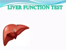
/
HISTOLOGY OF  L IVER LIVER HISTOLOGY OF  L IVER LIVER

HISTOLOGY OF L IVER LIVER - PowerPoint Presentation

pamela
pamela . @pamela
Follow
384 views
Uploaded On 2022-06-01

HISTOLOGY OF L IVER LIVER - PPT Presentation

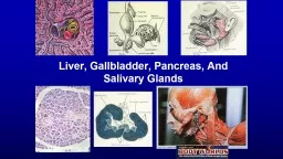
2 nd largest organ of the body Largest gland Location Receives blood from Portal vein Hepatic artery Drains to IVC FUNCTIONS OF LIVER Detoxification of metabolic waste products eg ID: 913472

portal amp liver hepatocytes amp portal hepatocytes liver bile cells bilirubin fibers vein hepatic synthesis medical application central blood

Share:

Link:

Embed:

Download Presentation from below link

Download Presentation The PPT/PDF document "HISTOLOGY OF L IVER LIVER" is the property of its rightful owner. Permission is granted to download and print the materials on this web site for personal, non-commercial use only, and to display it on your personal computer provided you do not modify the materials and that you retain all copyright notices contained in the materials. By downloading content from our website, you accept the terms of this agreement.


Presentation Transcript

Slide1

HISTOLOGY OF LIVER

Slide2

LIVER

2

nd

largest organ of the bodyLargest gland Location Receives blood from:Portal vein Hepatic artery Drains to:IVC

Slide3

FUNCTIONS OF LIVER

Detoxification of metabolic waste products e.g.

de-

amination of amino acids to produce ureaDestruction of spent RBC’s Synthesis & secretion of bileSynthesis of plasma proteins including albumin & clotting factorsSynthesis of plasma lipoproteins Metabolic functions e.g. glycogen synthesis, gluconeogenesis, storage of glycogen, some vitamins & lipids

Detoxification of various drugs & toxins e.g. alcohol

Slide4

STROMA OF LIVER

Connective tissue (

Glisson’s

) capsuleThick at hilumBlood vessels & ducts surrounded by CTReticular fibers surround & supports liver cells & sinosuoids

Slide5

HEPATIC LOBULE

Hepatocytes are

epithelial

cellsLobules –structural unit of liver (0.7×2mm)Portal spaces 3-6Lobule has:Portal area /portal triadCentral vein

Slide6

PORTAL TRIAD

A branch of:

Portal vein

Largest structureThin walled lined by endotheliumHepatic artery/ arterioleSmallest structureBile ductLined by cuboidal epitheliumLymphaticsDelicate, collapsedNervesLayer of hepatocytes immediately bordering the portal triad is

limiting plate

Slide7

HEPATIC LOBULE

Hepatocytes form interconnecting plates –branch & anastomose

Hepatocyte plates one cell thick

Directed from periphery to center forming sponge like pattern of liverSinosuoids

Slide8

HEPATIC SINUSOIDS

Dilated veins with INCOMPLETE basement membrane

Supported by reticular fibers

Cells:Endothelial cells- fenestrated without diaphragmKupffer cells (macrophages) present within

sinosuoids

Metabolize aged erythrocytes,

Secrete proteins

Digest bacteria

Slide9

PERISINOSOIDAL SPACE (of disse

)

A

subendothelial spacePresent between hepatocytes & sinosuoidsMicrovilli of hepatocytes project into the spaceFat storing (Ito) cells that stores Vit. A present hereSpace continuous with sinusoids

Space drains into

lymphatics

of portal

triad

Slide10

Slide11

MEDICAL APPLICATION

NORMAL ITO CELLS

:

Vitamin A rich lipid inclusionsUptake ,store , release of retenoidsSynthesis &Secrete ECM proteins and proteoglycansLocal immunityCHRONIC LIVER DISEASE – Ito cells acquire the features of myofibroblasts

Cells found close to damaged hepatocytes

Play a major role in fibrosis

Alcoholic liver disease

Slide12

BLOOD SUPPLY

Portal vein-70%

Portal venule- to portal space

Distributing venules-around periphery of lobule into sinosoidsCentral or centrolobular veinHepatic artery -30%Arterioles –lead to sinosoidsDirection of blood flow- from periphery to center of lobuleDifference in properties & functions of peripheral & centrolobular hepatocytes

Slide13

HEPATOCYTES

Polyhedral cells 20-30µm

Eosinophilic

Large nucleus /binucleateBile canaliculiGap junctions between hepatocytesHepatocyte surfaces related to:Perisinosoidal spaceAdjacent hepatocyteBile canaliculi

Slide14

HEPATOCYTES

Nucleus

Prominent nucleolus

Disperse chromatinPolyploidyCytoplasmGlycogen-lacey appearanceFat-spherical vacoulesRibosomes & RER- basophiliaSERMitochondria-eosinophilicLysosomesGolgi complexPeroxisomes

Slide15

FINE STRUCTURE OF HEPATOCYTES

Rough endoplasmic reticulum

– protein synthesis

Smooth endoplasmic reticulum – oxidation, methylationConjugation of bilirubin to gluronate – bilirubin glucoronideSynthesis of bile acidsGolgi apparatus – near nucleusFormation of lysomesSecretion of proteins, glycoproteins, lipoproteins into plasmaLysosomes –turnover & degradation of organelles Mitochondria Peroxisomes –Important for oxidation of excess fatty acid

Breakdown of hydrogen peroxide

Breakdown of purines to uric acid

Synthesis of cholestrol, bile acids & some lipids for myelin

Drug inactivation –by enzyme glucuronyle transferase

Slide16

Bile canaliculi

1-2µm diameter

Limited by plasma membrane of hepatocytes

Tight junctionsCanaliculi empty into bile ductules lined by cuboidal cells called cholangiocytesHering’s canalsBile ducts –portal spacesRight & left hepatic ductsDirection of bile flow – opposite

to that of blood

Slide17

MEDICAL APPLICATION

DISORDER OF PEROXISOME FUNCTION

Inherited disorder

Mutation of enzymes in peroxisomes.Adrenoleukodystrophy (X-ALD) results from failure to metabolize fatty acids properlyResult:Deterioration of myelin sheath of neurons

Slide18

RBC – heme – unconjugated bilirubin + plasma albumin - absrobed by hepatocytes – bilirubin released from albumin – bilirubin + glucoronic acid – bilirubin glucoronide (water soluble) – bile canaliculi

Conjugated bilirubin (bilirubin glucoronide) – urobilinogen – urine

Urobilinogen – stercobilinogen - intestines

Slide19

MEDICAL APPLICATION

When bilirubin or bilirubin glucuronide is not excreted properly various diseases characterized by JAUNDICE occur

Neonatal hyperbilirubinemia

– jaundice in newbornUnderdeveloped smooth ERTreatment – blue light exposureUnconjugated bilirubin transformed into water soluble photoisomer that can be excreted by kidneys

Slide20

BILE

Exocrine function of liver

Consists of:

Bile acids – emulsify fatsCholesterol Phospholipids Bilirubin Electrolytes

Slide21

MEDICAL APPLICATION

Gall stone formation (cholelithiasis)

Causes

Obstructs bile flowJaundice – presence of bilirubin in bloodRupture of tight junctions around bile canaliculi

Slide22

MEDICAL APPLICATION

Drugs inactivated by liver can induce increase in smooth ER in hepatocytes e.g. barbiturates

Barbiturates can increase synthesis of glucuronyltransferase synthesis

Use barbiturates in glucuronyltransferase deficiency

Slide23

LOBULATION

CLASSIC HEPATIC LOBULE

Central vein at center

Portal triads at peripheryArea near portal triad well supplied by oxygen & nutrientsArea near central vein not well supplied

Slide24

Slide25

LOBULATION (continued)

PORTAL LOBULE

Triangular area

Three central veinsPortal triad at centerConsists of tissues draining bile into bile duct

Slide26

LOBULATION (continued)

LIVER ACINUS

Diamond shaped area

Two portal triads Two closest central veinsBased on nature of blood supply & O2 gradientZONE 1- nearest arteriole- most O2 & nutrients, oxidative metabolismZONE III –near central vein – least O2 & nutrients, glycolysis, lipid formation,ischemic necrosisZONE II – intermediate range

Slide27

Slide28

COMPARISON

Slide29

Slide30

LIVER REGENERATION

Has strong capacity for regeration

Compensatory hyperplasia

Original mass of tissue restoredSurgical removal produces similar responseImportant in liver transplantLIVER STEM CELLS (oval cells)Present in initial epitetlium of bile ductules near portal areasCan give rise to hepatocytes & cholangiocytes

Slide31

MEDICAL APPLICATION

Continuous or repeated damage to liver cells

Increased amount of CT along with multiplication of hepatocytes

Nodules formed –central mass of disorganized hepatocytes surrounded by CTCirrhosis Causes of cirrhosisEthanol alters hepatic regeneration through unknown mechanism favoring development of cirrhosis

Slide32

Slide33

EXTRAHEPATIC BILIARY PASSAGES

Simple columnar epithelium

Subepithelial CT:

Elastic fiberLymphoid tissueMucous glands-tubuloalveolar BV, nervesCOMMON BILE DUCTSmooth muscle fibers-isolatedOblique & transverse fibers- near duodenum

Slide34

GALL BLADDER

Parts

Three layers

MUCOSAEpithelium- simple columnarStriated borderLamina propria has collagen fibers, muscle fibers, BV, tubuloacinar glands

Slide35

GALL BLADDER

Epithelium –absorptive cells

Microvilli

Intercellular spacesMitochondriaApical mucous granules

Slide36

GALL BLADDER

MUSCULAR LAYER

Interlacing smooth muscle fibers

Elastic & reticular fibers between musclesPerimuscular layer Adventitia / serosa

Slide37