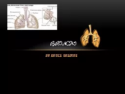
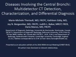
/
Diseases Involving the Central Bronchi: Diseases Involving the Central Bronchi:

Diseases Involving the Central Bronchi: - PowerPoint Presentation

lauren
lauren . @lauren
Follow
342 views
Uploaded On 2022-02-12

Diseases Involving the Central Bronchi: - PPT Presentation

Multidetector CT Detection Characterization and Differential Diagnosis MarieMichele Theriault MD FRCPC Kathleen Eddy MD Joy N Borgaonkar MD FRCPC Judith L Babar MRCP FRCR Daria Manos MD FRCPC ID: 908509

bronchial ddx fig airways ddx bronchial airways fig endobronchial lesion findings focal arrow wall diffuse tumor ancillary image multifocal

Share:

Link:

Embed:

Download Presentation from below link

Download Presentation The PPT/PDF document "Diseases Involving the Central Bronchi:" is the property of its rightful owner. Permission is granted to download and print the materials on this web site for personal, non-commercial use only, and to display it on your personal computer provided you do not modify the materials and that you retain all copyright notices contained in the materials. By downloading content from our website, you accept the terms of this agreement.


Presentation Transcript

Slide1

Diseases Involving the Central Bronchi: Multidetector CT Detection, Characterization, and Differential Diagnosis

Marie-Michele

Theriault

, MD, FRCPC, Kathleen Eddy, MD,

Joy N.

Borgaonkar

, MD, FRCPC, Judith L. Babar, MRCP, FRCR, Daria Manos, MD, FRCPC

Department of Diagnostic Radiology,

Université

de

Sherbrooke

,

Sherbrooke

, Canada (M.M.T.); Department of Diagnostic Radiology, Dalhousie University, 1276 S Park St, PO Box 9000, Halifax, NS, Canada B3H 2Y9 (K.E., J.N.B., D.M.); and Department of Radiology,

Addenbrooke’s

Hospital, Cambridge University, Cambridge, England (J.L.B.).

Address correspondence to D.M. (e-mail: daria.manos@nshealth.ca).

Presented as an education exhibit at the 2016 RSNA Annual Meeting (CH007-EB-X).

All authors have disclosed no relevant relationships.

Slide2

IntroductionOverview

Radiologists encounter a variety of bronchial diseases. An organized approach to lesions involving the central bronchi can help radiologists formulate an appropriate differential diagnosis. Information obtained from the radiologist is invaluable for directing further investigation and management.

Early disease can be difficult to detect and can be easily missed if the airways are not systematically evaluated as part of every computed tomographic (CT) examination.

This presentation provides a guide to the identification and differential diagnosis of central bronchial lesions, including wall thickening and endoluminal nodules. Diseases limited to small airways and characterized primarily by bronchiectasis are not included.

A

Fig. 1. Axial CT image (A) shows a solitary tiny endobronchial lesion (arrow) in the superior segment of the right lower lobe (RLL). The lesion was missed. CT image obtained 2 years later (B) demonstrates a larger lesion completely obstructing the bronchus (arrow).

B

Slide3

Introduction

Learning Objectives

Review the anatomy and function of the bronchial tree.

Discuss the ancillary CT signs that may improve detection and characterization of central bronchial lesions, including the double artery sign, the finger-in-glove sign, and

postobstructive bronchiectasis.Use a pattern-based approach to the differential diagnosis of central bronchial lesions, including focal abnormalities such as debris and tumors, as well as diffuse abnormalities, which usually result from inflammatory, infectious, or infiltrative processes.

Slide4

Anatomy

DDx

: Focal lesion

Conclusion

DDx: Diffuse or multifocal processAnatomy and function

Main bronchiUnlike the C-shaped cartilage in the trachea, cartilage rings encircle the entire lumen.

Normal main bronchial wall thickness: ≈1.3 mmWall thickness decreases as the caliber of the bronchial lumen decreases. For main, lobar, and segmental bronchi, wall thickness is normally about one-fifth (20%) of the internal bronchial diameter.

Calcification of cartilage is a normal finding in older patients (occurring more often in women than in men) and is often discontinuous.Lobar and segmental bronchi Cartilage found in incomplete rings

Ancillary CT findingsAssessment of the airwaysSubsegmental bronchi arise from the segmental bronchi and may further branch before dividing into bronchioles. Bronchioles, unlike bronchi, do not contain cartilage or glandular tissue in their walls. On CT images, it may be difficult to distinguish between subsegmental bronchi and bronchioles. The term small airways is used for airways with an internal diameter of 2 mm or less and a normal wall thickness less than 0.5 mm. Small-airways disease generally refers to any condition affecting the bronchioles. Small airways disease is not included in this review.

Fig. 2. Anatomy of the main airways with C-shaped cartilaginous rings in the trachea and circumferential rings in the main bronchi.

Subsegmental

bronchi and bronchioles

Slide5

In addition to facilitating gas exchange, the bronchial tree has defense functions.

The respiratory epithelium protects the respiratory system in three ways.

Physical barrier

: protecting the body against inhaled particles

Active barrier: removal of inhaled particles through the mucociliary system. Particles are trapped by mucus and then ejected through the synchronized motion of tiny cilia. Chemical defense: release of mediators involved in the control of inflammatory responseFunctionDDx: Focal lesionConclusion

DDx

: Diffuse or multifocal process

Anatomy and function

Cartilage

Fig. 3. Photomicrograph shows the normal anatomy of the bronchial wall.

Respiratory epithelium

This defense can be compromised by

Obstruction

: the exit of the

mucociliary

system is blocked, and mucus builds up distal to the obstruction.

Ciliary

dysfunction

: either congenital (

eg

, primary ciliary dyskinesia), infectious, or environmental (

eg

, smoking)

Ineffective mucus

: abnormal mucus impedes clearance (

eg

, in cystic fibrosis).

Ancillary CT findings

Assessment of the airways

Serous and mucin-secreting glands

Smooth muscle

Slide6

Ancillary CT findings

Assessment of the airways

DDx

: Focal lesion

Conclusion

DDx

: Diffuse or multifocal process

Anatomy and function

Modalities in the Assessment of the Airways

For basic evaluation,

multidetector

CT acquisition is made at end-inspiration with thin-section axial and orthogonal

multiplanar

reformation images in coronal and sagittal planes.

Intravenous administration of contrast material can be useful to characterize some lesions and for assessing associated mediastinal or lung abnormalities.

Multidetector

CT

Expiratory CT images can be obtained to assess for

Postobstructive

air trapping (see slide 9)

Tracheobronchomalacia

: Anteroposterior luminal diameter reduction of 70% in expiration is suggestive and can be seen as an isolated process, associated with chronic inflammation (

eg

, chronic obstructive pulmonary disease [COPD], relapsing

polychondritis

) or advanced age.

Dynamic expiratory CT is more sensitive than static end-expiratory imaging. Expiratory images can be acquired with low-dose imaging techniques.

A

B

Fig. 4. Bronchomalacia. Inspiratory (A) and expiratory (B) CT images

show collapse of the central bronchi in expiration (arrows).

High-spatial frequency (bone) reconstruction is used to assess bronchial wall thickness. Use a window width of −450 HU and a window mean of 1000–1400 HU.

Mediastinal windows are used to assess tissue density (calcification, fat, mucus, soft tissue) of the bronchial wall and endoluminal tissue.

Slide7

Fig. 5. Endobronchial hamartoma (arrows). Axial CT (A), coronal CT (B), virtual

bronchoscopic

(C) images.

Ancillary CT findings

Assessment of the airwaysDDx: Focal lesionConclusion

DDx

: Diffuse or multifocal process

Anatomy and function

More Advanced Multidetector CT Reconstructions

Fig. 6. Volume-rendered CT reformation shows bronchial narrowing in granulomatosis with

polyangiitis (arrow).

C

A

B

Advanced two- and three-dimensional reconstructions may improve diagnostic confidence and can provide more anatomically familiar information to the pulmonologist and surgeon.

CT techniques used in advanced visualization of the central airways include

Nonorthogonal

multiplanar

reformation (

eg

, coronal or sagittal oblique

multiplanar

reformation)

Maximum or minimum intensity projection

Curved planar reconstructions

Volume-rendered CT reformation

External (depicts external surface of airways and lungs)

Internal (virtual bronchoscopy)

Fig. 7. Minimum intensity projection

coronal image (10-mm-thick section) shows normal central airways.

Slide8

Fig. 8. Endobronchial hamartoma (arrow). Bronchoscopic images before (A) and after (B) treatment. Note the tip of the bronchoscope (*).

DDx

: Focal lesion

Conclusion

DDx: Diffuse or multifocal processAnatomy and function

*

Other Modalities

Bronchoscopy

Allows dynamic functional evaluation

Extraluminal

extent of disease not evaluated

Limited evaluation of peripheral lesions

Biopsy or washings possible

Treatment (laser) possible

Fig. 10. Endobronchial US image demonstrates a lower right

paratracheal

node just deep to the bronchial wall.

Ancillary CT findings

Assessment of the airways

Node

Bronchial wall

Endobronchial Ultrasonography (US)

Predominantly used for nodal staging in cancer

Radial endobronchial US can also help evaluate endobronchial lesions, including extraluminal extent.

Allows guided biopsy

Fluorine 18

Fluorodeoxyglucose

(FDG) Positron Emission Tomography (PET)/CT

Increased FDG uptake in malignancy

Helps distinguish

postobstructive

atelectasis and consolidation from tumor

A

*

Fig. 9. Axial fused FDG PET/CT image at the level of the right hilum (A) shows FDG uptake centrally within the RLL bronchus (blue arrow) corresponding to endobronchial squamous cell carcinoma. PET/CT image at the level of the lung bases (B) shows no uptake in the

opacified

RLL (*): this is

postobstructive

atelectasis. Yellow arrows outline the major fissure.

B

A

B

Slide9

DDx: Focal lesion

Conclusion

DDx

: Diffuse or multifocal process

Anatomy and function

Fig. 13. Coronal CT image shows focal tree-in-bud opacities (oval) confined to the posterior segment of the right upper lobe (RUL). Squamous cell carcinoma was found in the segmental bronchus at bronchoscopy.

Impaction and dilatation of bronchioles, often because of infection. However, when focal and isolated, check for a central obstructing lesion.

Tree-in-bud sign

When a

nondilated

bronchus is filled with mucus, the associated pulmonary artery will be accompanied by an apparently identical tubular structure. Look for a central obstructing lesion.


Double artery sign

Ancillary CT Findings

These seven CT signs are found in many different processes. However, all may occur secondary to an obstructing central bronchial process and should prompt the radiologist to review the supplying bronchus.

Ancillary CT findings

Assessment of the airways

Fig. 11. Axial paired inspiratory (A) and low-dose expiratory (B) CT images show

hyperlucent

lung distal to a carcinoid tumor (arrow) in the anteromedial basal segmental bronchus of the left lower lobe (LLL).

Gas may be trapped in lung distal to an obstructing lesion with

hyperlucent

lung on expiratory images. Occasionally,

hyperlucent

lung is visible on inspiratory images when the window width is narrowed.


Air trapping

Fig. 12. Axial CT image shows mucus plugs (yellow arrows) in the RLL bronchi adjacent to similar-appearing arteries. Compare with normal appearance of LLL bronchi (blue arrows).

A

B

Slide10

DDx: Focal lesion

Conclusion

DDx

: Diffuse or multifocal process

Anatomy and function

Fig. 15. Coronal CT image shows marked volume loss (oval) distal to a squamous cell carcinoma (arrow) in the LLL bronchus. Note elevation of the hemidiaphragm.

Atelectasis distal to an obstructing lesion


Postobstructive atelectais

Fig. 14. Axial CT image shows postobstructive consolidation (pneumonia in circle) in a 25-year-old male patient with a carcinoid tumor (arrow) obstructing the right main bronchus.

Consolidation distal to an obstructing lesion manifesting as alveolar opacities 

Postobstructive consolidation

Fig. 16. Sagittal oblique CT image shows bronchiectasis (circle) distal to an endobronchial carcinoid tumor (arrow) in a 69-year-old male patient.

Chronic obstruction may cause distal bronchial wall inflammation, leading to bronchiectasis.

Postobstructive

bronchiectasis usually occurs as a result of a chronic process or a slow-growing tumor.


Postobstructive

bronchiectasis

Ancillary CT Findings

Ancillary CT findings

Assessment of the airways

Finger-in-glove sign

Fig. 17. Coronal CT image demonstrates the finger-in-glove sign (arrow) in a 66-year-old female asthmatic patient with allergic bronchopulmonary aspergillosis (ABPA).

Impaction and dilatation of central bronchi manifesting as branching tubular opacities

Slide11

Differential Diagnosis

DDx

: Focal lesion

Conclusion

DDx: Diffuse or multifocal process

Anatomy and function

Focal lesion

Diffuse or multifocal process

NeoplasticNonneoplastic

Malignant tumor

(90%)

Benign tumor

(10%)

Nonneoplastic

Inflammatory and infiltrative

Infection

Mucus*

Blood

Broncholith

Granulomatous lesions

Foreign body

Neoplastic

Endobronchial hamartoma*

Squamous cell papilloma

Endobronchial lipoma

Leiomyoma

Pulmonary pleomorphic adenoma

Lung cancer*

Adenoid cystic carcinoma

Carcinoid

Mucoepidermoid

carcinoma

Metastasis

Viral*

Bacterial*

Tuberculosis

Fungal

COPD*

Aspiration*

Sarcoidosis

Granulomatosis with

polyangiitis

Amyloidosis

Inflammatory bowel disease (IBD)

Tracheobronchopathia

osteochrondroplastica

Relapsing

polychondritis

Mucopolysaccharidosis

Lymphoma

Tracheobronchial papillomatosis

Metastases*

Ancillary CT findings

Assessment of the airways

Most Common*

Slide12

Nonneoplastic

DDx

: Focal lesion

Conclusion

DDx

: Diffuse or multifocal process

Anatomy and function

Malignant tumor

Benign tumor

Common, especially in patients with chronic bronchial inflammation (eg, COPD, asthma, bronchiectasis) and in patients who have difficulty coughingCT findings

Fluid density (mediastinal window)

May see internal gasOften dependent within the bronchusCan also be seen peripheral to obstruction

BEWARE: Solitary plug may be an early sign of endobronchial neoplasm (Fig. 28)

.

A benign cause of persistent focal bronchial plugs includes bronchial atresia. The mucus plug (

mucocele

) forms peripheral to the atretic segment because of inability to clear secretions. The peripheral lung is usually

hyperlucent

because of air trapping.

Ancillary CT findings

Assessment of the airways

Mucus plug

Fig. 19. Bronchial

atresia

. Large branching endobronchial mucus plug (arrow) in the posterior segment of the RUL seen on coronal CT image (A). (B) Coronal minimum intensity projection (30 mm) in the same patient highlights the associated segmental air trapping.

B

A

Fig. 18. Mucus plug (arrow) in the posterior basal segmental LLL bronchus on paired axial enhanced CT images in lung (A) and mediastinal (B) windows. The endobronchial lesion had low opacity (−5 to 5 HU) consistent with fluid. Note the gas bubble.

A

B

Slide13

Nonneoplastic

DDx

: Focal lesion

References

DDx

: Diffuse or multifocal process

Anatomy and function

Malignant tumor

Benign tumor

Indistinguishable from other noncalcified

endobronchial material at CTUsually occurs in the setting of hemoptysis

May be aspirated from an upper tract source

Ancillary CT findings

Assessment of the airways

Fig. 20. Axial CT image shows a blood clot (yellow arrow) in the right intermediate bronchus in a 67-year-old man with pulmonary hemorrhage.

Endobronchial blood

Broncholith

Usually caused by

Extrusion of an adjacent calcified lymph node into a bronchus, usually in the setting of prior tuberculosis or histoplasmosis infection OR

In situ calcification of foreign material

Fig. 21.

Broncholith

. Axial CT image shows a peripherally calcified opacity (arrow) in the anterior segmental bronchus of the RUL.

Slide14

Nonneoplastic

DDx

: Focal lesion

Conclusion

DDx

: Diffuse or multifocal process

Anatomy and function

Malignant tumor

Benign tumorForeign body aspiration is most common in the 2nd year of life and in the elderly.

Predisposing factors includeIntubation

Neurologic deficitFacial trauma

Dental procedure

Fig. 22. Axial CT image (A) shows a calcified foreign body (yellow arrow) in the left main stem bronchus. Chest radiograph (B) better demonstrates the cause—a tooth (green arrow).

Ancillary CT findings

Assessment of the airways

B

A

Fig. 23. Aspiration in an 83-year-old man. Coronal oblique (A) and axial (B) CT images show right middle lobe (RML) collapse (blue arrows), obstruction of a 1.5-cm length of the RML bronchus (yellow arrow), and bronchiectasis. Photomicrograph of the transbronchial biopsy (C) in the same patient reveals degenerating exogenous material compatible with either aspirated adipose tissue or vegetable material.

B

A

C

Foreign body

Slide15

DDx

: Focal lesion

Conclusion

DDx

: Diffuse or multifocal process

Anatomy and function

Malignant tumor

Benign tumor

Nonneoplastic

Most common benign lung tumor

A

minority are endobronchial.

Most common benign endobronchial tumor

CT findings

Macroscopic fat (−40 to −120 HU).

Differential diagnosis: Lipoma (lipoma much more rare)

Calcification (>200 HU)

Can be

popcornlike

(cartilaginous)

Differential diagnosis: Carcinoid

Combination of fat and calcification

Considered diagnostic

Hamartoma

Ancillary CT findings

Assessment of the airways

Fig. 24. Endobronchial hamartoma. Axial CT image demonstrates a partially calcified nodule (yellow arrow) within the superior

lingular

bronchus. Note the peripheral mucus plugging (blue arrows).

Fig. 25. Paired axial CT images in lung (A) and mediastinal (B) windows demonstrate a low-attenuating endobronchial lesion in the LLL bronchus (arrows). This was persistent at serial CT examinations and therefore not consistent with mucus plug.

Bronchcoscopic

and endobronchial biopsy findings confirmed an endobronchial hamartoma. (Case courtesy of Joao Ignacio, University of Ottawa.)

Slide16

DDx

: Focal lesion

Conclusion

DDx

: Diffuse or multifocal process

Anatomy and function

Malignant tumor

Benign tumor

Nonneoplastic

Lipoma

Endobronchial location very rare

Arises from adipose tissue in the submucosal layer of bronchial wall

Most commonly appears as a focal endobronchial nodule of fat attenuation

Note: Hamartoma may give an identical appearance and is much more common.

May be asymptomatic or may present with cough, increased sputum production, or other symptoms of obstruction

Fig. 26. Axial CT image of a bronchial lipoma. Focal smoothly

marginated

fat-opacity nodule (arrow) in the left main stem bronchus.

Ancillary CT findings

Assessment of the airways

Slide17

DDx

: Focal lesion

Conclusion

DDx

: Diffuse or multifocal processAnatomy and function

Malignant tumor

Benign tumor

Nonneoplastic

Other benign neoplasms

These entities are rare with specific diagnosis generally requiring tissue biopsy.

Squamous cell papilloma

Solitary equivalent of tracheal papillomatosis (see slide 36)

Pulmonary pleomorphic adenoma

Slowly growing tumor; involves central airways; has salivary gland features

Airway leiomyoma

Two-thirds in the trachea, one-third in the bronchi

Arises from smooth muscle of bronchial wall

Other benign tumors

Hemangioma

Fibroma

Neurogenic tumors

Inflammatory

myofibroblastic

tumor

(also known as pulmonary inflammatory

pseudotumor

, plasma cell granuloma,

xanthogranuloma

); rare overall, but common pediatric primary lung tumor

Ancillary CT findings

Assessment of the airways

Fig. 27. Schwannoma in the RLL bronchus. Axial CT image (A) shows soft tissue at the origin of the RLL bronchus (yellow arrow). This could be misinterpreted as a lymph node. Coronal CT image (B) more clearly shows the endobronchial nature. Note the associated peripheral mucous plugging (blue arrows). The CT features are not specific, and

bronchoscopic

biopsy was required to make the diagnosis.

A

B

Slide18

DDx

: Focal lesion

Conclusion

DDx

: Diffuse or multifocal processAnatomy and function

Malignant tumor

Benign tumor

Non neoplastic

Lung cancer

Strongly associated with cigarette smoking

Squamous cell carcinoma is the most common subtype to affect the central bronchi.

When confined to bronchial tree, can be difficult to appreciate

Look for ancillary signs, including focal peripheral

postobstructive

tree-in-bud,

mucus plug, atelectasis, and consolidation.

Ancillary CT findings

Assessment of the airways

Fig. 29. Endobronchial squamous cell carcinoma. Axial CT image (A) shows a nodule in the LLL bronchus (yellow arrow). Coronal CT image (B) shows the same lesion (yellow arrow) causing

postobstructive

atelectasis (blue arrow).

B

A

Fig 28. Axial CT image shows endobronchial adenocarcinoma (arrow). Although mucus plugging is common in smokers, this lesion is isolated, in the upper lobe, and dilates the affected bronchi. These features are rare for the benign mucus plugging common in smokers and should raise concern for neoplasm.

Slide19

DDx

: Focal lesion

Conclusion

DDx

: Diffuse or multifocal processAnatomy and function

Benign tumor

Nonneoplastic

Fig. 31. Carcinoid. Axial

precontrast (A) and postcontrast (B) CT images demonstrate an enhancing lesion in the LLL bronchus (yellow arrows) with irregular calcification (blue arrow). Neuroendocrine neoplasmRanges from low-grade typical carcinoid to atypical carcinoid (aggressive course)

Not associated with smokingCT findings

80% arise centrally (main, lobar, segmental bronchi).May appear completely or partially endobronchial

Well-defined, round, slightly lobulatedOften strong enhancement due to

hypervasularity

Eccentric calcifications in 30%

A

Ancillary CT findings

Assessment of the airways

Fig. 30. Carcinoid. Axial CT image (A) shows endobronchial lesion in the anteromedial basal LLL bronchus (yellow arrow). Two years later, axial CT image (B) shows the tumor has enlarged and is now associated with

postobstructive

mucus plugging (blue arrow).

A

B

B

Carcinoid tumor

Malignant tumor

Slide20

DDx

: Focal lesion

Conclusion

DDx

: Diffuse or multifocal processAnatomy and function

Benign tumor

Nonneoplastic

Fig. 32. Carcinoid. Axial CT image (A) shows nodule (blue arrow) in the RML lobar bronchus. Note the associated complete atelectasis of the RML. (B) Photomicrograph from video-assisted

thoracoscopic surgical (VATS) wedge resection in the same patient demonstrates typical submucosal origin of tumor lifting the respiratory epithelium and protruding into the bronchial lumen.Ancillary CT findings

Assessment of the airways

Bronchial lumen

carcinoid

Cartilage in bronchial wall

Respiratory epithelium

B

A

Carcinoid tumor

Malignant tumor

Slide21

DDx

: Focal lesion

Conclusion

DDx

: Diffuse or multifocal process

Anatomy and function

Benign tumor

Nonneoplastic

Adenoid cystic carcinoma

Cancer of salivary gland origin

Tends to be central (trachea more common than main or lobar bronchi)

Tendency toward submucosal extension with circumferential and infiltrative growth

Ancillary CT findings

Assessment of the airways

Mucoepidermoid

carcinoma

Arises from minor salivary glands

Most involve lobar or segmental bronchus (trachea less common)

Intraluminal nodule adapting to the branching features of the airways

Fig. 33. Adenoid cystic carcinoma. Axial (A) and coronal (B) CT images in a 31-year-old man with a solitary endobronchial left main bronchus lesion (yellow arrow) extending through the bronchial wall.

Fig. 34. Mucoepidermoid carcinoma (arrows) in two patients. Curved coronal oblique CT image (A) in an 87-year-old woman shows a lesion in the left main bronchus with

postobstructive

atelectasis. Axial CT image (B) in a 28-year-old man shows a lesion in bronchus intermedius. Note that in both cases there is no evidence of extraluminal extension.

A

B

B

A

Malignant tumor

Slide22

DDx

: Focal lesion

Conclusion

DDx

: Diffuse or multifocal processAnatomy and function

Benign tumor

Nonneoplastic

Fig. 36. Endobronchial osteosarcoma metastasis. Photomicrograph demonstrates typical pattern of metastasis arising focally in the bronchial wall and then enlarging to fill the lumen. Note portions of the bronchial wall are preserved with intact epithelium

.Endobronchial tumor

Cartilage

Respiratory epithelium

Hematogenous

metastases to the bronchi are usually multifocal but can be solitary. (See also slide 37).

Ancillary CT findings

Assessment of the airways

Even in the presence of known

extrathoracic

malignancy, solitary metastases often require tissue confirmation to exclude primary endobronchial neoplasm.

Malignant tumor

Metastasis

Fig. 35. Axial CT images in two patients with solitary endobronchial metastasis: breast carcinoma (yellow arrow in A) and colorectal adenocarcinoma (orange arrow in B).

B

A

Slide23

Differential Diagnosis

DDx

: Focal lesion

Conclusion

DDx: Diffuse or multifocal process

Anatomy and function

Focal lesion

Diffuse or multifocal process

NeoplasticNonneoplastic

Malignant tumor

(90%)

Benign tumor

(10%)

Nonneoplastic

Inflammatory and infiltrative

Infection

Mucus*

Blood

Broncholith

Granulomatous lesions

Foreign body

Neoplastic

Endobronchial hamartoma*

Squamous cell papilloma

Endobronchial lipoma

Leiomyoma

Pulmonary pleomorphic adenoma

Lung cancer*

Adenoid cystic carcinoma

Carcinoid

Mucoepidermoid

carcinoma

Metastasis

Viral*

Bacterial*

Tuberculosis

Fungal

COPD*

Aspiration*

Sarcoidosis

Granulomatosis with

polyangiitis

Amyloidosis

IBD

Tracheobronchopathia

osteochrondroplastica

Relapsing

polychondritis

Mucopolysaccharidosis

Lymphoma

Tracheobronchial papillomatosis

Metastases*

Ancillary CT findings

Assessment of the airways

Most Common*

Slide24

DDx

: Focal lesion

Conclusion

DDx

: Diffuse or multifocal processAnatomy and function

Neoplastic

Inflammatory or infiltrative

Infection

Bacterial and Viral

Most community-acquired bronchitis is viral (85%–95%).

Smooth wall thickening and luminal narrowing are seen at CT.

Rhinoscleroma

is a unique bacterial infection uncommon in North America.

Chronic granulomatous infection affecting nose and larynx much more commonly than tracheobronchial tree

Caused by

Klebsiella

rhinoscleromatis

Causes wall thickening, nodules, and strictures

Fig. 37. Active bacterial bronchitis. Axial CT image (A) shows central bronchial wall thickening (arrow). Photomicrograph (B) from a VATS resection specimen in a different patient shows bronchial wall inflammation in active bronchitis.

cartilage

B

A

Inflammatory

exudate

(pus) in lumen

Thickened mucosa and

submucosa

Lymphoid follicle

Ancillary CT findings

Assessment of the airways

Slide25

DDx

: Focal lesion

Conclusion

DDx

: Diffuse or multifocal processAnatomy and function

Neoplastic

Inflammatory or infiltrative

Tuberculosis

Fig. 39. Active tuberculosis. Axial CT image shows RUL bronchial wall thickening (yellow arrow). The

subsegmental bronchial wall measures 1.8 mm and is almost as thick as the bronchial lumen. Note adjacent cavitary lung disease of tuberculosis (green arrow).

Central airway involvement mechanismSpread along peribronchial lymphatics or local extension from lymphadenitis

Less commonly, through spread of infected sputumTubercles in submucosa lead to ulceration, which may heal with or without stenosis.

CT findings of bronchial tuberculosis

Active disease

Irregular circumferential bronchial wall thickening with luminal narrowing

Lymphadenitis (may

cavitate

)

Associated

mediastinitis

possible; associated pulmonary disease typical

Fibrotic disease

Smooth concentric central airway narrowing

Minimal wall thickening

Fig. 38. Fibrotic tuberculosis 61 years after active infection. Coronal CT image shows narrowing of the left upper lobe bronchus (yellow arrow) with

postobstructive

atelectasis (green arrows). The

lingula

(*) is spared.

*

Ancillary CT findings

Assessment of the airways

Infection

Slide26

DDx

: Focal lesion

Conclusion

DDx

: Diffuse or multifocal processAnatomy and function

Neoplastic

Inflammatory or infiltrative

Fig. 41. ABPA. Unenhanced CT images in mediastinal (A) and lung (B) windows show

hyperattenuating mucus impaction (arrows) with associated complete atelectasis of the RUL (*). Fungal tracheobronchitisUsually in the setting of immunocompromiseMost common: Aspergillus and Candida speciesCT findings

Smooth or nodular tracheobronchial wall thickening with or without focal mucus and/or fungal plugs

APBAAffects patients with asthma or cystic fibrosis

Hypersensitivity reactionCT findingsBilateral central (segmental and subsegmental

) branching mucus plugs within

bronchiectatic

airways (finger-in-glove sign)

Hyperdense

mucus specific but not sensitive, seen in approximately 25% of patients

Fungal

Ancillary CT findings

Assessment of the airways

*

A

B

A

B

Fig. 40. Airway invasive fungal disease. Axial (A) and coronal (B) CT images in a 66-year-old immunosuppressed woman (liver transplant) with diffuse bronchial thickening and narrowing (yellow arrows) and

peribronchovascular

nodules (green arrows).

Infection

Slide27

DDx

: Focal lesion

Conclusion

DDx

: Diffuse or multifocal process

Anatomy and function

Neoplastic

Inflammatory or infiltrative

Infection

COPD and Asthma

Ancillary CT findings

Assessment of the airways

Fig. 43. Coronal CT image demonstrates bronchial diverticula (green arrows) in a smoker.

Diffuse airway wall thickening (AWT) is common in COPD and can affect both central bronchi and small airways.

AWT may occur independently of emphysema and reflects the chronic bronchitis COPD phenotype.

AWT in COPD is important to report and may reflect reversible disease.

Asthma typically affects smaller airways but can affect large bronchi.

Patients with asthma and COPD often have mucous plugs that tend to be basal, multifocal, and transient.

Tiny diverticula of the central bronchi are more common in smokers (from hyperplasia of the submucosal glands) but can also be seen in never smokers.

COPD can cause

tracheobronchomalacia

.

Fig. 42. Bronchial wall thickening (green arrows) on axial CT images in a patient who smokes one pack per day. Bronchial wall thickness is 2 mm or greater at the RUL anterior segmental bronchus (A), the LLL lobar and superior segmental bronchi (B), the medial and lateral segmental RML bronchi (C), and the LLL lateral and posterior segmental bronchi (D).

A

B

c

D

Slide28

DDx

: Focal lesion

Conclusion

DDx

: Diffuse or multifocal process

Anatomy and function

Neoplastic

Infection

Sarcoidosis

Multisystem inflammatory granulomatous disorder, lung involvement commonLarge airway stenosis underappreciated radiographically, often seen at bronchoscopy

Bronchial involvement more common than involvement of trachea or larynxIrregular or smooth narrowing caused by

Accumulation of granulomas in bronchial wallExtrinsic compression by lymph nodes

Distortion secondary to fibrotic parenchymal disease

Fig. 44. Bronchial wall thickening and luminal narrowing in sarcoid. Axial enhanced CT image (A) demonstrates severe narrowing of the LLL bronchus (yellow arrow) secondary to wall thickening. Coronal reformatted image viewed in bone windows (B) also shows the wall thickening of the LLL bronchus (yellow arrow) and reveals characteristic calcification of adjacent nodes (green arrow).

B

A

Ancillary CT findings

Assessment of the airways

Inflammatory or infiltrative

Slide29

DDx

: Focal lesion

Conclusion

DDx

: Diffuse or multifocal process

Anatomy and function

Neoplastic

Infection

Amyloidosis

Deposition of abnormal proteinaceous material (amyloid) in extracellular tissue

Tracheobronchial amyloidosis can be an isolated manifestation of amyloidosis or associated with systemic amyloidosis.

CT findings

Diffuse airway involvement is more common but can be focal (

amyloidoma

) or multifocal.

AWT, frequently calcified (no sparing of the posterior membranous trachea)

Luminal narrowing

Fig. 45. Bronchial amyloid. Coronal oblique CT images (A,B) show bronchial wall thickening with calcifications (yellow arrows) predominantly involving the large airways. Note peripheral atelectasis (green arrow) secondary to bronchial narrowing.

Fig. 46. Bronchial amyloid. Polarized light photomicrograph from a

transbronchial

biopsy shows characteristic apple green birefringence.

A

Ancillary CT findings

Assessment of the airways

B

Inflammatory or infiltrative

Slide30

DDx

: Focal lesion

Conclusion

DDx

: Diffuse or multifocal process

Anatomy and function

Neoplastic

Infection

Granulomatosis with Polyangiitis

Fig. 47. Bronchial involvement in granulomatosis with polyangiitis. (A,B) Axial CT images show bronchial narrowing and wall thickening (yellow arrows) and cavitating lung nodules and masses (green arrows). Virtual (C) and conventional (D) bronchoscopic images in the same patient show airway inflammation and narrowing (blue arrows).

Previously: Wegener granulomatosis

Idiopathic multisystem necrotizing granulomatous vasculitis

Subglottic involvement more common than bronchial involvement

CT findings of central airway involvement

Noncalcified

irregular circumferential wall thickening

Ulcerated wall thickening possible

Stenosis, obstruction

Endobronchial

pseudotumors

of inflammatory granulomatous tissue

Ancillary CT findings

Assessment of the airways

A

D

B

C

Inflammatory or infiltrative

Slide31

DDx

: Focal lesion

Conclusion

DDx

: Diffuse or multifocal process

Anatomy and function

Neoplastic

Infection

Inflammatory Bowel Disease

IBD only rarely affects the airway and is more common in ulcerative colitis than in Crohn disease.Manifestations include tracheobronchitis, bronchiectasis, and small airways disease.

CT findings of tracheobronchial involvementNoncalcified

tracheobronchial wall thickeningIrregular luminal narrowing and postobstructive findings

Fig. 48. Images in a 57-year-old man with ulcerative colitis. Axial (A) and coronal (B) CT images reveal circumferential thickening of the central airways, including the trachea and left and right main bronchi (yellow arrows). Coronal CT image with lung windowing (C) demonstrates

bibasal

bronchiectasis with bronchial wall thickening and mucus plugging (green arrows). Virtual CT

bronchoscopic

image (D) shows a featureless mucosal surface, with loss of the normal cartilaginous ring indentations.

Ancillary CT findings

Assessment of the airways

D

C

B

A

Inflammatory or infiltrative

Slide32

Tracheobronchopathia

Osteochondroplastica

Rare idiopathic process affecting the large airways of middle-aged adults, more often men than women

Multiple nodules, with or without calcification, arise from the cartilaginous rings and protrude into the airway lumen. Posterior trachea is spared as it contains no cartilage.

Involvement of the mid and distal trachea; main and lobar bronchi is typical.

DDx

: Focal lesion

ConclusionDDx: Diffuse or multifocal processAnatomy and function

Neoplastic

Infection

Fig. 49.

Tracheobronchopathia

osteochondroplastica

. Axial CT images (A,B) demonstrate multiple tiny nodules arising from the anterior and lateral walls of the trachea (yellow arrows). Axial CT images (C,D) in another patient demonstrate small endobronchial nodules (green arrows).

Ancillary CT findings

Assessment of the airways

B

D

A

C

Inflammatory or infiltrative

A

B

Slide33

DDx

: Focal lesion

Conclusion

DDx

: Diffuse or multifocal process

Anatomy and function

Neoplastic

Infection

Rare autoimmune disease of cartilageRecurrent episodes of cartilaginous inflammation results in destruction and fibrosisPeak prevalence 40–60 years, occurs in women more often than in men Airway involvement = poor prognosis

CT findingsCalcified thickening of cartilaginous portion of trachea and bronchi is specific.

Excessive tracheal and bronchial collapse in expiration (tracheobronchomalacia)More irregular and more thick than senile calcification of cartilage

Clinical diagnosis made when there are three or more of the following:Auricular chondritis

Nonerosive

seronegative polyarthritis

Nasal

chondritis

Ocular inflammation

Respiratory tract

chondritis

Audiovestibular

damage

Relapsing

Polychondritis

Fig. 50. Relapsing

polychondritis

. Axial (A) and coronal (B) CT images at the trachea show calcification and wall thickening (yellow arrows) that spare the posterior membranous (

noncartilaginous

) trachea (green arrow). Axial CT image (C) just below the carina shows similar findings at the main bronchi (yellow arrow). The wall is much thicker than in senile calcification. Technetium 99m–methylene diphosphonate bone scan (D) demonstrates exaggerated activity in cartilaginous structures, including the cartilaginous portion of the anterior ribs and larynx.

Ancillary CT findings

Assessment of the airways

B

D

A

C

Inflammatory or infiltrative

C

Slide34

Mucopolysaccharidoses are a group of disorders associated with malfunctioning of lysosomal enzymes leading to accumulation of glycosaminoglycans in tissues and progressive damage.

Hunter syndrome is one of the seven types of

mucopolysaccharidoses

(type 2) and is associated with airway involvement.

Most commonly affects distal trachea and central bronchiSevere airway stenosis possible and may be life threateningDDx: Focal lesionConclusion

DDx

: Diffuse or multifocal process

Anatomy and function

NeoplasticInfection

Mucopolysaccharidoses

Fig. 51. Hunter syndrome. Axial CT images viewed in lung windows (A) and mediastinal windows (B) show diffuse wall thickening of the right and left main bronchi (yellow arrow) resulting in severe luminal stenosis (green arrows). Sagittal CT image (C) demonstrates typical severe tracheal narrowing in the anteroposterior dimension (blue arrows).

C

A

B

Ancillary CT findings

Assessment of the airways

Inflammatory or infiltrative

Slide35

DDx: Focal lesion

Conclusion

DDx

: Diffuse or multifocal process

Anatomy and function

Neoplastic

Inflammatory or infiltrative

Infection

Lymphoma

Endobronchial involvement in lymphoma is rare.

More common with Hodgkin disease than with non-Hodgkin lymphoma

Typically occurs in the setting of advanced disease elsewhere

May manifest with symptoms of airway narrowing and obstruction

Endobronchial involvement may be nodular (solitary or multiple) or may manifest as diffuse wall thickening.

Fig. 52. Endobronchial involvement in non-Hodgkin lymphoma. Coronal CT image demonstrates a nodule in the right main stem bronchus (arrow). Nodal enlargement and lymphangitic

carcinomatosis

is also present. Multiple additional endobronchial nodules not shown.

Ancillary CT findings

Assessment of the airways

Slide36

DDx

: Focal lesion

Conclusion

DDx

: Diffuse or multifocal processAnatomy and function

Inflammatory or infiltrative

Infection

Tracheobronchial

Papillomatosis

Related to human papillomavirus

Multiple endobronchial nodules (squamous cell

papillomas

)

Most frequent in young people (transmission during vaginal birth or through sexual contact)

The larynx is most commonly affected, followed by the trachea and then the central bronchi. Occasionally there is spread to the lung parenchyma (nodules that usually

cavitate

).

Endobronchial obstruction by the nodules may result in:

Postobstructive

pneumonia, atelectasis, mucus plugging, and thin walled cysts

Typically treated with laser. May recur. May spread through airways. May undergo malignant transformation to squamous cell carcinoma.

Fig. 53. Tracheobronchial papillomatosis. Axial CT image (A) demonstrates bilateral endobronchial nodularity (yellow arrow) and

postobstructive

atelectasis (orange arrow). Note

postobstructive

mucus plugging (blue arrow). Another CT image (B) in the same patient demonstrates a cyst (green arrow).

B

A

Ancillary CT findings

Assessment of the airways

Neoplastic

Slide37

DDx: Focal lesion

Conclusion

DDx

: Diffuse or multifocal process

Anatomy and function

Inflammatory or infiltrative

Infection

Metastases

Fig. 54. Prostate metastases. Coronal minimum intensity projection shows endobronchial nodules (arrows) in the right main stem bronchus and the

interlobar

bronchus.

Transbronchial

biopsy was required to confirm the diagnosis.

Ancillary CT findings

Assessment of the airways

Hematogenous metastases to the bronchi

Most common primary malignancies are

Breast

*

Colorectal

*

Kidney

*

Stomach

Prostate

Melanoma

Thyroid

CT findings: Solitary or multiple nodules or eccentric wall thickening

BEWARE: Endobronchial renal cell carcinoma metastases, like the primary lesions, may grow slowly

.

Neoplastic

Slide38

DDx: Focal lesion

Conclusion

DDx

: Diffuse or multifocal process

Anatomy and functionConclusion

Both focal and diffuse central bronchial lesions may be easily overlooked.

CT provides excellent characterization of central bronchial lesions allowing evaluation of tissue density, extent of endobronchial involvement, assessment of the lung distal to the endobronchial abnormality, and evaluation of the surrounding tissues.

Ancillary CT signs improve detection of focal endobronchial lesions. Particularly when isolated and especially when persistent, ancillary signs should prompt careful evaluation of the supplying bronchus. Advanced reconstructions (eg, minimum intensity projection, three-dimensional rendering, virtual bronchoscopy) can improve diagnostic confidence, allow better anatomic assessment, and more clearly define airway compromise.

Functional impairment, symptoms, and uncertainty regarding diagnosis frequently lead to bronchoscopy for further evaluation of lesions demonstrated at CT. With the exception of transient processes such as mucus plugs and infection, most central bronchial lesions require tissue diagnosis. Management depends both on the pathologic finding of the abnormality and on the obstructive potential of the process.Ancillary CT findingsAssessment of the airways

Slide39

Bankier

AA, Fleischman D,

Mallek

R, et al. Bronchial wall thickness: appropriate window settings for thin section CT and radiologic-anatomic correlation. Radiology 2006;240:869–877.

Boisselle

PM, Lynch DA. Radiologic anatomy of the airways. In:

Boiselle

PM, Lynch DA, eds. CT of the airways. Totowa, NJ: Humana Press, 2008:25–48.

Criado

E, Sánchez M, Ramírez J

,

et al. Pulmonary sarcoidosis: typical and atypical manifestations at high-resolution CT with pathologic correlation. 

RadioGraphics

2010;30:1567–86.

Kang EY. Large airway diseases. J

Thorac

Imaging 2001;26:249–262.

Grenier

PA,

Beigelman-Aubry

C,

Brillet

PY.

Nonneoplastic

tracheal and bronchial

stenoses

Radiol

Clin

North Am 2009;47:243–260.

Hansell

DM,

Bankier

AA,

MacMahon

H, et al.

Fleischner

Society: glossary of terms for thoracic imaging. Radiology 2008;246:697–722.

Kim V, Desai P, Make BJ, et al. Airway wall thickness is increased in COPD patients with bronchodilator responsiveness.

Respir

Res 2014;15:84–93.

Lee KS,

Boiselle

PM. Update on

multidetector

computed tomography imaging of the airways. J

Thorac

Imaging.2010;25:112–124.

Litmanovich

D, O’Donnell CR, Banker AA, et al. Bronchial collapsibility at forced expiration in healthy volunteers: assessment with multidetector CT. Radiology 2010;257:560-567.

Marchioni

A,

Lasagni

A,

Busca

A, et al. Endobronchial metastasis: an epidemiologic and clinicopathologic study of 174 consecutive cases. Lung Cancer 2014;84:222–228.

Martinez F, Chung JH,

Digumarthy

SR, et al. Common and uncommon manifestations of Wegner granulomatosis at chest CT: radiologic-pathologic correlation.

RadioGraphics

2012;32:51–69.

Matsuoka S, Uchiyama K,

Sima

H, et al.

Bronchoarterial

ratio and bronchial wall thickness on high-resolution CT in asymptomatic subjects: correlation with age and smoking. Am J

Roentgenol

2003;180:513–518.

Miyara T, Oshiro Y, Yamashiro, et al. Bronchial diverticula detected by multidetector-row computed tomography; incidence and clinical features. J Thorac Imag 2011;26:204–208.

Mi-Young J, Gasser B, Afshin G, et al. Bronchial carcinoid tumor of the thorax spectrum of radiologic findings. RadioGraphics 2002;22:351–365.Montaudon M, P Desbarats P, Berger P, de Dietrich G, Marthan R, Laurent F. Assessment of bronchial wall thickness and lumen diameter in human adults using multi-detector computed tomography: comparison with theoretical models. J

Anat 2007;211:579–588.Munkholm M, Mortensen J. Mucociliary

clearance: pathophysiological aspects. Clin Pysiol

Funct Imaging 2014;34:171–177.Ngo AV, Walker CM, Chung JH, Takasugi JE

, et al. Tumors and tumorlike conditions of the large airways. Am J Roentgenol

 2013;201:301–313. 

Ngugyn

E. The gloved finger sign. Radiology 2003;227:453–454.

Nair

A, Godoy MC,

Holden

EL, et al.

Multidetector CT and postprocessing in planning and assisting in minimally invasive

bronchoscopic

airway interventions.

RadioGraphics

2012;32:E201–E232.

Palmucci

S,

Attina

G, Letizia M, et al. Imaging findings of

mucopolysaccharidoses

: a pictorial review. Insights Imaging 2013;4:443–59.

Park CM, Goo JM, Lee HJ, et al. Tumors in the tracheobronchial tree: CT and FDG PET features.

RadioGraphics

2009;29:55–71.

Prince JS, Duhamel DR, Levin DL, et al.

Nonneoplastic

lesions of the tracheobronchial wall: radiologic findings with

bronchoscopic

correlation.

RadioGraphics

2002;22:215–230.

Theriault MM, Manos D. Signs in cardiothoracic imaging: the double artery sign. J

Thorac

Imaging 2017;32:pW3.

Seo

JB, Song KS, Lee JS, et al.

Broncholithiasis

: review of the causes with radiologic-pathologic correlation.

RadioGraphics

 2002;2:199–213.

Solomonov

A, Zuckerman T,

Goralnik

L, et al. Non-Hodgkin′s lymphoma presenting as an endobronchial tumor: report of eight cases and literature review. Am J

Hematol

2008;83:416–419.

Yamamoto AK, Babar JL. Case 184: ulcerative tracheobronchitis. Radiology 2012;264:609–613.

References