/
Anomalies of the cardiac position: Anomalies of the cardiac position:

Anomalies of the cardiac position: - PowerPoint Presentation

leah
leah . @leah
Follow
343 views
Uploaded On 2022-06-15

Anomalies of the cardiac position: - PPT Presentation

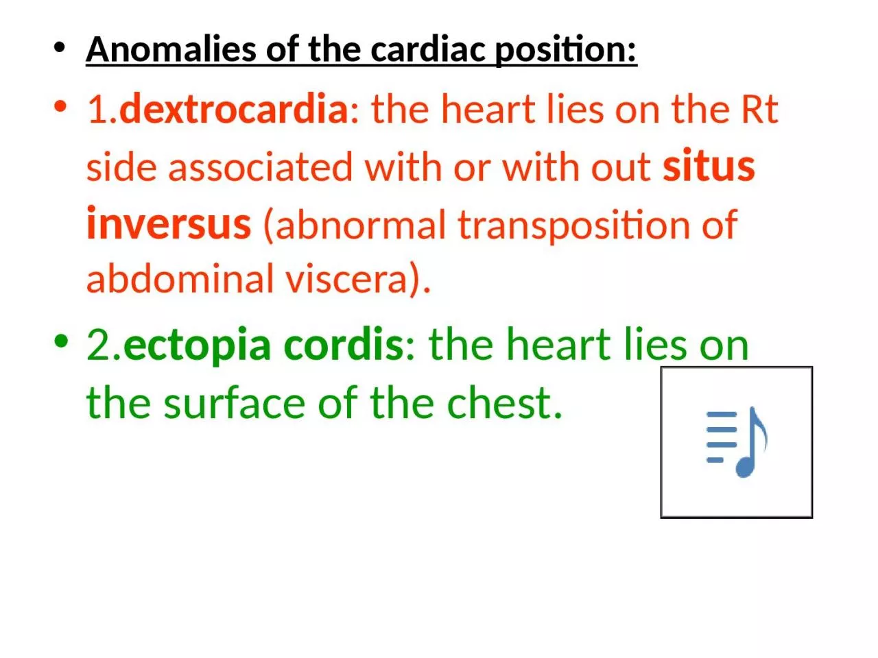
1 dextrocardia the heart lies on the Rt side associated with or with out situs inversus abnormal transposition of abdominal viscera 2 ectopia cordis the heart lies on the surface of the chest ID: 918949

artery aortic aorta arch aortic artery arch aorta forms arches carotid dorsal side internal disappears remnant arteries beginning common

Share:

Link:

Embed:

Download Presentation from below link

Download Presentation The PPT/PDF document "Anomalies of the cardiac position:" is the property of its rightful owner. Permission is granted to download and print the materials on this web site for personal, non-commercial use only, and to display it on your personal computer provided you do not modify the materials and that you retain all copyright notices contained in the materials. By downloading content from our website, you accept the terms of this agreement.


Presentation Transcript

Slide1

Anomalies of the cardiac position:1.dextrocardia: the heart lies on the Rt side associated with or with out situs inversus (abnormal transposition of abdominal viscera).2.ectopia cordis: the heart lies on the surface of the chest.

Slide2

Arterial system:Each of the pharyngeal arches has its own artery and cranial nerve. These arteries are called aortic arches that originate from the root of the aorta (the aortic sac), and end into the dorsal aorta. There are 6 pairs of aortic arches, in human embryo, the 5th arch often underdeveloped or

absent.

Slide3

Slide4

Slide5

The first aortic arch: largely disappears, remnant of it forms the maxillary artery.The second aortic arch: also disappears, remnant of it forms the hyoidal and stapedial arteries.The third aortic arch: forms the common carotid artery and the beginning of the of the internal carotid artery

.

Continuation of the internal carotid is derived from the dorsal aorta distal to the third arch.

Slide6

The forth aortic arch: forms the arch of the aorta on the left side between the Lt common carotid artery and the Lt 7th intersegmental artery (which is the primordium of the Lt subclavian artery). On the Rt side, the 4th aortic arch forms the beginning of the Rt subclavian artery

, the continuation of it is derived from the Rt dorsal aorta connecting the 4th, 5th, and 6th aortic arches and also by the 7th intersegmental artery.