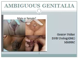
Gaurav Nahar DNB UrologyStd MMHRC INTRODUCTION Normal sexual differentiation Three steps establishment of chromosomal sex at fertilization46XX or 46XY development of undifferentiated gonads into testes or ovaries and ID: 525448
Download Presentation The PPT/PDF document "AMBIGUOUS GENITALIA" is the property of its rightful owner. Permission is granted to download and print the materials on this web site for personal, non-commercial use only, and to display it on your personal computer provided you do not modify the materials and that you retain all copyright notices contained in the materials. By downloading content from our website, you accept the terms of this agreement.
Slide1
AMBIGUOUS GENITALIA
Gaurav NaharDNB Urology(Std.)MMHRCSlide2
INTRODUCTION
Normal sexual differentiation: Three steps-establishment of chromosomal sex at fertilization(46,XX or 46,XY)
development of undifferentiated gonads into testes or ovaries, and
subsequent differentiation of internal ducts & external genitalia as a result of endocrine functions associated with the gonad present.Slide3
Genes determining testis or ovarian differentiation from
bipotential gonad.Slide4Slide5
Gonadal Stage of Differentiation
Urogenital ridge develops into adrenal cortex, gonads & kidneys.First 6 weeks : gonadal ridge, germ cells, internal ducts, & external genitalia are bipotential
in both.
Primordial germ cells
recognised
on posterior wall of secondary yolk sac- 3
rd
week.
Germ cells migrate to medial ventral aspect of
urogenital
ridge- 5
th
week.Slide6
SRY
(located on Chr Yp) initiates testicular organogenesis(via TDF).6-7th weeks : Germ cells transformation into
spermatogonia
&
oogonia
from differentiation of epithelial
gonadal
“testicular & ovarian cords”.
7-8 weeks:
Sertoli
cells
produce
MIS
(encoded by
chr
19p). MIS acts locally to produce
mullerian
regression.
8-9weeks: 2
nd
line of primordial cells –
Leydig
cells
differentiate & produce
testosterone
(T).Slide7
T causes
virilization of wolffian duct structures, urogenital sinus, and genital tubercle.Local source of T is imp. for wolffian
duct
development(
paracrine
effect);
doesnot
occur if T is
suppied
by peripheral circulation.
In tissues equipped with 5α-reductase (e.g., prostate,
urogenital
sinus, external genitalia), DHT is the active
androgen(endocrine effect).Slide8
In the
absence of SRY, ovarian organogenesis results.Estrogen synthesis in female embryo occurs just after 8 weeks of gestation.
Estrogen is
not required
for normal female
differntiation
.Slide9Slide10
Endocrine Effects on Phenotypic Development
Wolffian duct:R
equires
testosterone for development.
E
pididymis
, vas deferens, seminal vesicle with ejaculatory ducts.
M
üllerian
duct
:
D
oes
not require hormonal stimulation.
Requires absence of MIS.
Fallopian tubes, uterus, cervix, upper two-thirds of vagina.Slide11
Differentiation of Internal Genitalia(wolffian
and mullerian duct)and urogenital sinus in Male and FemaleSlide12
External Genitalia Diffentiation(8-16Wks)
Male (requires DHT):labioscrotal fold = scrotum
urethral fold / groove = urethra & penile shaft
genital tubercle =
glans
penis
Female (requires nil):
labioscrotal
fold = labia
majora
urethral fold = labia
minora
genital tubercle =
glans
clitorisSlide13
External Genitalia Differentiation in Male & FemaleSlide14
AMBIGUOUS GENITALIA
When the external genitalia do not have the typical anatomic appearance of normal male or female genitalia.Infants with ambiguous genitalia have genes of either a male or female, but with some additional characteristics of opposite sex.Incidence: 1 in 4000 infants.Slide15
Caused by various DSD.
Most cases present in newborn, not in adolescence.It is a social & medical emergency. It creates tremendous anxiety for the parents.
75% have life threatening salt wasting nephropathy
if unrecognized can cause vascular collapse & death(CAH).Slide16
Genitals may look either like a small penis or an enlarged clitoris.
Vagina may appear closed, resembling a scrotum, or scrotum may show some separation, resembling a vagina.Some infants have elements of both male and female genitals. The internal sex organs may not match the appearance of external genitalia. For example, a child who seems to have a penis may have ovaries, while a child with undescended testes may seem to have a vagina.Slide17
Micropenis
with hypospadias (arrowheads); scrotumis bifid with a midline cleft (arrow)Slide18Slide19
DISORDERS OF SEXUAL DIFFERENTIATION
(DSD or Intersex Disorders):Congenital conditions in which development of chromosomal, gonadal, or anatomic sex is atypical.Not all DSDs result in ambiguous external genitalia.Some DSD can have normal external genitalia (
eg
, Turner syndrome [45,XO] with female phenotype,
Klinefelter
syndrome [47,XXY] with male phenotype)Slide20
CLASSIFICATION OF DSD (Grumbach & Conte)Slide21
Female
pseudohermaphrodites (46,XX DSD): female genotype and two ovaries for gonads, but external genitalia show a variable degree of virilization.Male
pseudohermaphrodites
(46,XY DSD
): male genotype and two testes for gonads, but external genitalia show a variable degree of feminization.
True hermaphrodites (
ovotesticular
DSD
): both testicular and ovarian tissues in the gonads.
Pure
gonadal
dysgenesis
(PGD): both gonads are streak gonads (
ie
, dysfunctional gonads without germ cells).
Mixed
gonadal
dysgenesis
(MGD): a testis on one side and a streak gonad on the other.Slide22
CAUSES
46,XX DSD (Female Pseudohermaphrodite): includes I.CAH(21-α OH def.,11β
OH def., 3
β
HSD def.) II.
Virilising
maternal ovarian or adrenal tumor, III. Maternal ingestion of androgens &
progestins
, IV. Placental
aromatase
deficiency.
46,XY DSD
(Male
Pseudohermaphrodite
): I. Androgen receptor disorder with normal T- Partial androgen insensitivity II.
Inadequate testosterone production / defects in biosynthesis(17
β
HSD def., 3
β
HSD def., 17-
α
OH def.
a/w
17,20
Lyase
def.,
StAR
def.) III. 5
α
Reductase
def. IV.
Leydig
cell
aplasia
V. Drugs
True Hermaphrodite (
Ovotesticular
DSD
)
Partial/Mixed
Gonadal
Dysgenesis
Defects of testes maintenance
Abnormal
karyotypeSlide23
STEROID BIOSYNTHETIC PATHWAYSlide24
1- 46,XX DSD (female
pseudohermaphrodite) Gonads not palpable
Exposure
to excessive androgens in
utero
I-Congenital adrenal hyperplasia
(CAH)
The most common cause of ambiguous genitalia
60-70%
Results from enzymatic defect in the conversion of cholesterol to
cortisol
↑↑ ACTH ↑↑ adrenal androgens & steroid
precursors.
A
-21-
hydroxylase
deficiency
95% ↑
17-OH P
B
-11
β
-
hydroxylase
deficiency
↑11-deoxycortisol
+ ↑ BP
C-
3
β
-
hydroxysteroid
dehydrogenase
def.
↑
pregnelononeSlide25
I-CAH
A-21-
hydroxylase
deficiency
95%
↑
17-OH P
Autosomal
recessive.
↑↑adrenal androgens
musculinization
of
F
genitalia/
Mullerian
structures
unaffected.
Clitoral hypertrophy, labial fusion with
hyperpigmentation
, displacement of urethra, single
perineal
orifice (
urogenital
sinus)
Wolffian
derivatives
absent.
Androgen effect on the brain “Tom
boy”
behaviour
Puberty is delayed, menses is irregular & fertility is reduced in the salt wasting form & Pt not compliant with
Rx.Slide26
External genitalia of a patient with congenital
adrenal hyperplasia secondary to 21-hydroxylase deficiency,showing labioscrotal fusion and clitoromegalySlide27
A-
21- hydroxylase deficiency 1-Classical form
1: 10-15000
Cortisol
&
aldosterone
deficiency
Salt wasting &
virilisation
of varying degrees
2-Simple
virilising
form
Aldosterone
production is reduced but
not to
the point of salt wasting
3-Non-classic form
1:500
No ambiguous genitalia
Late onset premature
pubarche
&
advansed
bone age
menstrual disturbance &
hirsutism
in adult F Slide28
I-CAH
B- 11 β-hydroxylase deficiency
Autosomal
recessive
Musculinization
of F genitalia
cortisol
, ↑↑ adrenal androgens
↑↑11-deoxycortisol & 11-deoxycorticosterone ↑↑ BP, K
C-
3
β
-
hydroxysteroid
dehydrogenase
def.
V rare
Autosomal
recessive
↑↑
pregnelonone
cortisol
,
aldosterone
& androgens
Salt wasting
Undervirilised
M almost complete feminization
Partial def mildly
virilized
FSlide29
1-46XX, F Pseudohermaphrodites
II-Virilizing maternal ovarian/adrenal tumors Ovarian tumors
luteoma
of pregnancy,
arrhenoblastoma
,
hilar
cell tumor,
masculinizing
stromal
cell tumor &
krukenberg
tumor, lipoid cell tumor
Rare Adrenal tumors-
adrenocortical
carcinoma, adenoma.
III-Ingestion of androgens
V.
rare
progestogens
eg.19-nortestosterone,
danazol
, T,
norethinodrone
IV-Placental
aromatase
deficiency
defective conversion of androgens (T& ASD) to
estrogens(mutation of CYP19
aromatase
gene).Slide30
2. 46,XY DSD
(Male Pseudohermaphrodite)
Gonads are palpable
I-Androgen
receptor disorder with normal testosterone level/Partial androgen insensitivity
80%
(Complete androgen insensitivity “testicular feminization” unambiguous genitalia, F phenotype)
A wide spectrum of phenotypes
F with
clitoromegaly
-
--M
hypospadias
or
micropenis
.
↑↑ LH, T & estrogens
No
correlation
between the concentration of androgen receptors & the degree of
virilization
Slide31
2-46,XY DSD
(Male Pseudohermaphrodite)
II-Inadequate
testosterone production / defects in biosynthesis
1- 17
β
-
hydroxysteroid
dehydrogenase
def.
(testicular enzyme)
Rare
autosomal
recessive
F phenotype/ absent
mullerian
structures
Partial form ambiguous genitalia
Testis in inguinal canal or labia
Well differentiated
wolffian
duct structures
Virilization
at puberty with ↑↑ ASD ,
Normal
TSlide32
STEROID BIOSYNTHETIC PATHWAYSlide33
II
-Inadequate testosterone biosynthesis
Adrenal enzymes (V rare) CAH
2- 3
β
-
hydroxysteroid
dehydrogenase
def.
Salt wasting
F phenotype (complete form)/ambiguous genitalia (partial form)
3- 17
α
-
hydroxylase
def./ associate with 17-20-lyase def
Variable phenotype
Severe form
DX
at puberty with
water retention, ↑↑ BP &
hyperkalemia
.
4-
Congenital Lipoid
adrenal hyperplasia
Rare, caused by
StAR
enzyme deficiency.
Defect in the synthesis of 3 types of steroids
Severe salt wasting
F phenotype
Blind vagina without uterusSlide34
2-46,XY DSD
(Male Pseudohermaphrodite)
III-
5
α
-
reductase
def.
Wide range of phenotypes
All have differentiated
wolffian
ducts
Virilization
at puberty & male identity
↑↑ T : DHT ratio / N or ↑ M T level
Most are raised as females
IV-
Leydig
cell
hypoplasia
Impaired T production
Phenotype is usually F/ absent
mullerian
structures
V-
Drugs
Cyproterone
acetate block androgen receptors
Finasteride
Inhibit 5
α
-
reductase
Slide35
External genitalia of patient with 5α-reductase
deficiency; clitoromegaly with marked labioscrotal fusionand small vaginal introitusSlide36
3-True
hermaphroditism/Ovotesticular DSD
Very rare
90% present with ambiguous genitalia
2/3 raised as M
All have
urogenital
sinus & most cases have uterus
Chromosomal pattern 46,XX
75%
mosaic (XX/XY) > 46,XY
Has both ovarian & testicular tissue
1-Lateral
testis on one side & ovary on the other
2-Unilaterl
ovotestis
on one side & normal gonads on the other
3-Bilateral 2
ovotestis
Slide37
Infant with penile hypospadias
, chordee, andbilaterally undescended testes who was found to have truehermaphroditismSlide38
4-Partial/Mixed
gonadal dysgenesis
2
nd
most common cause of ambiguous genitalia in the newborn
45,XO/46,XY
M phenotype/ deficient
virilization
Testis on one side & streak gonads on the other
Testis is
dysgenetic
/non sperm producing
Unilat
.
unicornuate
uterus on the streak gonad side
Varying degrees of inadequate
musculinization
46XY
Bilateral
dysgenetic
testes
Uterus is present
Inadequate
virilization
&
cryptorchidism
Wide range of phenotypes
Sex of rearing FSlide39
5-Defects of testis maintenance /Bilateral vanishing testis
XYAbsent or rudimentary testisA spectrum of phenotypesSex of rearing M with T replacement (most of the time) 6-Abnormal
karyotype
Triploidy
69XXY
ambiguous genitalia 50%
Lethal
47XXY & 47XYY may present with ambiguous g.
Mosaic 46XX/46XY, 46XX/47XXY variable genitaliaSlide40
EVALUATION
HISTORY
Family
history
Ambiguous
genitalia, infertility or unexpected changes at puberty may suggest a genetically transmitted trait
-CAH ---
autosomal
recessive--occur in siblings
-Partial androgen insensitivity---X-linked
-XY
gonadal
dysgenesis
---sporadic
Consanguinity
↑↑the risk of
autosomal
recessive disorders
A
Hx
of neonatal death may suggest a missed diagnosis of CAH
Slide41
HISTORY
Pregnancy historyMaternal Hx of
virillization
-Placental
aromatase
def. allows fetal adrenal androgens to
virilize
both mother & fetus
-Maternal poorly controlled CAH
-Androgen secreting tumors
Medications
-
Progestins
-Androgens
-
Antiandrogens
Adolescent Pt
When ambiguity first noted?
Any pubertal signs? Slide42
PHYSICAL EXAMINATION
Overall assessment Abnormal facial appearance or other dysmorphic features suggesting a multiple malformation syndromeEvidence of salt wasting skin
turgor
, poor tone ,dehydration, low BP,
vomitting
, poor feeding
Hyper pigmentation of the skin due ↑↑ ACTH
Abdominal masses
In adolescent
evidence of
hirsutism
/
virilization
Tanner staging
Slide43
PHYSICAL EXAMINATION
Gonadal ExaminationPalpate labioscrotal tissue & inguinal canal for presence of gonadsNote No. of gonads, size, symmetry, position.Palpable gonads below the inguinal canal are almost always testicles
excluding
gonadal
female
eg
. CAH
Rectal exam
To palpate for presence or absence of the uterus Slide44
Examination
of external genitaliaPhallus
development may be in-between a penis & a clitoris
note size : length & breadth should be measured
(N mean stretched penile length 3.5 cm+_0.5)
the penis may appear bent buried or sunken
erectile tissue should be detected by palpation
Labioscrotal
folds
separated or fused fusion is an androgen effect
skin texture
rugosity
suggestes
exposure to androgens
color of the skin ↑↑ pigmentation may be evidence for CAHSlide45
Examination of external genitalia
Orificesshould be determined & recorded on a diagram
are there two openings ? or single
perineal
orifice (
urogenital
sinus)
urethral
meatus
on
glans
, shaft or perineum
vaginal
introitus
Slide46
Classification by Prader of
the various degrees of masculinization of the external genitalia in females with CAHSlide47
INVESTIGATIONS
Urgent U&E & glucoseHormonal assay17-OHP D3 & D6 (normally elevated in the 1
st
2 days of life)
N =82-400
ng
/dl
>400 CAH
200-300 ACTH
stim
test
Urinary 17-ketosteroids
N <= 1 mg/24 h
Testosterone, DHT,
androstenedione
D2 & 6
N T at birth 100ng/dl 50 in the 1
st
WK ↑T at
4-6Wk T at 6M barely detectable
↑ T:DHT 5
α
reductase
def
↑ ASD:T 17
ketosteroid
reductase
def.
Cortisol
, ACTH, DHEA Slide48
INVESTIGATIONS
Karyotyping several days/wksFISH (florescent in situ hybridization) for Y chromosome material PCR analysis of the SRY gene on the Y chromosome 1DRadiographic imaging
abdominal & pelvic U/S uterus &
gonadal
location
genitogram
single
perineal
orifice (
urogenital
sinus) detect the level of implantation of the vagina on the urethra
MRI
Cystoscopy
Rarely
laporoscopy
&
gonadal
biopsySlide49
Dx ALGORITHMSlide50Slide51
INTERPRETATION OF RESULTS
↑↑ 17-OHP + 46XX CAH due to 21-hydroxylase def.N 17-OHP + 46XX ACTH stim test check (11-deoxycorticosterone, 11-deoxycortisol, 17-hydroxypregnenolone ) to detect other adrenal enzymatic defects
N 17-OHP + N ACTH
stim
gonadal
dysgenesis
or true
hermaphroditism
46 XY + ↑↑T: DHT 5
α
-
reductase
def.
Basal T levels presence of functioning
leydig
cellsSlide52
INVESTIGATIONS OF PREPUBERTAL CHILDREN
Leydig cell function is active in the 1st 3M of life then quiescent till puberty
hCG
stimulation test
To determine the functional value of testicular tissue
hCG
1000 IU/D 3-5 D T
responce
< 3ng/ml
gonadal
dysgenesis
or inborn error of T biosynthesis (there will be also ↑↑ 17-OHP, DHEA, ASD)
T ↑↑ /N androgen insensitivity or 5
α
-
reductase
defSlide53
INVESTIGATIONS OF PREPUBERTAL CHILDREN
Under musculinized external genitalia + ↑↑ T
To test the adequacy of penile response to T
hCG
stim
may be prolonged 2-3
inj
/wk for 6 wks
T 25-100 mg IM Q 4 Wks 3M ↑↑ phallic length &diameter
An ↑↑ < 35mm is insufficient
Androgen receptor concentration < 300
fmol
/mg of DNA partial androgen insensitivity
Slide54
TREATMENT
It requires multidisciplinary team including: Ped
urologist
Gynecologist
Surgeon
Endocrinologist
Psychologist
Geneticist
Radiologist
Psychological support for the parents
Gender assignment
Medical treatment
Surgical treatmentSlide55
PSYCHOLOGICAL SUPPORT FOR THE PARENTS
Reassurance that a good outcome can be anticipatedProcess of investigation will take ? Around 3-4 Wk.To talk about their fears of future sexual identity & sexual orientation of their child
preferably
with psychologist or social
worker.Slide56
GENDER ASSIGNMENT
The choice must be the result of full discussion between parents & medical team.The decision is guided by Anatomical condition & functional abilities of the genitalia
Fertility potential (presence of F internal genital organs)
The etiology of the genital malformation
The f
amily’s cultural & religious background
Girls with CAH are fertile & must always be assigned a female
sex.Slide57
GENDER ASSIGNMENT
In cases which can not be fertile, gender assignment will depend on: The appearance of the external genitalia
The potential for unambiguous appearance
The
potential for normal sexual functioning
True
hermaphroditism
F since ovarian function may be preserved & may be fertile
Slide58
GENDER ASSIGNMENT
Great care should be taken in declaring a male sex considering:Potential for reconstructive surgeryProbability for pubertal
virilization
Response of the external genitalia to exogenous &
endog
. T
Pt with small phallus & poor response to androgens may be reared as F
Slide59
GENDER ASSIGNMENT
5α-reductase M sex assignement
pubertal
virilization
penile growth (subnormal) & male sexual identity
Inborn errors of T biosynthesis F effective M reconstructive surgery is highly unlikely
Complete Androgen Insensitivity F
Partial A I preferably F Slide60
MEDICAL TREATMENT
CAHMonitor electrolytes & glucoseHypoglycemia can appear in the first hours Serum electrolytes will become abnormal D 6-14Hydrocortisone 10-20 mg/m2/D PO
Fludrocortisone
acetate (
florinef
) 0.1 mg/D
Prenatal RX CAH
Mothers with family
Hx
Dexamethasone
is started at 5 wks
If confirmed DX of CAH in F fetus (by
chorion
villus
sampling/amniocentesis) continue Rx till term 90% N genitalia
Slide61
MEDICAL TREATMENT
Sex steroid replacement therapy at pubertyT enanthate 200-300 mg IM/ M:
M Pt with steroid enzyme def
M Pt with partial
gonadal
dysgenesis
, low
leydig
cell No,
truehermaphrodite
.
Estrogen
premarin
0.625 mg PO/D
for one year . Then cyclic estrogen progesterone or OCP (if there is uterus)
F Pt with enzyme def
3
β
-OH –steroid
dehydrogenase
& 17-hydroxylase
F 46 XY partial
gonadal
dysgenesis
, true
hermaphrodite,
M
pseudohermaphrodites
Slide62
SURGICAL TREATMENT
1-Genital surgery for FTiming of surgery …..controversial Clitoroplasty
3-6M of age for F with CAH
Vaginoplasty
delayed until the individual is ready to start sexual life
2-Genital surgery for M
More difficult & involves several stepsSlide63
SURGICAL TREATMENT
3-Gonadectomy to prevent cancerWhat are the facts?
XY Partial
gonadal
dysgenesis
Gonadolblastoma
55%
XY complete
gonadal
dysgenesis
Gonadoblastoma
30-66%
All
gonadoblastomas
progress to
seminoma
.? age
Seminoma
has a 95% 5-Y survival
Testicular enzyme defects, 5
α
-red, partial androgen insensitivity Risk of malignancy is negligible before adulthood
True
Hermaphrodite
risk is low in XX & higher
inXY
Slide64
SURGICAL TREATMENT
Pt raised as F gonadectomy must be performed in childhood
Pt raised as M
controversial
1-Gonadectomy is recommended by many physicians followed by HRT
2- Close medical surveillance
-Biopsy in childhood & excising gonads with
gonadoblastoma
-Repeated biopsy at puberty
-Follow up /palpation by experienced Dr every
6-12 MSlide65
SURGICAL TREATMENT
4-Gonadectomy to remove source of T in Pt raised as F to prevent progressive virilization especially at puberty
5-Laparoscopy
For evaluation of internal genitalia &
gonadal
biopsy
For excision of
mullerian
structures in pt raised as M or Pt raised as F with non communicating
mullerian
structures
For
gonadectomy
.Slide66
THANK YOU