/
RESEARCH Open Access An inducible CiliaGFP mouse model RESEARCH Open Access An inducible CiliaGFP mouse model

RESEARCH Open Access An inducible CiliaGFP mouse model - PDF document

liane-varnes
liane-varnes . @liane-varnes
Follow
427 views
Uploaded On 2015-05-15

RESEARCH Open Access An inducible CiliaGFP mouse model - PPT Presentation

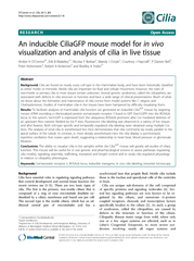
Motile cilia are important for fluid and cellular movement however the roles of nonmotile or primary cilia in most tissues remain unknown Several genetic syndromes called the ciliopathies are associated with defects in cilia structure or function an ID: 67058

Motile cilia are important

Share:

Link:

Embed:

Download Presentation from below link

Download Pdf The PPT/PDF document "RESEARCH Open Access An inducible CiliaG..." is the property of its rightful owner. Permission is granted to download and print the materials on this web site for personal, non-commercial use only, and to display it on your personal computer provided you do not modify the materials and that you retain all copyright notices contained in the materials. By downloading content from our website, you accept the terms of this agreement.


Presentation Transcript

RESEARCHOpenAccessAninducibleCiliaGFPmousemodelforinvivovisualizationandanalysisofciliainlivetissueAmberKO’Connor6† *Correspondence:byoder@uab.edu†Equalcontributors1DepartmentofCell,Development,andIntegrativeBiology,UniversityofAlabamaatBirminghamMedicalSchool,Birmingham,AL35294,USAFulllistofauthorinformationisavailableattheendofthearticle ©2013O'Connoretal.;licenseeBioMedCentralLtd.ThisisanOpenAccessarticledistributedunderthetermsoftheCreativeCommonsAttributionLicense(http://creativecommons.org/licenses/by/2.0),whichpermitsunrestricteduse,distribution,andreproductioninanymedium,providedtheoriginalworkisproperlycited.O’Connoretal.Cilia2013,2:8http://www.ciliajournal.com/content/2/1/8 resultinginperinatallethality,asseeninMeckel-Grüber Syndrome.Severalphenotypesarerelatedspecifically tomotileciliadefects,suchaschronicbronchiectasis, alteredleft-rightbodyaxisspecification,infertilityand hydrocephalus.Otherciliopathyphenotypesarelinked todefectsinciliarysignalingorsensoryrolesleading toblindness,anosmia,polydactyly,obesityorpolycystic kidneydisease[1-3]. Modelorganisms,suchas Caenorhabditiselegans and Chlamydomonasrheinhardtii, havebeenusedex- tensivelytostudyciliarybiology.Thesesystemshave theadvantagesofrapidgeneticscreensandbioche- micalanalysisofciliaproteins,butperhapsmostim- portantlytheciliainthesemodelsarereadilyvisible inlivingsamples.Assuch,theseorganismshavecon- tributedgreatlytoourgeneralunderstandingofcilia formationandmaintenance.However,understanding thephysiologicalrolesformammalianciliainmost tissuesandthemolecularmechanismsbehindcilio- pathyassociateddiseasesremainsachallengeinthe field.Unfortunately,todatetherehavebeennoindu- cible, invivo mammalianmodelstoeasilyvisualize cilia. Toaddressthislimitation,wegeneratedtheCilia GFP mousethatallowsdirectvisualizationofmammalian cilia invivo .CilialabelingisinducibleusingCrere- combinasefortissueorcelltypespecificanalysisor thelabelcanbeconstitutivelyexpressed.Weshow heretheutilityoftheCilia GFP mousefor invivo and exvivo detectionandanalysisofcilia.Importantly,we documenthereanovelfindingshowingsynchronized movementofprimaryciliainthekidneyofalive mousethatunderscorestheutilityofthismodel. Methods Constructgenerationandmouseengineering TogeneratetheSstr3::GFPROSA26targetingconstruct, Sstr3::GFPwasamplifiedfromanexpressionvector(akind giftfromDr.Pazour,UniversityofMassachusetts)with primersaddingattBsitestothe3  and5  ends,whichwas thenclonedintotheROSA26targetingvector(pRosa26- DEST,Addgene#21189)usingGatewaycloningtech- nology(LifeTechnologies,GrandIsland,NY,USA) (Figure1A)[5].Primers,forward:ggggacaagtttgtacaaaaaag- caggcttaaccatggccactgttacctatccc,andreverse:ggggacca- ctttgtacaagaaagctgggtattacttgtacagctcgtccatgcc.TheSstr3:: GFPconstructwaselectroporatedintoPrimogenixB6 (C57BL/6N-tac)embryonicstem(ES)cellsandG418re- sistantcolonieswereestablishedasdescribedpreviously ([6,7]).TheEScellcolonieswerescreenedbylongrange PCRusingforwardprimer:aaaagcagcagccatctgagatag,and reverse:cgagggacctaataacttcgtatagc,toyielda2.4kbprod- uct.Totestexpressionandciliarylocalizationpriorto generatingthemice,thetargetedEScellsweredifferenti- atedbyremovingleukemiainhibitingfactorandserum fromthemedia.Cellsweresubsequentlytransducedwitha CrelentivirustoremovetheNeocassetteandactivatethe expressionofSstr3::GFP(Figure1B).CorrectlytargetedES cellswereinjectedintoblastocyststogeneratemalechi- meras.Germlinepassagewasobtainedbycrossing chimericmalestoalbinoC57BL/6females.Genotyping primerareasfollow:acommonforwardwithinthe5  ho- mologyarm:ctcgtgatctgcaactccag;areverseinthe3  ho- mologyarm:gctgcataaaccccagatgactcc;areversenearthe 5  PKG-Neosite:gcgcatgctccagactgccttg;andareverseat the5  endofSstr3:gcggatgtgttccccagggtgg(Figure1A, arrows).Togethertheprimersyielda226bpwildtype Figure1 Sstr3::GFPROSA26targetedalleles. ( A )SchematicoftheSstr3::GFPconditional(Cilia GFP-OFF )andexpressing(Cilia GFP-ON )alleles.Before Cremediateddeletion(Cilia GFP-OFF ),theexpressionofSstr3::GFPisprecludedbythePGKpromotedNeocassetteflankedbyLoxPsites (bluetriangles).AfterCremediatedrecombination(Cilia GFP-ON )attheLoxPsitestheNeocassetteisdeletedallowingtheexpressionofSstr3::GFP. Spliceacceptor(SA),bluetrianglesrepresentLoxPsites,PKG-Neo:Phosphoglyceratekinasepromoterdrivinganeomycinresistancegene followedbythreepolyAtailsignals(3XpA,black).TheremainingattBsitesafterGatewayrecombinationarerepresentedbymaroonboxes flankingthecDNAofSstr3::GFP.( B )Toconfirmexpressionandciliarylocalization,embryonicstem(ES)cellswithcorrectlytargetedSstr3::GFP weredifferentiatedandtransducedwithaCreexpressinglentivirustoinduceexpressionofSstr3::GFP.( C )Afourprimercombinationwasused forgenotypingbyPCRtoamplifythewildtype(226bp),Cilia GFP-OFF (317bp)andCilia GFP-ON (423bp)alleles.Lane1:100bpladder,lane2: Cilia GFP-OFF /Cilia GFP-OFF ,lane3:Cilia GFP-OFF /WT,andlane4:Cilia GFP-ON /WT.Primerbindingsitesrepresentedin( A )assmallblackarrows,sequences inMethods. O ’ Connor etal.Cilia 2013, 2 :8 Page2of14 http://www.ciliajournal.com/content/2/1/8 band,a317bpproductfromthetargetedallelecontainingthefloxed-stop-sequence(CiliaGFP-OFF),anda423bpbandthatisamplifiedafterexcisionofthefloxed-stopcassette(CiliaGFP-ON)(Figure1C).SystemicandcelltypespecificdeletionoftheNeogenewasaccomplishedusingAdeno-virusEIIaearlypromoterCre(Tg(EIIa-cre)C5379Lmgd,[8]hereaftercalledEIIaCre);ProopiomelanocortinCre(POMC,Tg(Pomc1-cre)1Gsb,[9]hereaftercalledPOMCCre),andthePancreaticandDuodenalHomeoboxCreER(Tg(Pdx1-cre/Esr1*)1Mga[10],hereaftercalledPdx1CreER).Germline,systemicexpressionoftheCiliaGFP-ONallelewasaccomplishedcrossingfemaleEIIaCre;Sstr3::GFPwithwildtypeC57BL/6mice.ThisstudywascarriedoutinaccordancewiththerecommendationsintheGuidefortheCareandUseofLaboratoryAnimalsbytheNationalInstitutesofHealth.Theprotocolsusedwereap-provedandconductedaccordingtotheUniversityofAlabamaatBirminghamIACUC.TamoxifeninjectionsToactivatetheCrerecombinaseinPdx1CreERanimals,micewereinjectedwith6mgoftamoxifeneveryotherdayoverafive-dayperiodandthepancreatawereharvestedthreedaysafterthefinalinjection.Tamoxifen(Sigma-Aldrich,St.Louis,MOT5648)wasdissolvedincornoilat20mg/ml,andbolusinjectionsof300wereadministeredintraperitoneally.RenaltubuleisolationMicewereanesthetizedandeuthanized,andwholekidneyswereisolatedandcutintoapproximately2mmpieces.Thetissuewasincubatedat37°Cinthreechangesofcollagenasebufferfor30-minuteintervalswithgentleagitationevery10minutes(Collagenasebuffer:10mg/mlCollagenaseTypeIV(Sigma#C5138),50U/mLDNAseI(Sigma#D5025),and0.1mg/mlSoybeanTrypsinInhibi-tor(LifeTechnologies,GrandIsland,NY#17075-029)inDMEM/F12).Aftereach30-minuteincubation,thesuper-natantwasremovedandreplacedwithfreshcollagenasebuffer.Thesupernatants,containingthetubules,weregentlypelletedat100RelativeCentrifugalForce(RCF)foroneminute,thetubuleswerewashedthreetimesinphos-phatebufferedsaline(PBS)containing3%bovineserumalbumin(BSA)andpooled.Thetubuleswerekeptoniceandimagedliveorwerefixedin4%paraformaldehyde(PFA)forfiveminutesfollowedbyblockingandprocess-ingforimmunofluorescenceanalysis.ExvivoliveimagingBrainsfromCiliaGFPEIIaCremicewereremovedafteranesthetizationanddecapitation.Theexvivobrainswerecutsagittallydownthemidlineandplacedcutsurfacedownoncoverglassinroomtemperaturesterilefiltered,artificialcerebro-spinalfluid(125mMNaCl,2.5mMKCl,1.25mMNaH,2mMCaCl,1mMMgCl,25mMNaHCO,25mMglucose,pH7.3).CiliawereimagedliveusingaHamamatsuC9100-50EM-CCDcamera(HamamatsuPhotonicsK.K.,HamamatsuCity,Japan)onaninvertedNikonTE2000-Umicroscopeequippedwitha60×PlanApochromatoil-immersionTIRFMobjective(numericalaperture(NA),1.49;NikonInstrumentsInc.,Melville,NY),andaPerkinElmerUltraview-ERS6FEspinningdiskconfocalmodulecon-trolledbyVolocity6.2software(PerkinElmer,Shelton,CT,USA).VisualizationofSstr3::GFPwasaccomplishedusingafluoresceinisothiocyanate(FITC)laserfilterset(ChromaTechnology,Rockingham,VT,USA);thelightsourcewasa200mW488nmargonlaser(MellesGriot,Carlsbad,CA,USA).Forimmunocytochemistry,weim-agedAlexa-594conjugatedantibodystainingusingarhodamine/TRITCfilterset(Chroma)and20mW568nmargonkryptonlaser(MellesGriot),Alexa-647conju-gatedantibodystainingusingaCy5filterset(QioptiqInc.,Fairport,NY)and15mW640nmdiodelaser(MellesGriot),Hoechststainingusinga4',6-diamidino-2-pheny-lindole(DAPI)filterset(Chroma)and15mW405nmdiodelaser(Qioptiq).InvivoanalysisofrenalciliaAgroupof8-to16-week-oldmicewereanesthetizedbyintraperitonealinjectionof2.5%tribromoethanol(SigmaT48402).Adorsalincisionwasmadejustros-traltotheiliaccrestandthekidneywasgentlyteasedoutthroughtheopening,makingsurenottodamagetherenalartery,veinorureter.Themousewasplaced,incisionsidedown,insideaheatedchambermaintainedat37°C(LiveCellStageTopIncubationSystem#05-11-0035,PathologyDevices,Inc.,Westminster,MD,USA)withthekidneypositionedonacoverslipbathedinPBS.Themicewerecontinuouslyadministeredisofluraneat1to2%(VetOneBoise,ID#13985-030-60)throughouttheprocedureandwereeuthanizedafterimaging.TissueisolationandsectioningFreshtissueswereisolatedandprocessedasdescribedin[11].Forimmunofluorescencedetectionofneuronalcilia,animalswereanesthetizedwith2.5%tribromo-ethanol(SigmaT48402),andtranscardiallyperfusedwithPBS,followedby4%PFA.Thebrainwasthenisolatedandprocessedinthesamemannerasothertissues.Tis-suesweredissectedintoPBS,fixedin4%PFAovernightat4°C,saturatedwith30%sucroseinPBS,orientedinoptimalcuttingtemperaturecompound(FisherScien-tific,Pittsburgh,PA#14-373-65),andfrozeninadryice-ethanolbath.Sections10to15Mthickwerecutandmountedontoslides,fixedagainin4%PFAforfiveminutesatroomtemperature(RT)andthenusedforimmunofluorescencestaining.Spermwerecollectedbyetal.CiliaPage3of14http://www.ciliajournal.com/content/2/1/8 removingepididymidesandincubatinginWhittens-HEPESbuffer(100NaCl,4.4KCl,1.2KH2PO4,1.2MgSO4,5.4glucose,0.8pyruvate,4.8lactate,20HEPESinmM)for10minutesandthencapacitatedbyincuba-tioninWhittens-HEPESsupplementedwith15mMNaHCO3and5mg/mlBSAfor1hour.ImmunofluorescenceanalysisForimmunofluorescencedetectionofcilia,stainingwasperformedasin[12].Briefly,cryosectionswereblockedinPBS+(1%BSA,1%NormalDonkeySerum,inPBS+0.1%Triton-X100)for30minutes.Primaryandsecondaryantibodiesweredilutedinblockingso-lutionandappliedtosectionsforonehouratRTorovernightat4°C.Afterincubation,theslideswerewashedinPBSthreetimesforfiveminuteseachbeforebeingstainedwithHoechst,andmountedwith1,4-diazabicyclo[2.2.2]octane(DABCO).Thefollowingprimaryanti-bodieswereused:acetylated-tubulin(SigmaT7451,1mg/mlinput)pre-conjugatedtoAlexaFluor647(LifeTechnologies#A-20186)andusedat1:2,000,AdenylylcyclaseIII(ACIII,SantaCruzBiotechnology,SantaCruz,CA,USA,#sc-11617)usedat1:500,andArl13b(agiftfromDr.TamaraCasparyatEmoryUniversity)usedat1:2,000.Thefollowingsecondaryantibodieswereused:anti-goatandanti-rabbitAlexaFluor-594eachusedat1:1,000.Forisolatedtubules,immediatelyuponisolationthetubuleswerefixedin4%PFAfor5minutesfollowedbyblockinginPBS+for30minutes.Tubuleswereincubatedwiththedirectconjugatedacetylatedtubulinantibodyfor30minutesatRTandwashedinPBSfor5minutes.NucleiwerestainedwithHoechst(Sigma#33258),andimagedinPBSor50%glyceroldropletsoncoverglass.ResultsanddiscussionCiliamicewereengineeredtoexpressthecilialocalizedsomatostatinreceptor3proteinfusedwithgreenfluorescentprotein(Sstr3::GFP).Sincethecon-structwastargetedintotheubiquitouslyexpressedROSA26locusitshouldpermitlabelingofciliainnearlyeverytissueorcelltypeinthebodydependingontheCrelineusedtodeletethefloxedNeomycincassette(Figure1).TheutilityoftheCiliawasassessedusingubiquitousandcelltype-specificCrelinesinfixedtissue,inliveexvivosamples,andinsituinthecontextoflivinganimals.InCrenega-tivecontrolanimals(CiliaGFP-OFF/WT;Cre-)noGFPlabelingwasevident(Additionalfile1:FigureS1A-C).However,itshouldbenotedthatthelevelofactiva-tionoftheROSA26locushasbeenreportedtode-creaseinpostnatalanimals,andtoshowminimalactivationinafewcelltypes,suchasastrocytes[13],whichmayimpacttheutilityofthismousemodelincertaincircumstances.ThebrainTheependymalcellsliningtheventriclesareimportantformovementofcerebralspinalfluidanddefectsintheirmotilityareassociatedwithhydrocephalus[14,15].Interestingly,theclinicalimportanceofprimaryciliainthebrainhasrecentlybecomeapparent,despiteage-nerallackofunderstandingoftheirfunction[1,16].Onneurons,disruptionoftheprimaryciliumorciliarypro-teinsrevealedthattheyhaveanimportantroleinregu-latingsatiationandobjectrecognitionthroughunknownmechanisms[16-18].Inaddition,ciliaareknowntobeimportantforsightandsmell[16,19-21]andrecentlyitwasdemonstratedthattheyregulatepathwaysinvolvedinadultneurogenesisandmigrationofnewbornneurons[22].However,theubiquitousnatureofprimaryciliaonmostneuronsinthecentralnervoussystem(CNS)wasunexpected[23].Whilethemostcommonmethodofciliavisualizationinmammalsreliesonantibodiesagainstpost-translationallymodifiedformsoftubulin,suchasacetylated-tubulin,orpolyglutamylatedtubu-lin,thesemethodsproveespeciallychallenginginthebrainsincetubulinthroughoutneuronalprocessesandcellbodiesarehighlypost-translationallymodified[24].Thishasmadevisualizingciliaonneuronsdifficultwith-outspecialfixationandperfusionconditions,thusthisnewcilialabelmousemodelwillsimplifyandstreamlinestudiesofneuronalcilia.TodeterminewhethertheCiliamousecouldbeutilizedforvisualizingneuronalciliainliveexvivoples,weanalyzedEIIaCre;CiliamicewithmosaicactivationoftheSstr3::GFP(Cilia)allele[8].Pri-maryciliawereeasilydetectedoncellsthroughoutthebrain.Forexample,primaryciliawerefoundinthehippocampus(Figure2A)andmotileciliaweredetectedontheependymalcellswithintheventricles(Figure2B).Onthechoroidplexusepithelialcells,ciliaareusuallyfoundinsmallgroupsorasingleciliumpercell(Figure2Cinset)[14].Time-lapseliveimagingwasperformedontheepen-dymalcellstoexplorethepotentialutilityoftheCiliamouseinciliamotilitystudies.Imageswerecapturedat15framespersecond,andclearanddistinctciliacouldbeseen(Additionalfile2,seeMethods).Alloftheseciliawerevisualizedinlivesamplesimmediatelyafterisola-tionwithlittleornopreparationandnocounterstaining(Figure2A-C,andAdditionalfile2).Inadditiontoliveimaging,theGFPlabelwaseasilyvisibleafterfixationandsubsequentimmunofluores-cencestainingwithmarkersusedtoconfirmthatSstr3::GFPlabelscilia.Intheliningofthelateralven-tricleofCiliaGFP-ONCiliaGFPmice,Sstr3::GFPandetal.CiliaPage4of14http://www.ciliajournal.com/content/2/1/8 acetylated  -tubulinco-localizewithinmanyofthe ependymalcellsandintheprimaryciliaofneighbor- ingcells(Figure2D).Interestingly,theprimarycilia labelingissomewhatbrighterthanitisonthemotile ependymalcilia;thisislikelyduetotheincreased numberofciliaresultingingreatermembranesurface areawithinwhichtheSstr3::GFPlabelcanbecome diluted.Thishasbeenobservedforthehedgehog pathwaymediator,Smoothened,incellsinducedto formmultipleprimarycilia[25].Similarprimarycilia stainingwasobservedinthepituitary(Figure2E);and inthehindbrainregion(Figure2F)wheretheGFP signalco-localizedwithacetylated  -tubulinandthe neuronalciliamarkerACIII ,respectively.Together theseimages,aswellasthoseinthelivingsamples, demonstratethattheexpressionlevelsinthebrainare sufficienttofacilitatestudiesofneuronalcilia.For example,thismodelwouldfacilitatetheanalysisof theprevalenceofcilia,differencesinciliamorphology, orassessmentoftheefficiencyofciliaablationinspe- cificbrainregions(forexample,afterCredeletionofa floxedciliaryallele).Also,thismodelwouldenhance Figure2 AnalysisofSstr3::GFPexpressioninthebrain. ( A-C )Imagesfrom exvivo wholemountbrains.Sstr3 ::GFPsignal(green)in( A )hippocampal primarycilia,( B )ependymalmotileciliaand( C )choroidplexuscilia.( D-F )Sstr3:GFPco-localizationwithknownciliarymarkers.( D )Motileciliaofthelateral ventricle,( E )pituitaryprimarycilia,and( F )hindbrainprimarycilia.Ciliawereimmunolabeled(red)witheitheracetylated  -tubulin(AcetTub)in D and E or adenylatecyclaseIII(ACIII).Nucleiwerestaine dwithHoechst(blue).Arrowsindicateciliathata redouble-labeledandarr owheadsindicatecili athatareGFP negativeinagreementwithmosaicEIIaCreactivity. O ’ Connor etal.Cilia 2013, 2 :8 Page5of14 http://www.ciliajournal.com/content/2/1/8 studiesrequiringlivetissues,suchasthemeasurement ofligandinducedtranslocationofproteinsintoand outofthecilium,inelectrophysiologytopatchcili- atedneuronsoreventheciliumitself,orinpharma- cologicalstudiesoffactorsregulatingcilialength dynamicsinthebrain. Thekidney Theformationofcystsinthekidneyisacommon pathologicalfeatureassociatedwithmultiplehuman ciliopathies,includingformsofpolycystickidneydisease causedbymutationsinpolycystin1andpolycystin2, whereciliaarepresent[1,16].Althoughthecausesare notyetknown,significantefforthasgoneintodeter- mininghowthedisruptionofciliafunctionresultsin cystogenesis.Undernormalconditions,therenalcilium isthoughttobeamechanosensor,whereindeflectionsof theciliaryaxonemebyfluidflowelicitsacytosolic calciumresponse[26]. Invitro cellculturestudieshave determinedthatthismechanosensoryresponseis impairedinciliamutants,aswellasinmutantslacking polycystin-1orpolycystin-2[27].Thesedataleadtoa modelthatcystsdevelopthroughlossofthe mechanosensorysignal;however,todate, invivo studies validatingthishypothesishavenotbeenperformed.To evaluatewhethertheCilia GFP mousewillbeusefulin visualizingciliainthekidneyandtoaddressclinically importantquestions,suchaswhetherornotflowin- ducesciliarydeflection,weanalyzedtheexpressionof Sstr3::GFPinEIIaCrekidneys(Figure3).Infixedsam- ples,acutelyisolatedtissueandliveanimals,ciliacould easilybedetectedmakingstudiesofcilialength,orienta- tion,motilityandanalysisofthewholetubule/kidney practical.However,itshouldbenotedthatweare overexpressingacilium-localizedreceptorthathasbeen showntoalterthelengthandmorphologyofthepri- maryciliuminsomecases[28]. Inkidneysections,inliveisolatedtubules,and invivo , GFP-labeledciliawerereadilyidentifiable(Figure3A-C). Againthelabelwasseenwithoutfixationorstainingand persistedthroughouthandlingandimaging.Inisolated tubules,manyciliaremainedattachedthroughisolation, fixation,stainingandimaging(Figure3D).However, someGFPlabeleddebriswasobservedintheisolated tubulesthatarelikelytobeciliaryfragmentsbrokenoff duringisolation.Thesefragmentswerenotobservedin Figure3 Sstr3::GFPlabeledciliainthekidney. Sstr3::GFPsignal(green)in( A )fixedcryosectionsofthekidney,( B )isolatedEIIaCre; Cilia GFP tubules,( C ) insitu intheproximaltubuleofananesthetizedmouse.Immunofluorescencestainingof( D )isolatedtubulesand( E )kidney sectionsandSstr3:GFPlocalization.Acetylated  -tubulin(red)labelingwasusedtovalidatecilialocalizationoftheSstr3::GFPfusionprotein.Nuclei arelabeledwithHoescht(blue).Arrowsindicateexamplesofciliathataredouble-labeled. O ’ Connor etal.Cilia 2013, 2 :8 Page6of14 http://www.ciliajournal.com/content/2/1/8 theimagingofintactkidneys.Again,thespecificityof ciliarylocalizationwasconfirmedintubulesandin sectionsusingacetylated  -tubulin(Figure3D,E). Next,using insitu imagingtechniquesweevaluated ciliainthekidneysofliveEIIaCre:Cilia GFP mice.Pri- maryciliacouldbeclearlyobservedwithintheproximal tubulesofthecortex(Figure3C).Inthelivemice,the ciliadidnotsimplyprotrudeintothelumenperpendicu- lartothewallofthetubule;insteadtheyallbendinthe samedirectionnearlyparalleltotheapicalsurfacealong thelengthofthetubule.Theseciliaremainbentinthis positionwithanoccasionalciliumreversingitsposition (Additionalfile3).Thedeflectionoftheciliumislikely duetothelargeamountofprimaryfiltratepassing throughtheproximaltubules.Interestingly,mostofthe ciliaappeartobebendingataregularandspecificpoint abovethebaseandwhenoscillatingciliaareobserved, theygenerallymoveinanarcofapproximately106° (Figure4C).Similarobservationshavebeenmadeusing invitro flowstudiesandhavebeenattributedtotheri- gidityofthemicrotubuleswithintheciliaryaxoneme [29].Modelingtheciliumasanelasticcantileveredcol- umnfixedatthebaseresultsinthesamebendingprofile whensubjectedtoflowinducedshearstress[30-33].Al- ternatively,thisbendmaybeattributabletoamolecular domainsuchasthetransitionzone,theinversincom- partment[34,35],orbeingembeddedwithintheciliary pocket[36]. Figure4 Analysisofciliarymovementinthekidney. ( A )Sstr3::GFP-expressingciliainkidneyproximaltubulesinalivemouse.Imageisthe averagefluorescenceover40sdisplayingthefullrangeofmotionofthecilia.( B )Threedimensionalconfocalrenderingofthesweepofasingle ciliumovertime.Thewallofthetubuleisindicatedbyadashedlineanddirectionoffluidflowbythearrow.( C )Graphicrepresentationofthe ciliumsweepin( B )measuringthetotalangletraveledandtheproportionoftimetheciliumspentineach10 th ofthetotalpathduringan oscillationcycle.Calculationsbasedon( D )and( E ).( D )FastFourierTransformanalysisofthemotionofciliashowingauniform4.58Hzoscillation. ( E )Line-scanofthefluorescenceintensityalongthepathsweptoutbythetipoftheciliumin( B ). O ’ Connor etal.Cilia 2013, 2 :8 Page7of14 http://www.ciliajournal.com/content/2/1/8 Anotherinterestingobservationmadein5outof11miceanalyzedwasthattheciliawithinanephronwouldoscillatebackandforthwithinthetubularlumen(Figure4A,BandAdditionalfile4).Thisoscillationwasrapid(4.58±0.2Hz,Figure4D)andcouldbecapturedusingrelativelyhighspeedimageacquisition(approximately26fps).Thesweepoftheciliumduringoscillationwasir-regular,whereduringeachoscillationtheciliumwouldspendthemajorityofthetimebentalongthetubulewallcomparedtoanyotherpointofitssweep(Figure4C,E).Theseoscillationsaremostlikelypassiveandnotaresultofmolecularmotors,suchasdynein,asthereisnoevidencethattheseprimaryciliahavethemachinerynecessaryforactivemotility[37].Inaddition,thefre-quencyofoscillationissimilartothatdocumentedformouseheartratesunderanesthesia[38].Also,thepresenceofthisoscillatorymotionwouldchangeinthesameanimalovertime,eitherappearingordisappearingduringthecourseoftheexperiment,whichsuggeststhatthemovementmaybeelicitedbythedepthofanesthesia,heartrate,strokevolume,bloodpressureandtheirimpactonglomerularfiltra-tion;however,wewereunabletosimultaneouslymeasuretheheartrateofmicewhileimaging.Fur-thermore,mostoftheciliainawholefieldmoveinunison,suggestingregulationatthelevelofthewholekidney,notattheleveloftheindividualcells/tubules/glomeruli.Additionalevidencesupportingapassivemechanismisthatthemovementoftheciliuminthetubulesstopsalmostimmediatelyupondeathandtheciliaextendnearlyperpendicularlyintothenephronlumen(Additionalfile5,N=2).Togetherthesedatasuggestthattubularflowisnotconstant.Regularperi-odicoscillationintheflowrateofglomerularfiltratehasbeendocumentedusingfluorescentdextran[39]alongwithobservationsofoscillationinproximaltubularpressure[40].Thispulsatileflowrateintheproximaltubulesprovidesamechanismfortheoscil-lationofciliathatweobserveandalsoexplainswhytheciliaspendalargeproportionofeachoscillatorysweepbentinthedownstreamposition.Analternateexplanationisthatthecilia,atleastintheproximaltubuleimagescapturedhere,arenotpassivebutactuallyexhibitmotilebehavior.Itshouldbenotedthatsomestudieshavefoundthatciliainthenodeofthegastrulationstagemouseembryohavea9+0structure(similartothekidney)andhavearotationalmotilitythatisdistinctfromthewaveformmotionofciliaonependymalortrachealcells[41,42].However,othersstudieshavereportedthatthenodehasasecondformofciliathathasa9+2arrange-mentanditisnotclearwhichformisactuallyrespon-siblefortherotationalbeating[43].Follow-upstudieswillbenecessarytoconclusivelydeterminethecauseoftheciliaryoscillationwhichcouldimpactourun-derstandingofciliopathydisorders,suchaspolycystickidneydisease.TheeyeLossofvisionisalsoassociatedwithmultipleciliopathies,suchasSenior-Løkensyndrome,LebersCongenitalAmaurosisandBardetBiedlsyndrome[19,44,45].Thisisduetodysfunctioninthestructureortraffickingattheconnectingcilium(CC),ahighlymodifiedprimaryciliumintherodandconephotoreceptorsoftheretina(Figure5D,diagram)[19].Defectsintrafficking,proteinturnover,ciliaryassemblyorthedistributionofthesigna-lingcomponentsrequiredforvisionareallassociatedwithretinaldegeneration[1,19].Duetothestereotypicanat-omyoftheretina,andtheexaggeratedciliarystructure(Figure5D),therodcellsintheretinaareausefulmodelofciliaryfunctionandtrafficking[19,46,47],thusen-dogenousciliarylabelingwouldbebeneficialforlongitu-dinalinvivostudies.TodetermineiftheCiliaGFPmousewouldbesufficienttoanalyzethephotoreceptorCC,weevaluatedtheretinasofEIIaCre;CiliaGFPmice.Interestingly,theganglioncellsoftheretinacontainedmanyciliatedcells(Figure5B)asdidmanyofthecellsintheanteriorregionoftheinnernuclearlayer(INL,Figure5C).GFPwasconcentratedintheCCofthephotoreceptorsbutisdetectableintheoutersegments(Figure5D,Additionalfile1:FigureS1F).Inaddition,rhodopsinstainingindicatedthattheCiliaGFPlabeldoesnotovertlyinterrupttraffickingofrhodopsinoraffectthehealthoftheoutersegment(Additionalfile1:FigureS1F).Finally,attemptingtolabeltheciliaintheretinawithciliarymarkers,suchasArl13bandacetylated-tubulin,frequentlyrequiresantigenretrievalandcanbechallengingusingstandardimmunofluorescenceprotocols(Figure5,acetylated-tubulininpurple).Thus,theCiliaGFPmousewillbeusefulforidentifyingthecon-nectingciliaintheretinaoflivemice,andinsampleswithoutrelyingonantibodystainingapproaches.SpatialandtemporalcontrolofexpressionHavingtheabilitytolabelciliaonaspecificcelltypeinvivowillfacilitatestudiestoaddresswhatrolesciliahaveonthesecellsindifferenttissues.Ourpreviousworkhasshownthatprimaryciliaonasubsetofneu-ronsinthehypothalamusthatcontaintheproopio-melanocortinpeptide(POMC)haveavitalroleinthefunctionoftheseneuronscontrollingfeedingbehaviorinthemouse[17,48].Lossofciliafromtheseneuronscauseshyperphagiaandobesity.TodemonstratethefeasibilityofusingtheCiliaGFPmousetoaidinthestudyofciliafunctionintheseneuronsandtodemonstrateexpressionrestrictedtoaspecificgroupofcells,wecrossedtheCiliamousewiththePOMCCreline.etal.CiliaPage8of14http://www.ciliajournal.com/content/2/1/8 TheanimalsfromthiscrossshouldexpressSstr3::GFP specificallyinthePOMCneuronswithinthearcuatenu- cleus(ARC)ofthehypothalamus(Figure6A).Inthese POMCCre;Cilia GFP mice,cilialabelingwasdetected withintheARCbutnotinotherregionsofthebrainlike thehippocampus(Figure6A-D,N=2).Stainingsections ofthehypothalamuswithACIIIconfirmedthatSstr3:: GFPlabelingwasspecifictoprimarycilia(Figure6D). WedidnotquantifytheefficiencyofCrerecombination inthePOMCneuronsbut,qualitatively,thedistribution ofneuronsthathadundergonerecombinationbasedon Sstr3::GFPexpressionappearedsimilartoorgreater thanthatseenusingthemT/mGCrereportermouse (Gt(ROSA)26Sor tm4(ACTB-tdTomato,-EGFP)Luo ). Inadditiontocell-typespecif iccontrolofSstr3::GFPex- pression,wecouldalsoinduceexpressionataspecifictime pointinthelifeofamouse.Todemonstratetemporalcon- troloftheCilia GFP allele,weusedthetamoxifenresponsive Figure5 Sstr3::GFPlocalizationwithintheretina. ( A-D )Sstr3::GFPlocalizationinthemouseretina.Sstr3::GFPsignal(green)seen ( A )throughouttheretina,( B )inciliaofganglioncellsintheganglionlayer,( C )inciliaofcells(bipolar,horizontal,amacrineormüllercells)ofthe innernuclearlayer(INL),and( D )intheconnectingcilium(CC)ofrodcells.Fororientation,arodcellisdepictedintheschematicontheright. ThesectioninAwasimmunolabeledwithantibodiestoacetylated  -tubulin(purple),nucleiarelabeledwithHoescht(blue).CC,Connecting cilium;NL,innernuclearlayer;ONL,outernuclearlayer;OS,outersegment.ArrowindicatesanexampleofSstr3::GFPcontainingconnecting cilium,anteriorisup. O ’ Connor etal.Cilia 2013, 2 :8 Page9of14 http://www.ciliajournal.com/content/2/1/8 Pdx1CreERline.Inadultmice,Pdx1CreERisexpressed inthe  -cellsofthepancreas.Weharvestedpancreata frommicethreedaysafteraseriesoftamoxifeninjections (seeMethods)andprocessedthemforimmunofluores- cence.AsshowninFigure6E,manyoftheciliaintheislets werelabeledwithSstr3::GFP.Primaryciliaarepresenton isletcellsandintheductsofthepancreasasreportedpre- viously[49,50],andinagreementwithknownPdx1Creex- pression,onlytheisletswerelabeledinthesemice (Figure6E).Specificitywasconfirmedwithacetylated  - tubulinstaining(Figure6E)andtheabsenceofSstr3::GFP intheductsaswellasinanimalslackingtheCretransgene animalswasconfirmed(Additionalfile1:FigureS1A). GenerationoftheconstitutivelyexpressedCilia GFP-ON allele TogeneratealinewithconstitutiveexpressionofSstr3:: GFP,weutilizedEIIaCrethathasahighfrequencyof germlineCreactivityinfemalestoremovethefloxedstop sequence.Intheoffspring,CrenegativeCilia GFP-ON mice wereidentifiedtoestablishtheline.Asobservedinthein- ducibleCilia GFP line,ciliawerereadilydetectedwiththe germlineCilia GFP-ON mice.InheterozygousCilia GFP-ON females,wedidnotobserveanyovertdeleteriouseffects ofectopicexpressionofSstr3::GFPwiththecaveatthatno in-depthbehavioralanalyseswereperformed;however, maleCilia GFP-ON micearesterile,evenwhencarryingone copyoftheCilia GFP-ON allele.Themorphologyofthetes- tesinCilia GFP micelooksnormal(Figure7A)andthe malemicedomate,asconfirmedbyvaginalplugs,and theyproducesperm.However,isolationofspermfromthe epididymisrevealedtheyareimmotile(Figure7Band Additionalfiles6and7).Thematurespermflagellaex- pressSstr3::GFP(Figure7A,B)whichseemstobeinter- feringwiththeirmotility.Weobservedthatasmallsubset ofspermhadSstr3::GFPonlylocalizingtothemid-piece (Figure7B),whichmaycorrespondtothesmallfraction ofmotilespermatozoaobservedintheCilia GFP samples (Additionalfile7).Wedidnotspecificallyisolatemotile spermatozoafromCilia GFP micetoconfirmSStr3::GFP Figure6 CelltypespecificlabelingofciliausingCremediatedactivationintheCilia GFP mouse. ( A )Controlsectionofthedentategyrusin aPOMCCre;Cilia GFP mouseshowinglackofgreenSstr3::GFPcontainingcilia.CiliaareidentifiedbyimmunofluorescenceanalysiswithACIII(red). ( B-D )Sstr3::GFPcilia(green)areseenintheareaofthearcuatenuclei(ARC)ofthehypothalamussurroundingthethirdventricle(III)inPOMC Cre;Cilia GFP mice.CiliaintheARCwereidentifiedbyimmunofluorescenceanalysiswithACIII(red)in( C and D ).ExamplesofduallabeledACIII (red)andSstr3::GFP(green)positiveciliaareindicatedbyarrows,anexampleofaciliumlabeledwithACIIIbutnotSstr3::GFPthatislikelyon non-POMCneuronsisindicatedbyanarrowhead.( E )Sstr3::GFPexpressioninciliaonpancreaticisletcellsfromaPdx1CreER;Cilia GFP mouseafter tamoxifeninduction.Ciliaarelabeledwith(E)acetylated  -tubulin(AcetTub)inred.ManyciliawerepositiveforbothSstr3::GFPandacetylated  -tubulin(arrows)whileotherciliadidnotpossessSstr3::GFPandarelikelynon-  -cells(arrowhead). O ’ Connor etal.Cilia 2013, 2 :8 Page10of14 http://www.ciliajournal.com/content/2/1/8 localization;however,thesespermfrequentlydisplayeda hairpinbendrightattheendofthemid-piece.Thismay indicatethatthetail,withoutSstr3::GFP,ismotilewhile theSstr3::GFPcontainingthemid-pieceisnot.Itappears thatspermatiawithSstr3::GFPlocalizedcompletely throughouttheirflagellahavenomotility,whichwould indicatethatSstr3::GFPitselfmaybeinterferingwiththe molecularmachinerynecessaryforspermmotility. Conclusions Herewehavedevelopedanewtoolfor invivo and exvivo detectionandvisualizationofmammaliancilia, theCilia GFP mouse.Wehavedemonstratedthatthe Cilia GFP mouseisfunctional,ciliaspecific,andthatspatial andtemporalcontrolofexpres sionispossible.Inanimals withoutCreorwithouttamoxifen,nociliarylabelwas detected(Additionalfile1:FigureS1A-C).Inaddition, therewereafewtissueswhereciliarylabelingwasnoteasily observable,suchasthetracheaandthemotileciliainthe oviduct(Additionalfile1:FigureS1D,E)ofCilia GFP-ON mice.TheciliaintheseregionscontainedGFPbutata similarlevelastherestofthecellmembrane.Thereason thatciliaintheseregionsoftheCilia GFP mousewerenot highlylabeledisunknownbutitcouldbearesultof Figure7 Sstr3::GFPlocalizationintestesandsperm. ( A )Sstr3::GFPlocalizationinthemousetestes.Sstr3::GFPsignal(green)canbeseenin thespermflagellainthecenteroftheseminiferoustubulesofCilia GFP-ON mice(upperpanels),whicharealsopositiveforacetylated  -tubulin (red).Sstr3::GFPsignalisabsentinwild-typemousetestes(lowerpanels).( B )Sstr3::GFPsignalislocalizedtotheflagellumofCilia GFP mousesperm butnosignalisdetectedinwild-typemousesperm.ThearrowpointstotheexampleofasmallsubsetofspermwhereSstr3::GFPappearedto berestrictedtoonlythemid-pieceoftheflagellum.NucleiarecounterstainedwithHoescht(blue). O ’ Connor etal.Cilia 2013, 2 :8 Page11of14 http://www.ciliajournal.com/content/2/1/8 theoviductandtrachealepithelialcellshavingmanymoreciliapercell(approximately150,Additionalfile1:FigureS1DandE)[51]thanependymalcells(ap-proximately15percell,Figure2B)[52]orchoroidplexuscells(approximately5percell,Figure2C).WhichmightresultinthedilutionoftheGFPsignalamonganincreasednumberofciliaashasbeenshownforSmoothenedinmulti-ciliatedcells[25].AnothercaveattousingtheCiliamouseisthatwhentheCiliaalleleisexpressedinthetestes,spermfromCiliamaleshaveimpairedmotilitylikelyduetothelocalizationofSstr3::GFPintheflagella.ThisdoesraisethepossibilitythatSstr3maybealteringthefunctionormotilityofciliainothertissuesaswell.Thisisanimportantfactortoconsidersincearecentstudyhasshownthatover-expressingGPCRslikeSstr3inciliacouldaffectthenormaldistributionofciliaryproteinsandciliamorphology[28].Asidefromtheseex-ceptions,thismousewillfacilitatethestudyofciliaintissueswherestainingandimaginghavebeendifficult,suchasthebrainandeye,andforstudiesthatrequireliveorinsituTherearemultipleusesforthismousemodel:forex-ample,mammalianmutagenesisandinvivopharmaco-logicalscreensforfactorsthataffectciliogenesis,cilialengthcontrolandciliaryproteintrafficking;toassessre-coveryofciliaryproteinsbyFRAPanalysis;toanalyzeregulationofciliamotility;ortoscreenforsuppressormu-tationsusingknownciliamutantsandassessingtheres-torationofciliogenesis.Further,theCiliaGFPmousecanbeusedforassessingcilialosswhenusedinconjunctionwithfloxedallelesofgenesrequiredforciliogenesis,sinceCreexpressionwouldsimultaneouslydeletethefloxedgenerequiredforciliogenesisandremovethefloxed-stopcas-settetoinduceSstr3::GFPexpression.Lastly,wehaveshowntheutilityofthismodelwithinsitudocumentationofciliarymotioninthetubulesofthekidney.Indeed,thisobservationindicatestheCiliaGFPmousewillbeusefulforinvivomechanosensorystudiesthatmayprovideimport-antinsightsintohowciliadysfunctioncontributestocystdevelopment.AdditionalfilesAdditionalfile1:(A-C)ControlsectionsshowingnodetectableSstr3::GFPsignalinCre-animals.NoSstr3::GFPsignal(green)isseenin(A)pancreaticislets(B)theretina(C)andkidneytubulesinCilia;Crenegativeanimals.Thepresenceofciliawasconfirmedwithacetylated-tubulin(redorpurple).(D)ArepresentativeimageofthetracheainamouseshowingthatSstr3::GFPlabelinginthemotileciliatuftsisfaint(acetylatedtubulin,purple).Thelaminapropria(LP)showsstrongautofluorescence,alsoseeninSstr3::GFPnegativemice.Thechondrocytes(Ch)ofthetracheashowciliarylabeling(arrow).(E)ImageoftheoviductshowingfaintSstr3::GFPlabelinginthemotileciliatufts(acetylatedtubulin,red).(F)ImageoftheretinashowingcolocalizationofSstr3::GFPexpressionandrhodopsinlabelingintherodcalls.ArrowspointtoconnectingciliawhereGFPexpressionisstrongest(outernuclearlayer,ONL).Additionalfile2:Exvivoliveimagingofependymalcilia(sameanimalrepresentedinFigure2B).Synchronizedwave-likemovementsoftheciliaareeasilyobserved.Imageswerecapturedandshownat11framespersecond.Additionalfile3:Insituvideomicroscopyofakidneytubulecapturedatahighframeratedisplayingnociliaryoscillation.highvolumeofglomerularfiltrationciliainthetubuleslayparalleltothesurfaceoftheepitheliumwithacommondeflectionpointlocatednearthebaseofthecilium.Occasionally,aciliumwithinthetubulewillreverseitsposition(shownat26fps).Additionalfile4:Insituvideomicroscopyofakidneytubulecapturedatahighframeratedisplayingciliaryoscillation.Ciliainthetubuleoscillatesynchronouslybackandforthwithinthetubulesinthefield(shownat26fps).Additionalfile5:Insituimagingofciliarymovementwithinatubulebeforeandafterdeath.Synchronizedmovementoftheciliumterminateswithdeathoftheanimal.Furthertheseciliaextendintothetubulelumensupportingapassiveratherthanmotordrivenmechanismofmovement(shownat11fps).Additionalfile6:Livespermcollectedfromawild-typemouse.Differentialinterferencecontrasttime-lapsemovie(10fps)showingmovementofwild-typemousesperm.Additionalfile7:LivespermcollectedfromaCiliaDifferentialinterferencecontrasttime-lapsemovie(10fps)showinglackofmovementorabnormalmovementofspermfromCiliaAdenylatecyclaseIII;AcetTub:Acetylated-tubulin;ARC:Arcuatenucleus;BSA:Bovineserumalbumin;CC:Connectingcilium;CNS:Centralnervoussystem;DABCO:1,4-diazabicyclo[2.2.2]octane;DAPI:4',6-diamidino-2-phenylindole;EIIa:AdenovirusearlytranscriptionregionIIDNAbindingproteinpromoter;EScells:Embryonicstemcells;FITC:Fluoresceinisothiocyanate;GFP:Greenfluorescentprotein;INL:Innernuclearlayer;PBS:Phosphate-bufferedserum;Pdx1:PancreaticandDuodenalHomeobox1;PFA:paraformaldehyde;POMC:Proopiomelanocortin;RCF:RelativeCentrifugalForce;ROSA26:Reverseorientedspliceacceptorneomycin26;RT:Roomtemperature;Sstr3:Somatostatinreceptor3.CompetinginterestsTheauthorshavenofinancialinteresttodeclare.Authors'contributionsAKO,EBM,NFBandBKYdesignedandperformedexperiments.AKO,EBM,NFBandBKYwrotethemanuscript.MJC,CJHandPDBperformedexperiments.AKOandPHcreatedthetargetingconstruct.RAKcreatedthemousemodel.Allauthorsreadandapprovedthefinalmanuscript.AcknowledgementsWewouldliketoacknowledgetheUABTransgenicAnimal/EmbryonicStemCellFacilityforassistancewithgeneratingtheCiliamouse,andKevinJ.VanderwallforprovidingtheillustrationFigure5D.AKOwassupportedbyT32AR047512.ThisworkwassupportedbyNIHR01DK065655toBKYandbytheUABHepatorenalFibrocysticKidneyDiseaseCoreCenter(UABRPKDCC,P30DK074038).AuthordetailsDepartmentofCell,Development,andIntegrativeBiology,UniversityofAlabamaatBirminghamMedicalSchool,Birmingham,AL35294,USA.DepartmentofCraniofacialBiology,MedicalUniversityofSouthCarolina,Charleston,SC29425,USA.RalphH.JohnsonVeteransAdministrationMedicalCenter,DepartmentofMedicine,DivisionofNephrology,MedicalUniversityofSouthCarolina,Charleston,SC29425,USA.TheRoslinInstitute,UniversityofEdinburgh,EasterBushCampus,MidlothianScotland,UK.DepartmentofGenetics,UniversityofAlabamaatBirminghamMedicalSchool,Birmingham,AL35294,USA.CenterforTranslationalScience,sNationalMedicalCenter,Washington,DC20010,USA.etal.CiliaPage12of14http://www.ciliajournal.com/content/2/1/8 3Received:29April2013Accepted:14June2013References1.OhEC,KatsanisN(2012)Ciliainvertebratedevelopmentanddisease.Development139:443–4482.PazourGJ,WitmanGB(2003)Thevertebrateprimaryciliumisasensoryorganelle.CurrOpinCellBiol15:105–1103.DavenportJR,YoderBK(2005)Anincredibledecadefortheprimarycilium:alookataonce-forgottenorganelle.AmJPhysiolRenalPhysiol289:F1159–F11694.BerbariNF,O'ConnorAK,HaycraftCJ,YoderBK(2009)Theprimaryciliumasacomplexsignalingcenter.CurrBiol19:R526–R5355.HohensteinP,SlightJ,OzdemirDD,BurnSF,BerryR,HastieND(2008)High-efficiencyRosa26knock-invectorconstructionforCre-regulatedoverexpressionandRNAi.Pathogenetics1:36.NagyA(2003)ManipulatingtheMouseEmbryo:aLaboratoryManual,3rdedition.ColdSpringHarborLaboratoryPress,ColdSpringHarbor,NY,p7647.O’ConnorAK,KestersonRA,YoderBK(2009)Generatingconditionalmutantstoanalyzeciliaryfunctions:theuseofCre-loxtechnologytodisruptciliainspecificorgans.MethodsCellBiol93:305–3308.LaksoM,PichelJG,GormanJR,SauerB,OkamotoY,LeeE,AltFW,WestphalH(1996)Efficientinvivomanipulationofmousegenomicsequencesatthezygotestage.ProcNatlAcadSciUSA93:5860–58659.XuAW,KaelinCB,TakedaK,AkiraS,SchwartzMW,BarshGS(2005)PI3Kintegratestheactionofinsulinandleptinonhypothalamicneurons.JClinInvest115:951–95810.ZhangH,FujitaniY,WrightCV,GannonM(2005)Efficientrecombinationinpancreaticisletsbyatamoxifen-inducibleCre-recombinase.Genesis42:210–21711.BerbariNF,JohnsonAD,LewisJS,AskwithCC,MykytynK(2008)IdentificationofciliarylocalizationsequenceswithinthethirdintracellularloopofGprotein-coupledreceptors.MolBiolCell19:1540–154712.CroyleMJ,LehmanJM,O'ConnorAK,WongSY,MalarkeyEB,IribarneD,DowdleWE,SchoebTR,VerneyZM,AtharM,MichaudEJ,ReiterJF,YoderBK(2011)Roleofepidermalprimaryciliainthehomeostasisofskinandhairfollicles.Development138:1675–168513.AnthonyTE,KleinC,FishellG,HeintzN(2004)Radialgliaserveasneuronalprogenitorsinallregionsofthecentralnervoussystem.Neuron41:881–89014.BanizsB,PikeMM,MillicanCL,FergusonWB,KomlosiP,SheetzJ,BellPD,SchwiebertEM,YoderBK(2005)Dysfunctionalcilialeadtoalteredependymaandchoroidplexusfunction,andresultintheformationofhydrocephalus.Development132:5329–533915.NakamuraY,SatoKY(1993)Roleofdisturbanceofependyamlciliaryfunctionindevelopmentofhydrocephalusinrats.ChildsNervSyst9:65–7116.SharmaN,BerbariNF,YoderBK(2008)Ciliarydysfunctionindevelopmentalabnormalitiesanddiseases.CurrTopDevBiol85:371–42717.DavenportJR,WattsAJ,RoperVC,CroyleMJ,vanGroenT,WyssJM,NagyTR,KestersonRA,YoderBK(2007)Disruptionofintraflagellartransportinadultmiceleadstoobesityandslow-onsetcystickidneydisease.CurrBiol17:1586–159418.DohertyD(2009)Joubertsyndrome:insightsintobraindevelopment,ciliumbiology,andcomplexdisease.SeminPediatrNeurol16:143–15419.PazourGJ,BakerSA,DeaneJA,ColeDG,DickertBL,RosenbaumJL,WitmanGB,BesharseJC(2002)Theintraflagellartransportprotein,IFT88,isessentialforvertebratephotoreceptorassemblyandmaintenance.JCellBiol157:103–11320.KulagaHM,LeitchCC,EichersER,BadanoJL,LesemannA,HoskinsBE,LupskiJR,BealesPL,ReedRR,KatsanisN(2004)LossofBBSproteinscausesanosmiainhumansanddefectsinolfactoryciliastructureandfunctioninthemouse.NatGenet36:994–99821.McIntyreJC,DavisEE,JoinerA,WilliamsCL,TsaiIC,JenkinsPM,McEwenDP,ZhangL,EscobadoJ,ThomasS,SzymanskaK,JohnsonCA,BealesPL,GreenED,MullikinJC,ComparativeSequencingProgramNISC,SaboA,MuznyDM,GibbsRA,Attié-BitachT,YoderBK,ReedRR,KatsanisN,MartensJR(2012)Genetherapyrescuesciliadefectsandrestoresolfactoryfunctioninamammalianciliopathymodel.NatMed18:1423–142822.LouviA,GroveEA(2011)CiliaintheCNS:thequietorganelleclaimscenterstage.Neuron69:1046–106023.BishopGA,BerbariNF,LewisJ,MykytynK(2007)TypeIIIadenylylcyclaselocalizestoprimaryciliathroughouttheadultmousebrain.JCompNeurol505:562–57124.FukushimaN,FurutaD,HidakaY,MoriyamaR,TsujiuchiT(2009)Post-translationalmodificationsoftubulininthenervoussystem.JNeurochem109:683–69325.MahjoubMR,StearnsT(2012)Supernumerarycentrosomesnucleateextraciliaandcompromiseprimaryciliumsignaling.CurrBiol22:1628–163426.PraetoriusHA,SpringKR(2001)BendingtheMDCKcellprimaryciliumincreasesintracellularcalcium.JMembrBiol184:71–7927.NauliSM,KawanabeY,KaminskiJJ,PearceWJ,IngberDE,ZhouJ(2008)Endothelialciliaarefluidshearsensorsthatregulatecalciumsignalingandnitricoxideproductionthroughpolycystin-1.Circulation117:1161–117128.GuadianaSM,Semple-RowlandS,DaroszewskiD,MadorskyI,BreunigJJ,MykytynK,SarkisianMR(2013)Arborizationofdendritesbydevelopingneocorticalneuronsisdependentonprimaryciliaandtype3adenylylcyclase.JNeurosci33:2626–263829.HoeyDA,DownsME,JacobsCR(2012)Themechanicsoftheprimarycilium:anintricatestructurewithcomplexfunction.JBiomech45:17–2630.RydholmS,ZwartzG,KowalewskiJM,Kamali-ZareP,FriskT,BrismarH(2010)Mechanicalpropertiesofprimaryciliaregulatetheresponsetofluidflow.AmJPhysiolRenalPhysiol298:F1096–F110231.DownsME,NguyenAM,HerzogFA(2012)HoeyDA.Anexperimentalandcomputationalanalysisofprimaryciliadeflectionunderfluidflow.ComputMethodsBiomechBiomedEngin,JacobsCR[Epubaheadofprint.]32.SchwartzEA,LeonardML,BiziosR,BowserSS(1997)Analysisandmodelingoftheprimaryciliumbendingresponsetofluidshear.AmJPhysiol272:F132–F13833.YoungYN,DownsM,JacobsCR(2012)Dynamicsoftheprimaryciliuminshearflow.BiophysJ103:629–63934.ShibaD,YamaokaY,HagiwaraH,TakamatsuT,HamadaH,YokoyamaT(2009)LocalizationofInvinadistinctiveintraciliarycompartmentrequirestheC-terminalninein-homolog-containingregion.JCellSci122:44–5435.ShibaD,ManningDK,KogaH,BeierDR,YokoyamaT(2010)InvactsasamolecularanchorforNphp3andNek8intheproximalsegmentofprimarycilia.Cytoskeleton(Hoboken)67:112–11936.GhossoubR,Molla-HermanA,BastinP,BenmerahA(2011)Theciliarypocket:aonce-forgottenmembranedomainatthebaseofcilia.BiolCell103:131–14437.OstrowskiLE,DutcherSK,LoCW(2011)Ciliaandmodelsforstudyingstructureandfunction.ProcAmThoracSoc8:423–42938.HartCY,BurnettJC,Jr,RedfieldMM(2001)Effectsofavertinversusxylazine-ketamineanesthesiaoncardiacfunctioninnormalmice.AmJPhysiolHeartCircPhysiol281:H1938–H194539.KangJJ,TomaI,SiposA,McCullochF,Peti-PeterdiJ(2006)Quantitativeimagingofbasicfunctionsinrenal(patho)physiology.AmJPhysiolRenalPhysiol291:F495–F50240.DitlevsenS,YipKP,Holstein-RathlouNH(2005)Parameterestimationinastochasticmodelofthetubuloglomerularfeedbackmechanisminaratnephron.MathBiosci194:49–6941.NonakaS,TanakaY,OkadaY,TakedaS,HaradaA,KanaiY,KidoM,HirokawaN(1998)Randomizationofleft-rightasymmetryduetolossofnodalciliageneratingleftwardflowofextraembryonicfluidinmicelackingKIF3Bmotorprotein.Cell95:829–837.Erratumin:Cell1999,99:11742.EssnerJJ,VoganKJ,WagnerMK,TabinCJ,YostHJ,BruecknerM(2002)Conservedfunctionforembryonicnodalcilia.Nature418:37–3843.CasparyT,LarkinsCE,AndersonKV(2007)ThegradedresponsetoSonicHedgehogdependsonciliaarchitecture.DevCell12:767–77844.FanY,EsmailMA,AnsleySJ,BlacqueOE,BoroevichK,RossAJ,MooreSJ,BadanoJL,May-SimeraH,ComptonDS,GreenJS,LewisRA,vanHaelstMM,ParfreyPS,BaillieDL,BealesPL,KatsanisN,DavidsonWS,LerouxMR(2004)MutationsinamemberoftheRassuperfamilyofsmallGTP-bindingproteinscausesBardet-Biedlsyndrome.NatGenet36:989–99345.RoepmanR,LetteboerSJ,ArtsHH,vanBeersumSE,LuX,KriegerE,FerreiraPA,CremersFP(2005)Interactionofnephrocystin-4andRPGRIP1isdisruptedbynephronophthisisorLebercongenitalamaurosis-associatedmutations.ProcNatlAcadSciUSA102:18520–1852546.LiuX,UdovichenkoIP,BrownSD,SteelKP,WilliamsDS(1999)MyosinVIIaparticipatesinopsintransportthroughthephotoreceptorcilium.JNeurosci19:6267–6274O’Connoretal.Cilia2013,2:8Page13of14http://www.ciliajournal.com/content/2/1/8Published:July2013 47.FliegaufM,HorvathJ,vonSchnakenburgC,OlbrichH,MüllerD,ThumfartJ,SchermerB,PazourGJ,NeumannHP,ZentgrafH,BenzingT,OmranH(2006)Nephrocystinspecificallylocalizestothetransitionzoneofrenalandrespiratoryciliaandphotoreceptorconnectingcilia.JAmSocNephrol17:2424–243348.BerbariNF,PasekRC,MalarkeyEB,YazdiSM,McNairAD,LewisWR,NagyTR,KestersonRA,YoderBK(2013)Leptinresistanceisasecondaryconsequenceoftheobesityinciliopathymutantmice.ProcNatlAcadSciUSA110:7796–780149.HebrokM,KimSK,StJacquesB,McMahonAP,MeltonDA(2000)Regulationofpancreasdevelopmentbyhedgehogsignaling.Development127:4905–491350.ZhangQ,DavenportJR,CroyleMJ,HaycraftCJ,YoderBK(2005)DisruptionofIFTresultsinbothexocrineandendocrineabnormalitiesinthepancreasofTg737(orpk)mutantmice.LabInvest85:45–6451.KunimotoK,YamazakiY,NishidaT,ShinoharaK,IshikawaH,HasegawaT,OkanoueT,HamadaH,NodaT,TamuraA,TsukitaS,TsukitaS(2012)CoordinatedciliarybeatingrequiresOdf2-mediatedpolarizationofbasalbodiesviabasalfeet.Cell148:189–20052.NishimuraY,KurisakiA,NakanishiM,OhnumaK,NinomiyaN,KomazakiS,IshiuraS,AsashimaM(2010)InhibitorySmadproteinspromotethedifferentiationofmouseembryonicstemcellsintoependymal-likeciliatedcells.BiochemBiophysResCommun401:1–6doi:10.1186/2046-2530-2-8Citethisarticleas:O’Connoretal.:AninducibleCiliaGFPmousemodelforinvivovisualizationandanalysisofciliainlivetissue.Cilia20132:8.Submit your next manuscript to BioMed Centraland take full advantage of: € Convenient online submission€ Thorough peer review€ No space constraints or color “gure charges€ Immediate publication on acceptance€ Inclusion in PubMed, CAS, Scopus and Google Scholar€ Research which is freely available for redistribution Submit your manuscript at www.biomedcentral.com/submitO’Connoretal.Cilia2013,2:8Page14of14http://www.ciliajournal.com/content/2/1/8