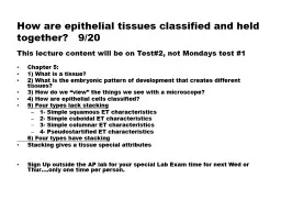
920 Review the origins of the four primary tissue types What extracellular fibers are found in connective tissue What is the function of connective tissue What are characteristics of the ten different connective tissues ID: 684857
Download Presentation The PPT/PDF document "What are the other three primary tissue ..." is the property of its rightful owner. Permission is granted to download and print the materials on this web site for personal, non-commercial use only, and to display it on your personal computer provided you do not modify the materials and that you retain all copyright notices contained in the materials. By downloading content from our website, you accept the terms of this agreement.
Slide1
How are epithelial tissues classified and held together? 9/20
This lecture content will be on Test#2, not Mondays test #1
Chapter 5:
1) What is a tissue?
2) What is the embryonic pattern of development that creates different tissues?
3) How do we “view” the things we see with a microscope?
4) How are epithelial cells classified?
5) Four types lack stacking
1- Simple
squamous
ET characteristics
2- Simple
cuboidal
ET characteristics
3- Simple columnar ET characteristics
4-
Pseudostartified
ET characteristics
6) Four types have stacking
Stacking gives a tissue special attributes
Sign Up outside the AP lab for your special Lab Exam time for next Wed or
Thur
….only one time per person.Slide2
How do we classify/describe the different cell types that make up tissues, organs, organ systems and the body? “Tissue”: a set of cells with similar appearance and function together in an organ.
How do we consider tissues?
1) Embryonic Origin
:
what cells were the embryonic precursors?
2) Shape/Appearance:
are cells round, flat or cube etc?
3) Things leaving cells
: Secretion vs. Excretion:
S: material has physiological function
E: waste removal from cell/body
4) Activity:
Do cells use contraction, exocytosis, endocytosis or phagocytosis?
5) Matrix:
What extracellular material is observed around the cells?
Collagen, Elastin, Cartillage, Interstitial Fluids
These factors let us classify the “tissue type”!
Tissues classification lets us understand the cause/effects of diseases.Slide3
How does the angle of observation affect what we observe?Two dimensional interpretation of three dimensional objects can be very difficult.
Terms used to describe orientation of tissue section:
Longitudinal View vs.
Cross Sectional View
Oblique View: Mix of the two for cells/tissue
Histological fixing and sectioning of tissue
Object on Slide
Histological Staining of tissue “Colors” on Slide
Sometimes fixation removes a part of the original tissue and what is “seen” on the slide is the remnant of where the cell used to be.
The color we observe on a slide is usually created by the stains the tissue was prepared with.
Try to interpret what you “see” on a slide as what it “was”.Slide4
What are our 3 Primary Germ Layers?Why are germ layers improtant?
#1: Life begins as a fertilized egg (zygote; Week 1) that begins to divide.
#2: Life progresses to a “hollow” ball of cells with a yolk sac underneath (blastoceol; Week 2)
Outside surface=“ecto”derm
Inside surface next to yolk sac= “endo”derm
#3: Week2-3
The ball of cells forms a neural tube (CNS). Cells squeeze between the edges and migrate into the middle space between the yolk sac and the outside.
Slide5
How do the three primary germ layers create the four primary classes of tissue?
1) Ectoderm:
Epithelial Tissues
Becomes: epidermis, hair, glands of skin, nerves, brain, a few blood vessels, and spinal cord
Basement membrane holds them to underlying base!
2) Endoderm:
Epithelial Tissues
Becomes lining of lungs and glands of the gut
3) Mesoderm:
Middle Tissues
Becomes: dermis of skin, muscle, blood and connective tissues
Remember that cells can migrate in the body during embryonic development.
These 3 germ layers create the 4 Primary Tissue Classes:
1) Epithelial 2) Connective 3) Nervous 4) MuscularSlide6
The most fundamental way to organize epithelial cell classes is by layer and cellular shape (Know Table 5.2).
Four types have a single epithelial cell layer:
“Simple Squamous”
“Simple Cuboidal”
“Simple Columnar”
“Psuedostratified Columnar”
Four types have layered epithelial cells:
“Stratified Squamous”
1)-with the protein “keratin”
2)-without the protein “keratin”
3)“Stratified Cuboidal”
4)“Transitional”
Why is the basement membrane so important?
This is cellular super-glue for all epithelial tissues!
Why are underlying connective tissues required?Slide7
Why is the shape of simple squamous epithelium important for its function?
Where is SSE: alveoli, blood vessels, serous membrane of organs, glomerular capsule
Key Characteristic: FLAT and THIN!
Typical packing and “desmosomes”:
Why do we need a smooth surface?
Why do we need a thin barrier/membrane?
Where does gas exchange occur?
Why does filtration occur?
SSE tissues lining all blood vessels consist of “endothelial cells”
Additional Endothelial Functions
:
Hormonal functions-
Contraction, inflammation and permeability-
Clot Prevention-Slide8
Simple cuboidal epithelium helps with: protection, secretion and reabsorption.
Key characteristic: CUBE SHAPED!
Tight packing:
Desmosomes:
Provides Glandular Functions (Secretions): ovary, salivary, thyroid, pancreas, and liver
Kidney has dual functions:
Absorption and Secretion-
Slide9
.Slide10
How do we protect cells and underlying tissues in places, such as the stomach? How do we prevent acids, abrasives, or pathogenic organisms from damaging underlying tissues?
Simple Columnar Epithelium
:
Longer than they are wide to put nuclei by basement membrane for extra safety:
Tough basement membrane:
SCEs secrete digestive enzymes:
SCEs help with nutrient absorption:
Some have cilia (uterine tubes):
Huge intestinal surface area with villi (cells) and microvilli (cell structures)= nutrient/water absorption
Specialized
Goblet cells
make mucus to lubricate the passage of materialsSlide11
Pseudostratified Columnar Epithelium (PCE) has cilia that move materials embedded in mucus in a single direction.
Why is it called pseudostratified?
Nuclei are at several different levels in cell-
All cells still reach basement membrane-
It only “appears” multi-layered
Goblet cells are important!
Importance of cilia for the airways?
Importance of cilia in male reproductive tract?
Importance of basement membrane?Slide12Slide13
Stratified Epithelial cells (Table 5.3) can be: 1) Squamous with keratin 2) Squamous without
keratin
, 3) Cuboidal
or 4) Transitional.
Callus on Sole of Foot: SSE-K...creates a nasty water insoluble place where bacteria can’t live and water can’t evaporate from the body!…why?
Mucosa of vagina, mouth, anus, esophogus are covered by: SSE-NoK…why?
Ovarian follicles and seminiferous tubules of the testes: SCE…why?
Transitional Epithelium: Lower urinary tract and part of umbilical cord- it stretches!
Why transitional (partly rounded/partly flattened)?
WHAT IS EPITHELIAL CELL “EXFOLIATION”?
Do you treat the exfoliation on your scalp?Slide14
The Eight Kinds of Epithelial Tissue:Describe the key features of each and where they are found in the body:
Four Single Layer Epithelial Tissues:
Simple Squamous Epithelial
Simple Cuboidal Epithelial
Simple Columnar Epithelial
Pseudostratified Columnar Epithelial
Four Layered Epithelial Tissues:
Stratified Squamous Epithelium-Keratinized
Stratified Squamous Epithelial-Nonkeratinized
Stratified Cuboidal Epithelial
Transitional Epithelial