/
 HEPATITIS DR : RAMY A. SAMY  HEPATITIS DR : RAMY A. SAMY

HEPATITIS DR : RAMY A. SAMY - PowerPoint Presentation

lindy-dunigan
lindy-dunigan . @lindy-dunigan
Follow
346 views
Uploaded On 2020-04-02

HEPATITIS DR : RAMY A. SAMY - PPT Presentation

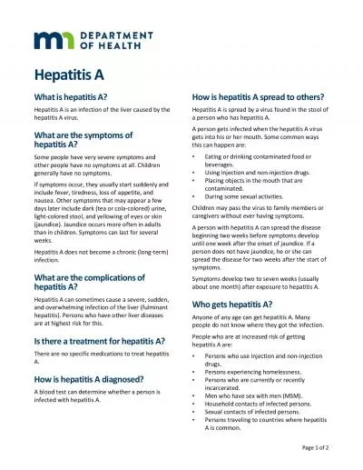
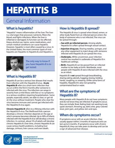
MD lecturer of internal medicine Drramisamifmedbuedueg Introduction Hepatitis Inflammation of Liver viral alcoholic immune Drugs Toxins biliary obstruction gall stones ID: 774729

hepatitis chronic liver aih hepatitis chronic liver aih viral acute disease type hcv autoimmune anti hepatic ribavirin amp patients

Share:

Link:

Embed:

Download Presentation from below link

Download Presentation The PPT/PDF document " HEPATITIS DR : RAMY A. SAMY" is the property of its rightful owner. Permission is granted to download and print the materials on this web site for personal, non-commercial use only, and to display it on your personal computer provided you do not modify the materials and that you retain all copyright notices contained in the materials. By downloading content from our website, you accept the terms of this agreement.


Presentation Transcript

Slide1

HEPATITIS

DR : RAMY A. SAMY

M.D. / lecturer of internal medicine

Dr_ramisami@fmed.bu.edu.eg

Slide2

Introduction

Hepatitis:

Inflammation of Liver

viral

alcoholic

immune

Drugs

Toxins

biliary obstruction - gall stones

Types :

Acute

Chronic

Fulminant

Viral Hepatitis

:

Hepatitis

A

,

B, C, D

,

E

, & other

systemic

specific

CMV, EBV& other

Slide3

Pattern of Viral Hepatitis

Carrier state

asymptomatic phase

Acute hepatitis

Chronic Hepatitis

Fulminant hepatitis

Cirrhosis

HCC

1- Chronic Persistent Hepatitis (CPH)

2- Chronic Active Hepatitis (CAH)

Slide4

ACUTE HEPATITIS

Def

:

Acute inflammation of the liver.

Etiology:

1-

Viral: A, B, C, D, E, F, G.

2-

Non viral: (inf.)

- Toxoplasma. -

Leptospira

hegica

.

3-

Drugs:

-

Halothan

. -

Paracetamol

.

- Mono Amino oxidase Inhibitor (MAOI).

4-

Poisons

:

-

Aflatoxins

-

Amanitaphaloids

.

Slide5

5- Misscellaneous: - pregnancy. - Wilson’s disease. - Circulatory insufficiency. - Alcohol.Pathology:Hepatocytes  swollen and become granular with variable.distribution:  * Centrizonal. * Focal (spotty). OR * Massive. Dead hepatocytes become shrunken. Lobules: portal tract infiltration with lymphocytes. N.B.: in Pregnancy Alcoholism Fatty changes S.Hepatitis C

Slide6

Causes:

Hepatitis A

HAV

Infective hepatitis

Hepatitis B

HBV

serum hepatitis

Hepatitis

D

HDV

Hepatitis C

HCV

Post transfusion hepatitis

Hepatitis E

HEV

Epidemic/ Entral

virus

27 nm RNA

42 nm DNA

Hepa virus

35 nm

Incomplete

RNA+HBsAg

30 – 60 nm

RNA flavi firus

32 nm RNA

transmission

Feco

-oral

Paraentral and post transfusion

Sexual low risk 1-5 %

Intrauterine low risk <5%

Feco-oral

Incubation p.

2 – 6 weeks

2 – 6 months

2 – 6 months

2 – 6 months

2 – 6 weeks

Chronicity

&liver cancer

no

Yes

Yes

Yes

No

Immunization

-passive

Non specific

Ig

Specific Ig

IgM

Non specific

Ig

-active

HAV vaccine

danger

Heptavax HBV vaccine

Heptavax HBV vaccine

Slide7

CLINICAL PICTURE OF ACUTE HEPATITIS (VIRAL):

Variable from asymptomatic

to fluminant hepatitis

A-

Pre-icteric

:

2 weeks

Anoraxia, Nausea, vomiting, diarrhea.

Fever, headache, malaise, abdominal pain.

Pruritis.

In Hepatitis B:

*

Immunological manifestations as:

*

maculopapular rash, polyarthritis,

vasculitis,

*

glomerulonephritis.

B-

Icteric phase

(may not occur):

Jaundice: dark urine, pale stool.

Hepatomegally, splenomegally.

Tender lymphadenopathy.

Rarly

fulminant hepatic failure

Slide8

C-

Convalescent stage:

- Improvement occur (clinically and laboratory).

- Complete cure in A,E, chronecity in B,C, D,G

.

Investigations:

1-

Liver function tests:

-

serum enzymes. -

serum bilirubin.

2-

Haematological:

- Leucopenia. - Lymphocytosis.

- Haemolytic anaemia. -Aplastic anaemia.

3-

Immunological:

- HAV

:

IgM

acute infection.

IgG (alone)

*

previous exposure.

*

non infectivity.

*

immunity.

Slide9

- HBV: * HBs Ag  infection, infectivity if persist after acute phase  carrier state or chronicity. * HBcAg  not detected in the serum. (in the liver). * HBeAg  - viral replication - Infectivity after 3 months * HBV Ab: anti HBs +ve Clearance of HBsAg. After successful vaccination. Recovery and non infectivity. * Anti HBC: IgM  persists for 3-6 months. appears shortly after HBsAg (acute infection) HBsAg –ve (HbsAb not get +). Pt. is infective +ve anti HBC IgM.

Slide10

* Anti-HBC IgG: - Appear during acute illness - Persists indefinitely. * Anti-HBe  after anti HBC ( infectivity).* HBV DNA: The most sensitive index of viral replication and infectivity (by PCR).-HCV: Anti HCV: Detection of Anti HCV using C100antigen (1st generation) (ELIZA). 2nd generation  using C22, 223 (RIBA). PCR: detect HCV Ag qualitative and quantitative.

Slide11

HDV: anti-HDV PCR (HDV –RNA( .HEV: Ag, Ab

Slide12

CHRONIC HEPATITIS

Definition

:

It is a chronic inflammatory reaction for 6 months or more.

Etiology:

1-

Viral:

B, C, D

2-

Autoimmune

.

3-

Drugs:

INH, ketoconazol,

Nitrofurantoin.

4-

Heridetary:

*

1 antitrypsin deficiency.

*

Wilson’s disease.

Slide13

Pathology:

Inflammatory cells infiltrate of the portal tracts (lymphocytes, plasma cells, macrophages)

piece meal necrosis

collagen deposition

bridging necrosis

cirrhosis.

Slide14

Classification: According to the etiology.Pathological classificationChronic persistant according to grade stage of fibrosisChronic lobular - Portal Chronic active - Peripheral - Non - Lobular - Moderate inflammation - Severe (minimal, moderate, severe)

Slide15

CHRONIC VIRAL HEPATITIS

Clinical Picture:

Symptoms

:

Anorexia, vague abdominal pain, past history of the cause and may be nothing at all.

On Examination

:

Liver is enlarged and tender with smooth surface.

Spleen usually not enlarged or intercostally enlarged.

Slide16

INVESTIGATION:

Serum enzymes

[SGOT, SGPT, ALT] are all normal or slightly elevated,

bilirubin

is normal.

Viral markers

may be +ve.

Liver biopsy

: normal hepatic architecture with portal tract, infiltration with chronic inflammatory cells.

Slide17

Treatment:

Symptomatic ttt only + Reassurance.

Avoid hepatotoxic drugs

.

Slide18

CHRONIC AUTOIMMUNE HEPATITIS

Slide19

c/o:

Easy fatiguability, pruritis. jaundice, arthralgia ± Ascites, etc....

Multisystem manifestations:

Polyarthopathy.

Pallor, skin rashes,

Glomerulonephntis

D.M.

Autoimmune Haem.An., neutropenia.

Myxoedema, Thyrotoxicosis, Hashimoto's thyroiditis.

0/E:

Enlarged firm liver (usually smooth surface), tender.

Manifestations of LCF and portal hypertension.

Slide20

AIH subtypes

AIH type 1

AIH type 2

AIH type 3

on the basis of immunoserologic markers.

The International Autoimmune Hepatitis Group has not endorsed this subclassification.

Used mainly for descriptive value

Slide21

AIH type 1

Most common form worldwide

characterized by the

presence or absence of SMA and/or ANA in serum

Surrogate markers:

Perinuclear antineutrophil cytoplasmic

antibodies which occur in PSC and chronic UC, are found in 90% of patients who have type 1 AIH.

Bimodal age distribution (10-20; 45-70)

Female:male 3.6:1

Risk factors for type 1 AIH

in whites of northern European descent [HLA-DR3 (DRB1*0301)] and –DR4 (DRB1*0401)]

Slide22

AIH type 1

Associated with concurrent extrahepatic diseases

Autoimmune thyroiditis (12%)

Graves disease (6%)

Chronic UC (6%) * (cholangiography to exclude PSC)

<1% RA, pernicious anemia, systemic sclerosis, Coomb’s test-positive HA, ITP, symptomatic cryoglobulinemia, leukocytoclastic vasculitis, nephritis, erythema nodosum, SLE, fibrosing alveolitis.

40% of AIH type 1 presents with an acute onset of symptoms/signs indistinguishable from that of acute viral or toxic hepatitis and the disease may appear fulminant in fashion.

target autoantigen is unknown, but ASGPR (asialoglycoprotein receptor) found on hepatocyte surface is a candidate

Responds well to glucocorticoids

Slide23

AIH type 2

Characterized by presence of

anti-LKM1

(liver/kidney microsome type 1) in serum

P-ANCA is not found

Mainly

children

(2-14 yo) but also seen in adults (in Europe, 20% of pts are adults; in US, 4% of pts are >18 yrs)

Only AIH with an identified target autoantigen:

cytochrome monooxygenase P-450 IID6 (CYP2D6)

found in the cytosol of hepatocytes.

Recognized

homologies

b/w epitopes of CYP2D6 and genome of

HCV

.

<10% of Europeans with chronic Hep C have detectable anti-LKM1

Suggests molecular mimicry and antibody crossreactivity, multiple exposures to virus mimicking self may be a way to break self-tolerance and induce AIH type 2.

Anti-LKM1 + pts with chronic Hep C in US pts is rare – differences in indigenous virus or host susceptiblity?

Acute or fulminant presentation is possible

Thus essential to screen all pts who have an acute decompensation for type-specific autoantibodies.

Associated with HLQA-B14, -DR3, -C4A-QD

Susceptibility factor in German and Brazilians: DRB1*07

Like AIH type 1, also responds well to glucocorticoids

Slide24

AIH type 2

Distinct form of anti-LKM positive AIH

Occurs in association with

autoimmune polyendocrinopathy disorder (APECED)

aka

Polyglandular autoimmune syndrome type I (APS1)

rare autosomal recessive disorder

Caused by a signal gene mutation of the APS1 gene which encodes a transcription factor, autoimmune regulator (AIRE) which is expressed in epithelial and dendritic cells within the thymus where it regulates clonal deletion of autoreactive T cells, thus can affect self tolerance

Features of this disease are

ectodermal dystrophy, mucocutaneous candidiasis, multiple endocrine gland failure

(parathyroids, adrenals, ovaries)

Marked by the presence of numerous organ and non-organ specific autoantibodies and multiple concurrent autoimmune diseases.

most common among Finns, Sardinians, and Iranian Jews

Pts with APECED and AIH have an

aggressive liver disease

that does not respond well to standard immunosuppressive regimens.

Slide25

AIH type 3

Least established form of the disease

Designation largely abandoned

Characterized by presence of antibodies to soluble liver antigen and liver/pancreas (

anti-SLA, anti- LP

)

30-50 yo

Target autoantigens:

thought

to be Glutathione S-transferase, but a transfer ribonucleoprotein (tRNP) 50-kd protein was described in 2000 as the more likely target.

Clinical and laboratory features that are indistinguishable from AIH type 1

Also responds well to glucocorticoids

Slide26

AIH subtypes

Slide27

DRUG INDUCED LIVER DAMAGE

1-

Hepatocellular damage:

Associated with history of drug intake.

Indistinguishable from acute or chronic hepatitis.

Induced either by:

*

Toxic metabolite.

or

*

Immune response to drugs:

as

-

Thiazide

-

Rifampicine

-

Halothan

-

Acetaminophan

-

NSAIDs

-

INH

Slide28

2-

Cholestatic

liver damage:

as

*

Estrogen

*

Phenothiazine

*

Tricyclic antidepressants.

*

Erythromycin

*

Sulphasalazine

*

Tolbutamide

3-

Drugs causing hepatic fibrosis:

Methotrexate,

amiodarone

Slide29

4- Drugs causing chronic active hepatitis: Methyldopa, nitrofurantoins, sulphonamides. 5- Drugs causing hepatic tumours Benign Malignant oral contraceptive pills synthetic androgens

Slide30

Fulminant

Hepatic Failure

Slide31

Fulminant Hepatic Failure

Definition

-

Altered mental status with

coagulopathy

in setting of acute liver disease. Hepatic encephalopathy occurring within 8 weeks of onset of illness defines FHF.

Slide32

Etiology

Viral hepatitisHepatitis A - rarelyHepatitis B - appx 1% of hep BHep C -- probably not, but ??Hep D -- delta agent coinfects with Hep BHep ECMV, HSV

Toxins Carbon tetrachloridePhosphorusAmanita phalloides (antidote penicillin and silybin)Industrial cleaning solvents

Slide33

Etiology

DrugsAcetaminophenAcetaminophen in Tx doses with alcoholIdiosyncratic reaction -- halothane, sulfonamides, phenytoin, and others.

Vascular

Heart failure --

centrolobular

necrosis

Sinusoidal obstruction secondary to

metastates

Budd

Chiari

Veno

-occlusive disease

Slide34

Pathology

Viral, toxic

--

hepatocellular

necrosis, diffuse, extensive, resulting in

caseating

necrosis.

Cardiac

--

similar, with particularly severe

centrolobular

necrosis from higher intravascular

pressure.

Blood flows from portal triad (hepatic artery and portal vein) to central veins to hepatic vein. With poor cardiac output,

splanchnic

flow markedly decreased, resulting in markedly decreased portal

flow.

Slide35

Pathology

Fatty liver of pregnancy and Reye’s show

microvesicular

collections of fat in

hepatocytes

and much less necrosis.

Special stains -- iron in

hemochromatosis

, copper in Wilson’s, etc.

Slide36

Clinical Presentation

Typically -- nonspecific symptoms, nausea, malaise, jaundice, altered mental status, coma -- all over a few days.

The altered mental status occasionally precedes clinical jaundice.

Mental status changes often start with agitation, delusions, irritability before progressing to lethargy, stupor, and coma.

Slide37

Clinical Presentation

Laboratory --

Transaminases

usually high (>1000)

Bilirubin

-- usually mixed

hyperbilirubinemia

Ammonia -- usually elevated

Coagulopathy

with prolonged PT, PTT, decreased factors

Low level DIC

Low level

fibrinolysis

Respiratory alkalosis

Metabolic acidosis, increased lactate

Slide38

Chronic viral hepatitis

Etiology:- This syndrome is induced by :

> 70% of HCV

5 – 10% of HBV

& 80% of HDV super infection

HAV & HEV infection never lead to chronic hepatitis

N.B. :

Slide39

Clinical features & presentation

Patient may present an a variety of ways :

- Clinical symptoms may range from non to mild fatigue or patient may present with complication of cirrhosis

- there may be no signs but jaundice or signs of chronic liver disease may be present

Slide40

chronic viral hepatitis

Chronic

HCV

Chronic

HBV

Slide41

Patterns of viral hepatitis C

1-

most patient with hepatitis C are asymptomatic

( suspicious often with screening of risk factors which include :-

I.V. drug use

sharing of straws to snort cocaine

heamodialysis

& Bl. transfusion

in contrast to HBV

sexual transmission

is rare ,

vertical transmission

is uncommon )

2-

70-80% of all patients infected with HCV will develop chronic hepatitis in the 10 years following infection, within 20 years 30% of those will develop cirrhosis & over 30 years of those with cirrhosis may develop HCC .

Slide42

Patterns of hepatitis B infection

Recovery

Transient subclinical infection

65%

Acute hepatitis

25 %

death

Acute HBV

10%

Chronic HBV infection

Healthy HBsAg carier

70-90%

HCC

Chronic hepatitis 10-30%

cirrhosis

100 %

99 %

1 %

Slide43

Table 3: Interpretation of HCV Assays

Slide44

Utility of the Liver Biopsy

It provides helpful information on the

current

status

of the liver injury (Grade, Stage).

It identifies features useful in the decision to embark on therapy (steatosis and excess hepatocellular iron).

It may reveal advanced fibrosis or cirrhosis that necessitates surveillance for hepatocellular carcinoma (HCC) and/or screening for varices.

Slide45

Comparison of Scoring Systems for Histological Stage

Slide46

Characteristics of Persons for Whom Therapy Is Widely Accepted

Age

18

years or

older

, and

HCV RNA

positive

in serum, and

Liver biopsy showing chronic hepatitis with

significant fibrosis

(bridging fibrosis or higher), and

Compensated

liver disease (total serum bilirubin

1.5

g/dL; INR

1.5

;

serum albumin _

3.4

, platelet count

75,000

mm with

no evidence

of hepatic decompansation (hepatic encephalopathy or ascites), and

Acceptable hematological and biochemical indices (Hemoglobin

13

g/dL for

men

and

12

g/dL for

women

; neutrophil count

1500

/mm3 and serum creatinine _

1.5

mg/dL, and

Willing to be treated and to adhere to treatment requirements, and

No

contraindications

Slide47

Characteristics of Persons for Whom Therapy Is Currently Contraindicated

Major uncontrolled depressive illness

Solid organ transplant

(renal, heart, or lung)

Autoimmune hepatitis or other

autoimmune

condition known to be

exacerbated

by peginterferon and ribavirin

Untreated

thyroid

disease

Pregnant or unwilling to comply with adequate contraception

Severe concurrent medical disease such as severe hypertension, heart failure, significant coronary heart disease, poorly controlled diabetes, chronic obstructive pulmonary disease

Age less than 2 years

Known hypersensitivity to drugs used to treat HCV

Slide48

The Adisory Committee meeting report (28 December 2006, modified on 25 June 2007)

Routine blood tests: CBC, LFTs (AST, ALT, T. Bil, Albumin, ALP, Prothrombin time), S.creatinine, FBS, PCR for HCV.

Other blood tests: HB sAg, ANA, TSH,

α

FP, ABA.

Abdominal U/S.

Fundus examination.

ECG.

Liver biopsy.

BMI.

Slide49

A liver biopsy may be unnecessary in:

Persons with genotypes

2

and

3

HCV infection, since more than

80%

of them achieve a sustained virological response (SVR) to standard of care treatment.

Medical & paramedical staffs.

Extra-hepatic manifestations.

Compansated liver disease

(Child A)

will be treated if there is

no

varices.

Liver biopsy

>F0 &>A1

with

elevated

liver enzymes

(Metavir score).

Liver biopsy

≥ A2 & ≥ F2

with

normal

liver enzymes.

BMI

<35 %

Slide50

The Optimal Treatment of Chronic HCV:

Peginterferon Alfa and Ribavirin

There are two licensed pegylated interferons in the United States, peginterferon

alfa-2b

(Peg-Intron,

Schering

Plough Corp., Kenilworth, NJ), with a 12-kd linear polyethylene glycol (PEG) covalently linked to the standard interferon alfa-2b molecule, and peginterferon

alfa-2a

(Pegasys, Hoffmann-La

Roche

, Nutley, NJ) with a 40-kd branched PEG covalently linked to the standard interferon alfa-2a molecule

(Zeuzem et al, 2003).

Slide51

The optimal dose of peginterferon

alfa-2b

, based on the original registration trial is

1.5 µg/kg/week

dosed according to body weight.

Weight-based ribavirin (

800 mg

for patients

< 65 kg; 1,000 mg

for patients weighing

65 to 85 kg; 1,200 mg

for patients weighing

85 to 105 kg; and 1,400 mg for patients weighting >105

kg but

<

125 kg) was more effective

(Jacobson et al, 2007).

Peginterferon

alfa-2a

is administered at a fixed dose of

180

µg/week given subcutaneously together with ribavirin

1,000 to 1,200

mg daily,

1,000

mg for those who weight

< 75 kg

and

1,200

mg for those who weight

> 75 kg

.

Slide52

Adverse Events:

The most common adverse events in these trials were influenza- like side effects such as fatigue, headache, fever and rigors, which occurred in more than

half of the patients

.

Psychiatric side effects (depression, irritability, and insomnia), which occurred in

22% to 31%

of patients.

Laboratory abnormalities are the most common reasons for dose reduction. Among these,

neutropenia

(absolute neutrophil count [ANC] of 1500 mm3) was a frequent laboratory abnormality, occurring in

18% to 20%.

The dose was reduced 50% for an ANC of 750 mm3 and permanently discontinued for an ANC of <500

Despite the decline in the neutrophil count, serious infections are uncommon

(Suza et al 2002)

and

Granulocyte colony stimulating

factor is rarely necessary except in patients with advanced cirrhosis.

Anemia

was observed in approximately

one-third

of patients, Dose modification for anemia (hemoglobin level <10 g/dL) was required in 9% to 15%

Slide53

Growth factors, such as

erythropoietin

, have been used to counter the anemia associated with peginterferon and ribavirin. Although growth factors improve a patient’s sense of well-being and have reduced the requirement for ribavirin dose reduction, their use has not been shown to improve SVR rates

(Shiffman et al, 2007).

In one analysis, the use of a hematological growth factor nearly

doubled the cost

of treatment for chronic hepatitis C

(Del Rio et al, 2006).

Although generally safe, erythropoietin use has been associated with

serious side effects

including cardiovascular and thromboembolic events, pure red cell aplasia, progression of certain cancers, and death

(Bennett et al, 2008).

Therefore,

routine use

of growth factors

cannot be recommended

at this time and dose reduction is the primary mode for managing cytopenias.

Slide54

Neuropsychiatric

side effects include depression, anxiety, insomnia, emotional lability, mood disorders, frank psychosis, suicidal ideation, actual suicide, and homicide

The most consistent

risk factors

for developing depression are the presence of mood and anxiety symptoms prior to therapy. A past history of depression and of receiving higher doses of interferon, as well as being female, have been identified as risk factors, but are less reliable ones

(Raison et al, 2005).

Interferon-induced depression appears to be composed of two overlapping syndromes, a

depression-specific syndrome

characterized by mood, anxiety, and cognitive complaints, and

neurovegetative symptoms

, characterized by fatigue, anorexia, pain and psychomotor slowing

(Capuron et al, 2002).

Depression-specific symptoms are highly responsive to

serotonergic antidepressants

whereas neurovegatative symptoms are not. These symptoms may be more effectively treated with agents that modulate catecholaminergic function. When selecting an agent, consideration should be given to drug– drug interactions, underlying hepatic function, the possibility of drug-induced hepatotoxicity and other adverse side effects.

Consultation and follow up with a psychiatrist is advised.

Slide55

Pegylated interferon may

induce autoimmune

disorders, such as autoimmune thyroiditis, or may

worsen pre-existing

autoimmune disorders. Therefore, the presence of autoimmune conditions prior to treatment is a

relative contraindication

to therapy.

With regard to

ribavirin

, the most common side effect is

hemolytic anemia.

Since ribavirin is cleared by the

kidney

, the drug should be used with

extreme caution

in patients with renal disease and renal failure.

Other side effects associated with ribavirin include mild lymphopenia, hyperuricemia, itching, rash, cough and nasal stuffiness.

Ribavirin is reported to cause

fetal death

and

fetal abnormalities

in animals and thus it is imperative for persons who receive the drug to use

strict contraceptive

methods both during treatment and for a period of

6 months

thereafter. The education of patients and caregivers about side effects and their management is an integral component of treatment and is important for a successful outcome.

Slide56

Follow up pannel:

To detect

response

:

PCR

at 4, 12, 24, 36, 48 and

72 weeks

.

To monitor

complications

:CBC (ANC), AST, ALT, Creatinine, T.bilirubin after each injection in the first month, then monthly till end of treatment.

Slide57

Table 5: Virological Responses During Therapy and Definitions

Slide58

Premanant Discontinuation (INF & Ribavirin)

Dose Reduction by 50 %

<8.5 g/dl

< 10 g/ dl

(only ribavirin)

HB Level

< 1000

< 1500

(Only INF)

WBC

< 500

< 750

(Only INF)

Neutrophils

< 25,000

< 50,000

(Only INF)

Platelets

> 1.5 UNL

N/A

Creatinine

2× Baseline OR >10 UNL

N/A

ALT-AST

> 4 mg % (For >4 weeks)

>5 mg %

(only ribavirin)

Indirect Bil.

> 2.5 UNL

N/A

Direct Bil.

Slide59

NOVEL HCV

ANTIVIRAL

Slide60

SOFOSBUVIR

(

sovaldi

)

Slide61

THANK YOU