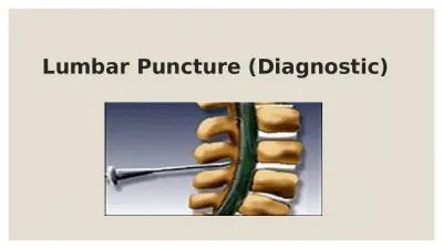
/
Diagnostic Lumbar Puncture is one of the most commonly performed invas Diagnostic Lumbar Puncture is one of the most commonly performed invas

Diagnostic Lumbar Puncture is one of the most commonly performed invas - PDF document

lois-ondreau
lois-ondreau . @lois-ondreau
Follow
384 views
Uploaded On 2017-11-21

Diagnostic Lumbar Puncture is one of the most commonly performed invas - PPT Presentation

TABLE 1 Indications for Lumbar Puncture To exclude subarachnoid haemorrhage in acute severe To investigate or exclude meningitisBacterialViral Tuberculous The Ulster Medical Society 2014wwwumsac ID: 606957

TABLE Indications for Lumbar

Share:

Link:

Embed:

Download Presentation from below link

Download Pdf The PPT/PDF document "Diagnostic Lumbar Puncture is one of the..." is the property of its rightful owner. Permission is granted to download and print the materials on this web site for personal, non-commercial use only, and to display it on your personal computer provided you do not modify the materials and that you retain all copyright notices contained in the materials. By downloading content from our website, you accept the terms of this agreement.


Presentation Transcript

Diagnostic Lumbar Puncture is one of the most commonly performed invasive tests in clinical medicine. Evaluation of an acute headache and investigation of in�ammatory or infectious disease of the nervous system are the most common indications. Serious complications are rare, and correct technique will minimise diagnostic error and maximise TABLE 1: Indications for Lumbar Puncture To exclude subarachnoid haemorrhage in acute severe To investigate or exclude meningitisBacterialViral Tuberculous The Ulster Medical Society, 2014.www.ums.ac.uk Carolynne M. Doherty MRCP, Neurology RegistrarRaeburn B. Forbes MD(Hons) FRCP, Consultant NeurologistDepartment of Neurology, SHSCT, Craigavon Area Hospital, Portadown, County Armagh. BT63 5QQCarolynne M Doherty, Raeburn B Forbes 94 to obtain indirect measurements of intracranial pressure, although non-invasive methods of intracranial pressure estimation are undergoing validation.CONSENT AND DOCUMENTATIONConsent should include the risk of Post-Lumbar Puncture Other risks to discuss include failure to obtain CSF, localised bruising, bleeding and local discomfort at the injection site. Iatrogenic meningitis and nerve root injury are exceptionally The procedure should be documented in the patient’s notes. The position and vertebral space selected, local anaesthesic CSF appearance (clear, cloudy, blood-stained or pigmented) and number of samples collected should be documented, allowing another physician to retrospectively interpret the investigative �ndings accurately. A proforma (Figure 1) can serve as both aide memoire and audit tool. It is also advisable to document that post Lumbar Puncture advice has been given, and a patient information lea�et is one way to provide ANATOMYfor anyone performing Lumbar Puncture. The Lumbar Puncture needle pierces in order: skin, subcutaneous tissue, supraspinous ligament, interspinous ligament, ligamentum �avum, epidural space containing the internal vertebral venous plexus, dura, arachnoid, and �nally the subarachnoid If you are able to visualise the anatomy it will allow re-positioning of the needle, should the initial pass be unsuccessful - Figure 3 demonstrates the relationship between The most important bony landmark is the L4 spinous process, which is located at the intersection of the ‘intercristal’ or ‘Tuf�er’s’ line (the line between the top of the iliac crests) and the lumbar spine midline (Figure 4). Radiological studies have shown that this clinical landmark is accurate in over although in females or obese people Tuf�er’s Line tends to be found at a higher level than L4.The approximate distance from the skin to the epidural space is 45 - 55mm and the dura mater may be up to 7mm beyond Typically, a standard 90mm Whitacre needle (Vygon UK) will need to be inserted to two thirds of its length before it reaches the ligamentum �avum, with CSF obtained CSF SPECIMEN HANDLING TABLE 2: Tests frequently performed on CSF Test VolumeTransport in opaque Viral PCRVirologyPaired serum ACE 95 . The lumbar puncture proforma currently in use in Craigavon Area Hospital Ward …………………………..Yes Yes Other Acute Headache (CT normal)Yes Yes Yes Yes Yes CT Brain PerformedYes Yes Yes Local Anaesthetic Amount used ……… mL(NB: Maximum dose in adult is c. 2mg/kg) Was sedation required Yes Gauge of LP needle ……G Atraumatic needle Yes Level of LP (L3/4 is recommended)Yes If Yes – which leg Right Stylet re-inserted prior to needle withdrawal Yes Opening pressure (to be recorded in ALL cases of acute headache) ……cmYellow top clotted – Biochemistry Lie down if post-LP headache occursContact GP if persistent post-LP headacheLP performedDate: Time: 96 CSF samples are collected and where necessary paired serum (Table 2). Some samples require larger volumes (e.g. Cytology), and others require rapid transport to the laboratory (Cytospin testing for lymphoproliferative cells). Testing for CSF Xanthochromia (to detect bilirubin from CSF sample to the laboratory. To protect from light the CSF bottle should be wrapped in foil or placed in an envelope. Samples should be correctly labelled with patient identi�ers, time and date. Immediate transport by hand to the laboratory by an emergency porter is preferred to the use of vacuum tube delivery systems, as excessive movement of blood specimens has caused increased rates of haemolysis,to in vitro CSF oxyhaemoglobin synthesis which would affect Isotope dilution studies show that an average adult makes about 500 ml of CSF every 24 hours, and that CSF is replaced about 4 times daily i.e. there is approximately 20 ml of CSF manufactured each hour. About 30 ml of CSF resides in the Lumbar Cistern, so taking about 10 ml of CSF to facilitate diagnostic testing is not likely to endanger the patient. The patient is more likely to come to harm from inadequate CSF Diagnostic Lumbar Puncture is an aseptic procedure, but as there is no direct injection into the spinal canal, the procedure can be done in the ward setting and does not need to be done in an operating theatre. It should be noted that The Association of Anaesthetists of Great Britain and Ireland recommends that injections into the spinal canal, such as epidural blood patching, should only be performed using aseptic techniques in a theatre environment.Lumbar Puncture, standard bedside aseptic procedures apply with no-touch technique, sterile drapes and use of There has been wide variation in what clinicians, particularly anaesthetists, feel constitute aseptic technique for spinal procedures. Clinicians’ views regarding hand-washing technique, donning of gowns or masks, and wearing of jewelry when placing an epidural catheter have been shown to vary. Wearing of masks may be associated with reduced bacterial transfer. A cluster of four cases of streptococcal meningitis was caused by a physician who had chronic Bacteria in the ori�ces of sebaceous glands and hair follicles Fig 2: Cadaveric sagittal section through lumbar spine showing proper needle trajectory (from Boon et aland lumbar puncture needle (11). Fig 3. Correct position of tip of Lumbar Puncture needle in centre 97 are protected by the stratum corneum from disinfectants.The overlying skin should therefore be cleaned with a solution that breaches this layer such as povidone-iodine or 0.5% chlorhexidine and 70% alcohol. Traditionally, chlorhexidine had not been recommended for procedures with meningeal exposure due to a possible association with arachnoiditis, but chlorhexidine does not seem to be associated with an increased incidence of neurological complications in spinal and has been recommended for anaesthetic We routinely use chlorhexidine to prepare the skin for the less hazardous procedure of diagnostic Lumbar Puncture. The Anaesthetic Literature supports use of 0.5% We suggest that the evidence for reduced frequency of PLPH from use of atraumatic needles mandates a change in practice, and physicians in training should no longer be taught to use classic needles routinely. A classic ‘Quincke’ needle has a cutting, bevelled tip, but an atraumatic needle has a pencil point tip with a side aperture (Figure 6). The recommended needle is a 22 gauge atraumatic needle. In our own practice we use a 22 gauge Whitacre needle, which is now the standard stock Lumbar Puncture needle in our institution. The biggest adjustment for people changing from traumatic to atraumatic needle is that the skin must be punctured �rst prior to insertion of the Lumbar Puncture needle, as an atraumatic needle will not pierce the epidermis. Options include using the introducer needle provided to pierce the skin, or using the puncture site made by the green 19G local anaesthetic needle Passage of atraumatic needle is a completely different sensation to a classic needle, as the practitioner experiences greater resistance when traversing the tissue planes to the CSF. A good analogy is the difference in sensation felt when cutting through a banana (analogous to the standard In 2005, The American Academy of Neurology recommended but this practice has to date been poorly adopted. However, with appropriate training and support, medical staff will adopt use of atraumatic needles and reproduce the bene�ts demonstrated Atraumatic needles have even been shown to be more cost-effective than classic cutting needles. Patients with PLPH can require in-patient care, which makes atraumatic needles both effective and cost-effective options for diagnostic Needles of smaller diameter have been demonstrated to be associated with a reduction of risk of PLPH from 24.4% The smallest diameter needle with which the practitioner can con�dently perform the procedure avoiding an increased number of attempts should be chosen,is known that larger bore atraumatic needles allow adequate pressure measurement and collection of suf�cient CSF. Fig 4: Surface Markings for Lumbar Puncture from Training Intercristal Line (eponymously Tuf�er’s Line). Red Dots are L3/4 interspinal space. A diagnostic Lumbar Puncture should be . The distance from surface to Ligamentum Flavum is . Classic (Quincke, or Bevelled-tip) and Whitacre Atraumatic 98 PATIENT POSITIONINGA right-handed practitioner should position the patient in the left lateral decubitus position, with the vertebrae in line in the horizontal plane and the head in a neutral position and the knees �exed. Opening Pressure at Lumbar Puncture is a surrogate measurement of Intracranial Pressure. An accurate the same level as the midline of the spine (Figure 4), which should also be at the same level as the patient’s head. A few centimetres of ‘head up’ bed tilt or more than one pillow could Always ensure that the patient is comfortable, and that the bed height is appropriate for the operator, as the practitioner risks compromising aseptic technique if the patient has to be Lumbar Puncture can also be performed in the seated position, providing pressure measurement is not required. There are occasions when pressure measurement is sacri�ced in order to obtain a CSF sample, for example in elective Lumbar Puncture for neuro-in�ammatory disease. If the procedure is to be performed in the upright position, seated with the chin comfort and optimise positioning. This position widens the interspinous distance.20 Remember that an elevated Opening Pressure may be the only sign of Cerebral Venous Sinus or Idiopathic Intracranial Hypertension, so in acute headache a Lumbar Puncture should be performed in the lateral decubitus position, whenever possible, to allow a HOW TO GET CSF AND MEASURE OPENING The best location to perform a Lumbar Puncture will depend on local facilities, but a treatment room, or somewhere calm and quiet is preferred to a ward bay. The attitude of operator and assistant should be one of calm con�dence and reassurance. An experienced operator will have the patient consented, positioned and comfortable, and from the moment of washing hands until the �rst CSF sample appears should Aseptic technique is required, and a pre-prepared epidural pack contains all the equipment needed other than your needle, lidocaine and manometer (Figure 7). The manometer (Figure 8) should be prepared prior to commencing the spinal injection, by connecting the two tubes and loosening the tap (which is always slightly stiff and is dif�cult to open if you do Once skin is sterile, local anaesthetic can be administered. Our practice is to raise a small intradermal bleb of lidocaine, which produces almost immediate cutaneous anaesthesia (Figure 9). A small amount of lidocaine can be in�ltrated into deeper tissues, but care must be taken not to distort local anatomy by administering too much local anaesthetic. If you anaesthetise the skin and have a correct trajectory, there is little bene�t to in�ltrating large volumes of anaesthetic.the lumbar cistern is diamond shaped and surrounded by bony structures. The Lumbar Puncture needle should be inserted at an angle that will allow it to pass between the spinous processes (Figure 10). The most common problem encountered by operators is their needle impacting on a bony structure - either the superior surface of the L4 spinous process, or the inferior surface of the L3 spinous process. If the needle tip is advanced beyond 50mm and the needle hits bone, then you have probably impacted on bone around the intervertebral space. Consider the anatomy and needle angle, and thus which bone is likely to have been reached. Withdraw the needle and adjust the trajectory, gradually proceeding until a “give” is felt on passing through the ligamentum �avum (Figure 2), remembering that the distance to ligamentum 11-12 19G and 25G hypodermic needles to draw up and inject 13 Whitacre 22G spinal needle (atraumatic needle) 99 �avum is approximately 2/3rds of the length of a standard needle in a non-obese patient. Whichever needle type has insertion commences, as there have been case reports of implantation of epidermal plugs resulting in the growth of Once the needle is suf�ciently advanced, withdraw the stylet slowly and wait about 5 seconds to see if CSF emerges. If it 2 or 3 mm and check again for CSF. Once CSF is obtained, connect the manometer and measure Opening Pressure (unless will take approximately one minute for CSF pressure to be measured and it is normal to observe the meniscus of CSF at the top of the manometer oscillate with respiration. Like all �uid measurements the pressure is the height of the lowest part of the meniscus at the top of the �uid column (Figure 11). Distracting the patient with conversation or other relaxation techniques may be used to ameliorate anxiety and allow a falsely elevated opening pressure to fall. CSF pressure measurements, from large series of adults indicate that CSF pressures between 60mm and 200mm are regarded as although in overweight individuals pressures as high as 250mm can be asymptomatic. Patient anxiety, incorrect head position and excessive �exion of the patient’s legs against the abdomen can all arti�cially elevate CSF pressure.Samples should ideally be taken either using sterile bottles by the individual performing the procedure, or by an assistant holding an open specimen container underneath the �ow of CSF from the end of the spinal needle. Twenty drops per bottle is suf�cient for most commonly performed tests (which provides about 2ml of CSF). A minimum volume of 5ml of CSF is required for cytopathological examination for example in suspected malignant meningitis. Replacement of the stylet is associated with a reduced incidence of post Lumbar Puncture headache, and failure to replace the stylet has been associated with nerve root herniation. After the needle has been removed, a sterile dressing should be placed IF YOU ARE UNABLE TO GET CSF…If you are unable to obtain CSF, having optimised position and needle trajectory, consider whether there is another suitably quali�ed physician available to attempt the procedure. Low CSF pressure, or viscous CSF in cases of malignant or tuberculous meningitis may mean that CSF will not appear despite perfect positioning and technique. Aspiration of CSF is not recommended, as it may cause spinal cord injury. people suspected of Idiopathic Intracranial Hypertension. Ultrasound identi�cation of the interspinous space should become routine practice, now that evidence shows a reduced risk of complications and a higher success rate in obtaining CSF with ultrasonic identi�cation of the inter-spinal space.A small sterile dressing is placed on the site: a pressure dressing is not required. The patient can mobilise as soon as it is comfortable to do so. Written information should be doses of regular analgesics such as paracetamol or NSAIDs are reasonable. Bed rest is more comfortable than being ambulant, but prolonged bed rest does not reduce the . Three way tap attached to end of Manometer . An intradermal bleb of 0.5ml of 1% lidocaine will produce 100 COMPLICATIONSThe most common complication is Post Lumbar Puncture Headache (PLPH). In one meta-analysis, the frequency of An important feature in the diagnosis of PLPH is the postural component; the patient will report a background headache which worsens within a few minutes of adopting upright posture and improves within a few minutes of lying �at.gender, and headache before or at the time of the procedure. The volume of �uid removed is not a risk factor, and the pathophysiology of PLPH is more plausibly explained as a loss of compliance of the spinal compartment, rather than due Relief of PLPH by adopting a supine posture means that the brain and its supporting dura are not mechanically stretched by the loss Lumbar Puncture, and the natural history is for symptoms to resolve by about 10 days. The pain is usually diffuse, global or bitemporal headache, which can be accompanied by nausea, altered hearing, tinnitus, photophobia or neck stiffness. Low pressure may produce diplopia due to traction reducing the risk of PLPH is needle selection, and a 22G atraumatic needle is now recommended for diagnostic Lumbar Puncture. In our own practice we reduced the risk of PLPH from 50% to 10% with the introduction of a guideline that required initial attempts with an atraumatic This observation is consistent with the experience There is anecdotal evidence that a straightforward procedure may increase the risk of PLPH due to the low levels of trauma and therefore clotting factors that might promote closure of the defect thus preventing CSF leakage. Directing the needle with the bevel parallel to the dural �bres (which run craniocaudally) has been shown to be associated with a reduction in PLPH. This advice only applies to classic ‘traumatic’ needles so is irrelevant when practitioners transition to using atraumatic needles due to the pencil point shape of the tip (Figure 5). Replacing the stylet has been shown to reduce the risk of PLPH, theoretically because a strand of arachnoid may splint the dural defect open if the A Cochrane Review has suggested that there is no bene�t to routine bed rest or intravenous �uids in prevention of PLPH after Lumbar Maintaining a supine posture, oral or intravenous �uids and symptomatic management with analgesia and antiemetics are logical �rst steps to conservative management of PLPH. There is some evidence for the use of intravenous caffeine or intravenous theophylline, but the de�nitive treatment if conservative management fails is epidural blood patching. The optimum timing of blood patching remains one of clinical judgement. The natural history is for resolution of PLPH by 10-14 days, and unless symptoms are completely disabling, it may be prudent to delay epidural blood patching for this length of time. However, epidural blood patching has been demonstrated to be effective and the prospect of near-immediate relief may be dif�cult to deny a patient in distress. An epidural blood patch is commonly viewed as causing a tamponade which seals the CSF leak; although it may be more correct to state that epidural blood injection increases spinal Epidural Blood Patch should Prophylactic epidural blood patching is not currently recommended, as it is not proven to be effective and would require all diagnostic Lumbar Punctures to take and reversible cerebral vasoconstriction syndrome have been reported as very rare complications of low CSF pressure states. These may present with worsening headache following Lumbar Puncture, and require additional neuro-imaging to con�rm their presence. Imaging studies cannot completely exclude raised intracranial pressure, but they will exclude mass lesions which pose a risk of Tentorial Herniation. Tentorial Herniation is preceded by lateral brainstem shift, so a unilateral mass lesion poses of patients with suspected meningitis indicate that Lumbar Puncture without prior brain imaging is safe in people with normal conscious level, no focal neurological signs and no Severe thrombocytopenia, bleeding diathesis or anticoagulant therapy are contraindications to Lumbar Puncture, although it can be performed safely at platelet counts of 50 x 10 A survey showed that 45% of physicians and paediatricians perform coagulation studies and platelet Aspirin therapy is not associated with a high risk of bleeding in spinal anaesthetic , but data on clopidogrel and other GP IIa/ Fig 10. Insertion of needle at an angle to allow passage between spinous processes of L3 and L4 - note anatomical landmarks. 101 anaesthetic procedures suggest that procedures should be avoided until platelet function has recovered. Warfarin should be stopped 5-7 days in advance of procedures and the INR should be less than 1.2. Low molecular weight heparin can be used in the interim but treatment dose heparin should Diagnostic Lumbar Puncture is an essential skill for emergency medicine and neurology services. Historically, specialists used to be critical of the unthinking use of Lumbar Puncture, but more recently specialists have been critical of underuse, especially in suspected meningitis. It is important that Lumbar Puncture is performed by practitioners who do them frequently enough to maintain their skills. Lumbar Puncture mannequins (as seen in Figure 4) have been shown to useful for skill development in trainee doctors, and could Fluoroscopically-guided Lumbar Puncture is an option if a for repeated procedures. A recent systematic review of the use of ultrasound guidance for both Lumbar Puncture and epidural anaesthesia concluded that compared with standard palpation methods, ultrasound imaging reduced the number of insertion attempts, of needle redirections, and failed or traumatic procedures. Pre-procedural scanning is used to identify vertebral levels, the midline, and the depth to the spinal canal. Dynamic or real time scanning can be used to visualise the progression of the needle. In modern medical practice there is a growing trend toward ultrasound guidance It is likely that ultrasound guidance will become a routine part of Lumbar Puncture practice in future, particularly in the context of increasing rates of obesity. However, this will probably require the procedure to be concentrated into the hands of people who are both competent in ultrasound Department as a result of the fundraising efforts of the late Helen Annesley.The Lumbar Puncture Proforma was written by Elaine Johnston who Pearce JM. Walter Essex Wynter, Quincke, and lumbar puncture. Neurol Neurosurg PsychiatryHasbun R, Abrahams J, Jekel J, Quagliarello VJ. Computed tomography of the head before lumbar puncture in adults with suspected meningitis. 3.Kneen R, Solomon T, Appleton R. The role of lumbar puncture in suspected CNS infection--a disappearing skill? ArchDis ChildHospital Episode Statistics, Admitted Patient Care - England, 2011-12. Ragauskas A, Matijosaitis V, Zakelis R, Petrikonis K, Rastenyte D, Piper I, et al. Clinical assessment of noninvasive intracranial pressure NeurologyDepartment of Health, Social Services and Public Safety, Northern Ireland. NPSA Alert 21. Safer practice with epidural injections and infusions. London: National Patient Safety Agency; 2007.Armon C, Evans RW. Addendum to assessment: Prevention of post-lumbar puncture headaches: report of the Therapeutics and Technology Assessment Subcommittee of the American Academy of Neurology. NeurologyFishman RA. Cerebrospinal �uid in diseases of the nervous system. 2nd ed. Philadephia: W.B. Saunders Co.; 1992.Boon JM, Abrahams PH, Meiring JH, Welch T. Lumbar puncture: Ievins FA. Accuracy of placement of extradural needles in the L3-4 interspace: comparison of two methods of identifying L4. Br J Anaesth11.Chakraverty R, Pynsent P, Isaacs K. Which spinal levels are identi�ed by palpation of the iliac crests and the posterior superior iliac spines? Hogan QH. Epidural anatomy: new observations. Can J Anaesth13.Hatfalvi BI. The dynamics of post-spinal headache. Headache Fig 11 102 Vakharia VN, Lote H. Introduction of Sprotte needles to a single-centre acute neurology service: before and after study. JRSM Short ReportsTung CE. Education research: changing practice. Residents’ adoption of NeurologyThomas SR, Jamieson DR, Muir KW. Randomised controlled trial of atraumatic versus standard needles for diagnostic lumbar puncture. Dakka Y, Warra N, Albadareen RJ, Jankowski M, Silver B. Headache rate and cost of care following lumbar puncture at a single tertiary care Neurology. 2011;Straus SE, Thorpe KE, Holroyd-Leduc J. How do I perform a lumbar puncture and analyze the results to diagnose bacterial meningitis? Carson D, Serpell M. Choosing the best needle for diagnostic lumbar NeurologySandoval M, Shestak W, Sturmann K, Hsu C. Optimal patient position for lumbar puncture, measured by ultrasonography. Emerg Radiol.Cumurciuc R, Crassard I, Sarov M, Valade D, Bousser MG. Headache as the only neurological sign of cerebral venous thrombosis: a series J Neurol Neurosurg PsychiatryInfection control in anaesthesia. Association of Anaesthetists of Great Sellors JE, Cyna AM, Simmons SW. Aseptic precautions for inserting an epidural catheter: a survey of obstetric anaesthetists. Hebl JR. The importance and implications of aseptic techniques during Reg Anesth Pain Med:311-23.Sato S, Sukeyuki S, Dan K. Human skin �ora as a potential source of Scott M, Stones J, Payne N. Antiseptic solutions for central neuraxial blockade: which concentration of chlorhexidine in alcohol should we Br J AnaesthMcDonald JV, Klump TE. Intraspinal epidermoid tumors caused by Arch Neurol28.Corbett JJ, Mehta MP. Cerebrospinal fluid pressure in normal NeurologyStrupp M, Brandt T, Muller A. Incidence of post-lumbar puncture syndrome reduced by reinserting the stylet: a randomized prospective J NeurolSiddiqi TS, Buchheit WA. Herniated nerve root as a complication of J NeurosurgShaikh F, Brzezinski J, Alexander S, Arzola C, Carvalho JC, Beyene J, et al. Ultrasound imaging for lumbar punctures and epidural catheterisations: Ellis G. An episode of increased hemolysis due to a defective pneumatic Cruickshank A, Auld P, Beetham R, Burrows G, Egner W, Holbrook I, et al. Revised national guidelines for analysis of cerebrospinal �uid for bilirubin in suspected subarachnoid haemorrhage. Ann Clin BiochemThe International Classi�cation of Headache Disorders: 2nd edition. Levine DN, Rapalino O. The pathophysiology of lumbar puncture J Neurol SciThomke F, Mika-Gruttner A, Visbeck A, Bruhl K. The risk of abducens palsy after diagnostic lumbar puncture. NeurologyPeukert T, Gray O, Forbes R. Lumbar puncture audit: do atraumatic needles matter? Abstracts from the ABN Annual Meeting Gateshead. J Neurol Neurosurg Psychiatry38.Rizzoli P. Taking the sting out of lumbar puncture. Arevalo-Rodriguez I, Ciapponi A, Munoz L, Roque i Figuls M, Bon�ll Cosp X. Posture and �uids for preventing post-dural puncture headache. Bezov D, Ashina S, Lipton R. Post-dural puncture headache: Part II--prevention, management, and prognosis. Headache. 2010;50(9):1482-98.Boonmak P, Boonmak S. Epidural blood patching for preventing and treating post-dural puncture headache. v. Laverse E, Cader S, de Silva R, Chawda S, Kapoor S, Thompson O. Peripartum isolated cortical vein thrombosis in a mother with postdural puncture headache treated with an epidural blood patch. Case Rep MedSchievink WI, Maya MM, Chow W, Louy C. Reversible cerebral vasoconstriction in spontaneous intracranial hypotension. Fisher CM. Brain herniation: a revision of classical concepts. Neurol SciBritish Committee for Standards in Haematology. Blood Transfusion Task Force. Guidelines for the use of platelet transfusions. Waldman W, Laureno R. Precautions for lumbar puncture: a survey of Neurology47.Horlocker TT, Wedel DJ, Schroeder DR, Rose SH, Elliott BA, McGregor DG, et al. Preoperative antiplatelet therapy does not increase the risk of spinal hematoma associated with regional anesthesia. 48.Layton KF, Kallmes DF, Horlocker TT. Recommendations for anticoagulated patients undergoing image-guided spinal procedures. AJNR Am J NeuroradiolDripps RD, Vandam LD. Hazards of lumbar puncture. J Am Med Assoc:1118-21.Uppal V, Kearns RJ, McGrady EM. Evaluation of M43B Lumbar puncture simulator-II as a training tool for identi�cation of the epidural . 2011;Anonymous. Guidance for the implementation of local Trust policies for the safe insertion of chest drains for pleural effusions in Adults, following the NPSA Rapid Response Report –NPSA/2008/RR003. London: British Thoracic Society, 2008.Hind D, Calvert N, McWilliams R, Davidson A, Paisley S, Beverley C, et al. Ultrasonic locating devices for central venous cannulation: 327(7411) The Ulster Medical Society, 2014. www.ums.ac.uk The Ulster Medical Society, 2014.Diagnostic Lumbar Puncture www.ums.ac.uk