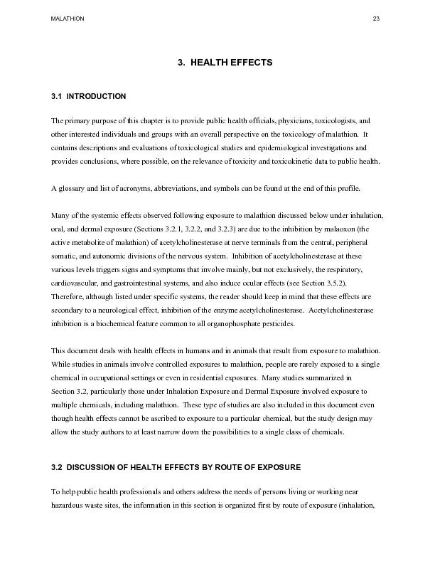
/
MALATHION HEALTH EFFECTS mediating the toxic effects as a consequence MALATHION HEALTH EFFECTS mediating the toxic effects as a consequence

MALATHION HEALTH EFFECTS mediating the toxic effects as a consequence - PDF document

lois-ondreau
lois-ondreau . @lois-ondreau
Follow
471 views
Uploaded On 2016-06-24

MALATHION HEALTH EFFECTS mediating the toxic effects as a consequence - PPT Presentation

MALATHION HEALTH EFFECTS There are no adequate data to evaluate whether pharmacokinetics of malathion in children are different from adults There is no information to evaluate whether metabolism o ID: 375418

MALATHION HEALTH EFFECTS There are

Share:

Link:

Embed:

Download Presentation from below link

Download Pdf The PPT/PDF document "MALATHION HEALTH EFFECTS mediating the t..." is the property of its rightful owner. Permission is granted to download and print the materials on this web site for personal, non-commercial use only, and to display it on your personal computer provided you do not modify the materials and that you retain all copyright notices contained in the materials. By downloading content from our website, you accept the terms of this agreement.


Presentation Transcript

��MALATHION HEALTH EFFECTS mediating the toxic effects as a consequence of oxidative stress, and (3) the persistence of DNA damage induced by inhibition of repair enzymes. The cells used will be splenocytes and thymocytes from C57B1/6 mice. The research is sponsored by the U.S. Department of Agriculture. ��MALATHION HEALTH EFFECTS There are no adequate data to evaluate whether pharmacokinetics of malathion in children are different from adults. There is no information to evaluate whether metabolism of malathion is different in children than in adults since the specific P-450 enzymes involved in the metabolism are not known. Only one report was found regarding levels of malathion (or metabolites) in human milk (Roggi et al. 1991). There is evidence in animals that it (or its metabolites) can be transferred via breast milk to the offspring acenta (Machin and McBride 1989b; Mathews and Devi 1994). Further information on the dynamics of malathion and metabolites during pregnancy and lactation would be useful. Biomarkers of exposure need to be further studied in order to better estimate human exposure at all age levels following acute or chronic exposure to malathion. There are no data on the interaction of malathion with other chemicals in children. Studies in animals have suggested that malnutrition, as may occur among some sectors of the general population, may exacerbate the toxicity of malathion (Bulusu and Chakravarty 1984; Prabhakaran and Devi 1993). Further studies on children from undernourished populations should be conducted to explore this issue. The information available indicates that methods to reduce peak absorption of malathion and to interfere with the mechanism of action used for intoxication in adults are applicable to children. Child health data needs relating to exposure are discussed in 6.8.1 Identification of Data Needs: Exposures of Children. The following ongoing studies concerning health effects associated with malathion have been identified in the Federal Research in Progress (FEDRIP 2002) database. Dr. R.L. Carr at the Mississippi State University has proposed to determine the effects on behavior and neurochemical function in animals exposed to chronic low levels of malathion and other insecticides. Laboratory rats will be exposed at birth and continue until weaning, a critical time of central nervous system development. The research is sponsored by the U.S. Department of Agriculture. Dr. H.P. Misra at the Virginia Polytechnic Institute plans to investigate the mechanism of potential genotoxicity of malathion and pesticide mixtures in immune cells . Specific studies include induction of apoptotic cell death by pesticides, (2) the role of p38 MAP kinase and NF-kappa B in ��MALATHION HEALTH EFFECTS information is available on dealing with long-term, low-level exposures. This may be due, in part, to the limited information on toxic effects associated with such exposures. If additional information becomes term exposures, then studies examining methods for mitigating the effects of such exposures would become a data need. Children’s Susceptibility. Information on the effects of malathion in children is derived mainly from case reports of accidental ingestion of high amounts of commercial formulations (Ekin 1971; Healy 1959; Juši 1978; Tuthill 1958) and cases of dermal exposure (Parker and Chattin 1955; Ramu et al. 1973). In all of these cases, exposure to malathion resulted in the characteristic signs and symptoms eased salivation and lacrimation, miosis, nausea, vomiting, abdominal cramps and diarrhea, excessive bronchial secretions and dyspnea, bradycardia and low blood pressure, and muscle fasciculations. One fatality occurred among the cases described by Ramu et al. (1973). A case report of aplastic anemia in a 12-year-old child following inhalation of malathion fumes after fumigation of a home was described by Reeves et al. (1981), but this case seems to be unique; besides, there is no evidence that malathion was the causal agsignificant differences in the responses between children and adults. Studies in animals have shown that young animals are more susceptible to the toxicity of high doses of malathion and that this is related to and DuBois 1967; Lu et al. 1965; Mendoza 1976; Mendoza and Shields 1976, 1977). Data on developmental effects of malathion in humans are limited. Two studies conducted in California after aerial spraying of malathion did not find consistent or significant developmental effects in the offspring from women who were pregnant during the spraying (Grether et al. 1987; Thomas et al. 1992). An additional study of paternal exposure to malathion found no significant association between exposure and congenital malformations (García et al. 1998). Modevelopmental toxicant when administered at doses that are not maternally toxic (Khera et al. 1978; Lochry 1989; Machin and McBride 1989a; Schroeder 1990; Siglin 1985). However, some uncertainty still remains, largely because of limitations of experimental design or reporting in some studies. Therefore, a well-designed developmental study would be useful. Also, as previously mentioned, a developmental neurotoxicity study in rats in which pups are tested at various ages after being exposed and/or via maternal milk would fill a data gap. There is no evidence that malathion has hormone­ ��MALATHION HEALTH EFFECTS Defining the source of malaoxon responsible for the acute neurotoxicity of malathion will be critical to our understanding of human hazards. The source may not be a single organ (as was discovered for parathion) (Nakatsugawa 1992). Although the detoxification of malathion is mainly set by carboxylesterase, other biotransformation enzymes assume a much greater role when carboxylesterase is suppressed. Since the suppression occurs to varying degrees with all commercial formulations, there is also a need to define the nature and significance the metabolism of malathion and malaoxon. Human hazards from malathion will be better understood through further studies of absorption of malathion. Here again, studies of absorption in isolation from the influence of carboxylesterase may reveal clearer data. In addition, relationships between physical properties such as partition coefficients and absorption of chemicals were explored in early literature without clear conclusions. The reported lack of clear correlation in early literature (Ahdaya et al. 1981; Shah et al. 1981), for example, may need to be reexamined. The technique employed seems inadequate to estimate such physical parameters accurately, relying on near-background level counts to estimate the concentration in the oil phase. More reliable partition coefficients have been published. Meaningful distribution studies may only be possible when better metabolic data and techniques have been secured. When the levels of malathion and malaoxon are obscured by the rapid metabolism, more data on just the distribution of both the parent insecticide and metabolites would reveal little additional information. Comparative Toxicokinetics. Absence of carboxylesterase in human blood is a conspicuous departure from the rat model, which has most often been employed. Comparative studies involving volunteers, rats, mice, and perhaps other mammals may reveal useful patterns. Interestingly, fairly wide differences between rats and mice are already apparent. Umetsu et al. (1977) tested the acute oral toxicity of malathion samples in female Sprague-Dawley rats and Swiss white mice fasted 6 hours before dosing. For a technical malathion, the LD50 for the rat was 1,500 mg/kg and for mice was similar at 1,850 mg/kg. For a 99.3% pure malathion, the LD50 values were 9,500 mg/kg for rats and 3,000 mg/kg for mice. For a sample purified by recrystallization, the respective values were 12,500 and 3,600 mg/kg. Comparison seems valid since all tests were done during the course of a single study using the same source of animals. Comparative data for metabolism, particularly for carboxylesterases in various tissues in various species against malathion and malaoxon will contribute greatly to our understanding of potential toxic effects on humans. Methods for Reducing Toxic Effects. There is good information on the procedures used to limit absorption and to interfere with the mechanism of action of organophosphates, including malathion, after acute exposures (Aaron and Howland 1998; Carlton et al. 1998; Osmundsen 1998). However, no ��MALATHION HEALTH EFFECTS response relationships, to test other organophosphates to determine specificity, and to test cells from occupationally and accidentally (or intentionally) exposed subjects. There are no biomarkers of effect specific for malathion. As an organophosphate pesticide, malathion in sufficient amounts, produces typical signs and symptoms of cholinergic stimulation. Plasma and RBC cholinesterase levels are widely used as biomarkers of exposure to organophosphates, but alone, their levels do not predict whether adverse health effects will occur except in cases of significant inhibition (Maroni 2000). Because baseline data for plasma and RBC cholinesterase are not usually available for nonoccupationally exposed individuals, additional studies of normal values by age and sex are needed for assessing potential adverse effects. Absorption, Distribution, Metabolism, and Excretion. Among the areas of absorption, distribution, metabolism, and excretion, the greatest data needs seem to lie in metabolism. The most unique feature of malathion toxicokinetics (i.e., the extremely rapid hydrolysis at the carboxylester linkages) is still poorly understood. Since carboxylesterase activities are the major determinant of the malathion and malaoxon levels , many aspects of this enzyme need to be studied. Although it is known that carboxylesterase exists in more than one form, details remain unknown. The question of whether the same carboxylesterase hydrolyze both malathion and malaoxon is still unansunknown whether the reported variable ratio of monocarboxylic acid metabolites is due to the variation in the enzyme or to variable contributions of different isozymes. re of the enzyme that yields dicarboxylic acid from either of the monocarboxylic acids. Beyond the isozyme question, kinetic data on these enzymes would also contribute to our understanding of toxicokinetics. Knowledge of Kmax values of isozymes in ilitate construction of useful PBPK models. Though the importance of malaoxon in the acute toxicity of malathion is unquestionable, generation, distribution, and metabolism of malaoxon has been little studied. This likely reflects the technical difficulty imposed by the overwhelmingly active carboxylesterase that prevents quantitation or even detection of malaoxon. Such difficulties may be overcome by inhibiting carboxylesterase as was demonstrated by the increase in cytochrome P-450 binding of malathion and malaoxon in the presence of bis-(p-nitrophenyl)phosphate, a carboxylesterase inhibitor (Stevens and Greene 1973). studies of distribution and metabolism may also benefit from similar strategies. Suppression of carboxylesterase in Sprague-Dawley rats has been accomplished with TOTP. ��MALATHION HEALTH EFFECTS Epidemiological and Human Dosimetry Studies. Information on the health effects of malathion in humans is derived from case reports of accidental or intentional exposure to malathion (Choi et al. 1998; Crowley and Johns 1966; Dive et al. 1994; Faragó 1967; Juši 1978; Monje Argiles et al. 1990; Morgade and Barquet 1982; Namba et al. 1970; Ramu et al. 1973; Tuthill 1958; Zivot et al. 1993), epidemiological studies (Grether et al. 1987; Kahn et al. 1992; Thomas et al. 1992), studies of exposure to multiple pesticides including malathion (Brown et al. 1990, 1993; Cantor et al. 1992; Ernest et al. 1995; Hermanowicz and Kossman 1984; Peedicayil et al. 1991; Rupa et al. 1991b; Stålberg et al. 1978; Zahm 1997; Zahm et al. 1993), and controlled exposure studies (Golz 1959; Milby and Epstein 1964; Moeller and Rider 1962). The most likely identifiable subpopulations exposed to malathion are pesticide applicators, farm workers, individuals involved in the production of malathion, and individuals exposed in homes after residential application. Well-designed epidemiological studies of exposed workers and follow-up evaluations of cohorts from the general population who may have been exposed during aerial application of the pesticide are needed. Specific assessment of cancer risks and examination of the effects of malathion on the nervous system and immune systems are needed. The nervous system is a known e long-term effects of acute exposure. Malathion seems to have modulatory effects on the immune system of animals at noncholinergic dose levels. Therefore, evaluation of the immune status of exposed humans would be important. Studies in animals have shown that impurities in malathion formulations play an important role in the toxicity of malathion and have inherent toxicity themselves. Identification of impurities and inert ingredients in commercial formulations as well as potential levels of exposure seems indicated. Biomarkers of Exposure and Effect. Malathion metabolites (MCA, DCA, DMPT, DMthe general U.S. population and from workers exposed to the pesticide (Fenske 1988; Krieger and Dinoff 2000; Kutz et al. 1992; MacIntosh et al. 1999b; Warren et al. 1985). In the general population, MCA was the most abundant and was found in only a small percentage of the samples (Kutz et al. 1992; MacIntosh et al. 1999b). Since malathion does not seem to accumulate in the body, the presence of malathion metabolites in the general population probably reflects continuous background exposure via the food ounts. Additional studies of the general population correlating malathion metabolite levels with health status as well as with dietary habits would provide useful information for risk characterization and risk assessment. Suggestive evidence of malathion-induced specific mutations in human T-lymphocytes exposed was presented by Pluth et al. (1996, 1998). Further studies on this issue are needed to establish dose­ ��MALATHION HEALTH EFFECTS Lee and Tai 2001; Matsukawa et al. 1997; Monje Argiles et al. 1990; Namba et al. 1970; Ramu et al. 1973). As an organophosphate pesticide, malathion inhibits the activity of the enzyme acetylcholinesterase as well as that of plasma cholinesterase. The inhibition of acetylcholinesterase at various levels within the nervous system produces a characteristic set of signs and symptoms including respiratory distress, bradycardia, increased bronchial secretions, excessive salivation, lacrimation, pupillary constriction, fasciculations, abdominal cramps, and diarrhea (Aaron and Howland 1998; Carlton et al. 1998; Osmundson 1998). Most of these signs and symptoms have been observed in the cases listed above. In addition, abnormal electromyographic findings were reported in some studies of documented ingestion of malathion (Crowley and Johns 1966; Dive et al. 1994; Monje Argiles et al. 1990). A study of controlled administration of malathion in capsules to volunteers identified a NOAEL and LOAEL for inhibition of plasma cholinesterase of 0.23 and 0.34 mg/kg/day, respectively (Moeller and Rider 1962), and these findings were used to derive an intermediate oral MRL for malathion. Studies of workers exposed to several pesticides have also documented inhibition of plasma and RBC cholinesterase, and in some cases, the degree of enzyme inhibition has been correlated with the presence or absence of clinical signs (Ernest et al. 1995; Peedicayil et al. 1991; Stlberg et al. 1978). Information is lacking on potential effects of long-term, low-level exposure to malathion as well as on potential long-term effects of acute high exposure to malathion. This information can only be obtained from evaluation of cohorts exposed only to malathion, but data from subjects exposed to a few organophosphates would also be helpful. Studies in animals support the findings in humans. In addition to measurements of cholinesterase activity, a few studies have examined the effects of malathion on neurobehavioralstudy that tested a functional observation battery in rats reported increased motor activity 21 days after a single gavage dose of 2,000 mg/kg of malathion (88% pure) (Ehrich et al. 1993), whereas a similarly designed study found decreased motor activity 14 days after dosing with 2,000 mg/kg malathion (96.4% pure) (Lamb 1994a). Similar inconsistencies were seen between two 90-day feeding studies (Desi et al. 1976; Lamb 1994b). Further studies are needed to clarify these inconsistencies. Chronic-duration studies have reported inhibition of plasma and RBC cholinesterase activity in both rats (Daly 1996a) and mice (Slauter 1994) and body tremors in female mice after 70 weeks of dosing with approximately 2,980 mg/kg/day of malathion (NCI 1978). Should additional chronic studies be conducted, microscopic examination of nervous tissues from both peripheral and central nervous system may be conducted. Also, a subgroup of animals could be tested for possible subtle neurobehavioral alterations of long-term, low-level exposure. Finally, pilot studies should be designed to evaluate possible neurodevelopmental effects of gestational and lactational exposure to malathion. ��MALATHION HEALTH EFFECTS application of malathion in Southern California (Schanker et al. 1992). No delayed hypersensitivity was seen among these cases. Another study of subjects in Southern California following aerial spraying of malathion found no significant increases in the number of visits for allergic problems to hospital emergency departments during the application period compared with the prespray period or the corresponding period the previous year (Kahn et al. 1992). No information was located regarding immunological effects in humans following oral exposure to malathion. Studies in animals suggest that malathion can induce both immune enhancement and immune depression at noncholinergic dose levels. A number of studies conducted by Rodgers and colleagues showed that malathion stimulated immune cell function. For example, relatively low (1–2 mg/kg) single oral or dermal doses �of malathion (99% pure) increased serum histamine levels in rats and mice (Rodgers and Xiong 1997b). Daily oral dosing for 14 days with 1 mg/kg/day of malathion stimulated macrophage function in mice (Rodgers and Xiong 1997d). Immunosuppressive effects were reported mostly in repeated dosing studies. Mice administered malathion in the diet for 3–12 weeks showed alterations in both humoral immune function and cell mediated immunity (Banerjee et al. 1998). Immune suppression was also seen in rats and rabbits exposed to malathion for intermediate-duration periods (Banerjee et al. 1998). Cell mediated suppression was also observed in mice administered malathion for 90 days at 1 and 10 mg/kg/day, but cell mediated enhancement was seen at 0.1 mg/kg/day (Rodgers and Xiong 1997c). Enhancement of antibody production was reported in female mice treated with 0.018 mg/kg/day in a 28-day study (Johnson et al. 2002). The available studies in animals indicate that malathion modulates immune parameters at relatively low doses, but further research is needed to determine whether these observed responses place the animals at increased riskEllefson (1992) suggested that malathion-induced mast cell degranulation may be due to inhibition of an esterase on the cell surface; therefore, further research should explore this possibility. The immune enhancing properties of malathion may be relevant for people with autoimmune disease as their condition may aggravate if exposed to malathion. As suggested by Rodgers and Xiong (1997b), it is also possible that the immune enhancement by malathion may be related to reports of possible contact hypersensitivity (Milby and Epstein 1964) or skin rashes and irritation after malathion applications for pest control (CDHS 1991; Schanker et al. 1992). Neurotoxicity. Information in both humans and animals indicates that the nervous system is the main target of malathion-induced toxicity following acute exposure by any route. This is particularly evident after exposure to high doses of malathion, as has occurred, for example, in cases of accidental or intentional ingestion of malathion formulations (Choi et al. 1998; Dive et al. 1994; Juši and Mili 1978, ��MALATHION HEALTH EFFECTS who discussed a possible association between the use of a hair lotion containing malathion by a woman during weeks 11 and 12 of pregnancy and the birth of a severely malformed child. However, a causal link is difficult to establish. No information was located regarding developmental effects in animals following inhalation or dermal exposure to malathion. Transfer of malathion (and/or metabolites) to the fetus through the placenta and via maternal milk has been demonstrated indirectly (Chhabra et al. 1993; Mathews and Devi 1994). Oral studies in rats and rabbits have not shown embryotoxicity (Khera et al. 1978; Lochry 1989; Machin and McBride 1989a; Siglin 1985) or showed embryotoxicity at doses that also caused maternal toxicity (Prabhakaran et al. 1993). However, reduced fetal weight and crown-rump length were seen in mice in a study that provided no information on maternal effects (Asmatullah et al. 1993). A study by Kalow and Martin (1961) found increased neonatal mortality in rats treated for at least 5 months before pregnancy, but again, no information was presented on maternal effects, which seriously weak2-generation study, the only developmental effect noticed was a decrease in body weight gain in pups from the F1A and F2B litters during the lactation period at parental doses of 394 mg/kg/day of malathion for males and 451 mg/kg/day for females, with corresponding NOAELs of 131 and 153 mg/kg/day (Schroeder 1990). No teratogenic effects of malathion were reported in the studies evaluated (Khera et al. 1978; Lechner and Abdel-Rahman 1984; Lochry 1989; Prabhakaran et al. 1993; Schroeder 1990; Siglin 1985). Although most of the data suggest that malathion is not a developmental toxicant, there is still uncertainty regarding whether effects could occur at doses not causing maternal toxicity, largely because of limitations of experimental design or reporting in some studies. Therefore, a well-designed study is warranted. In addition, a developmental neurotoxicity study in rats in which pups are tested at various ages after being exposed in utero and/or via maternal milk would fill a data gap. The oral route should be preferred to complement the existing information and because there is no evidence that the toxicity of Immunotoxicity. There is some information suggesting that exposure to malathion may affect the immune system in humans. Albright et al. (1983) described a case of possible immune complex ansient renal insufficiency with massive proteinuria after spraying intensively with malathion. Impairment of neutrophil chemotaxis was observed among a group of workers exposed to organophosphate pesticides, including malathion, for 0.1–29 years (Hermanowicz and Kossman 1984). Malathion was shown to be a contact sensitizer in a laboratory study with volunteers (Milby and Epstein 1964). A case of immediate IgE reaction to malathion and another of irritant reaction to malathion and to the bait was described among ��MALATHION HEALTH EFFECTS No information was available regarding reproductive effects in animals following inhalation exposure to malathion. Acute studies in male animals, mainly rats, administered malathion orally have reported transient alterations in spermatogenesis and alterations in the seminiferous tubule epithelium (Krausse 1977; Krausse et al. 1976; Ojha et al. 1992; Piramanayagam et al. 1996), but none of these studies assessed reproductive function. A 12-week gavage study also reported reversible testicular alterations in rats given 45 mg/kg/day malathion (Balasubramanian et al. 1987b), but no morphological alterations were seen in the reproductive organs from male rats or mice in chronic bioassays (NCI 1978, 1979a). An intermediate-duration dermal study in male and female rabbits did not microscopic alterations in the reproductive organs following application of up to 1,000 mg/kg/day of malathion 6 hours/day, 5 days/week for 3 weeks (Moreno 1989). Malathion was not a reproductive toxicant when administered to females at doses that did not induce maternal toxicity (Lechner and Abdel-Rahman 1984; Mathews and Devi 1994; Prabhakaran et al. 1993; Siglin 1985). A 2-generation study in rats found no significant effects on reproductive performance, fertility indices, and gestation length (Schroeder 1990). The only significant finding in a chronic bioassay was an increased incidence of cystic endometrial hyperplasia in mice treated with approximately 1,490 mg/kg/day of malathion for 80 weeks (NCI 1978). The overall evidence in animals suggests that malathion is not a reproductive toxicant, but the less than optimal quality of some studies do not allow drawing a firm conclusion. The issue of testicular toxicity should be further explored in studies in animals exposed at various ages including exposure in utero and later tested for reproductive performance. Also, studies examining standard semen and sperm parameters in adult animals would provide valuable information. These studies should be conducted by the oral route of exposure since this route is the most relevant for exposure of the general population and there is no evidence suggesting that the toxicity of malathion is route-specific. Developmental Toxicity. Data on developmental effects of malathion in humans are limited. Two studies conducted in California after aerial spraying of malathion did not find consistent or significant developmental effects in the offspring from women who were pregnant during the spraying (Grether et al. 1987; Thomas et al. 1992). Some positive associations were found in the former study between exposure and some anomalies, but no biologically consistent pattern was observed. Thomas et al. (1992) found a significant association for gastrointestinal anomalies and second trimester exposure, but the gastrointestinal tract is completely developed by the second trimester. An additional study of paternal exposure to malathion found no significant association between exposure and congenital malformations (García et al. 1998). No information was found regarding developmental effects in humans following oral exposure to malathion. The only data after dermal exposure are from Lindhout and Hageman (1987), ��MALATHION HEALTH EFFECTS were reported in mouse spermatogonia by Degraeve and Moutschen (1984) and tests for dominant lethal mutation also were negative in that study. In contrast, Salvadori et al. (1988) reported chromosomal aberrations in mouse spermatogonia and Hoda et al. (1993) noticed a decrease in meiotic index in primary spermatocytes of mice treated with malathion. A study in Drosophila also showed a positive dominant lethal mutation (Kumar et al. 1995). In general, the results from standard gene mutation tests in bacteria did not show mutagenicity with or without activation or gave a weak positive response (Pednekar et al. 1987; Shiau et al. 1980; Wong et al. 1989). Results from studies in mammalian cells, including human lymphocytes, showed that malathion appeared to produce cytogenetic damage as evidenced by chromosomal aberrations and sister chromatid exchanges (Balaji and Sasikala 1993; Garry et al. 1990; Nicholas et al. 1979; Nishio and Uyeki 1981; Pluth et al. 1996, 1998; Sobti et al. 1982; Walter et al. 1980). There is weak evidence of interaction of DNA bases by malathion (Wiaderkiewicz et al. 1986). Although it appears that malathion has been tested for genotoxicity in a wide variety of systems, the results have been mixed largely because of the different experimental designs and different malathion formulations tested. Studies in vitro should always monitor cell viability to clearly distinguish genetic effects from cytotoxicity. Also, it would be important to know the composition of the malathion mixture to be tested and to test each one of the components since humans are likely to be exposed to technical mixtures rather than to pure malathion. Additional studies are also necessary to examine the interaction of malathion and DNA. Continued studies on genomic sequence analysis combined with reverse transcription polymerase chain reaction (PCR) amplification of lymphocytes from malathion exposed subjects may be able to identify a mutation spectrum that may be fairly specific for malathion and that could eventually be used as a biomarker of exposure. Reproductive Toxicity. Limited information was located regarding reproductive effects in humans following exposure to malathion. A study of pregnant women exposed during periods of malathion spraying in California found a moderate association between stillbirths and certain exposure variables, but these associations were not consistent or statistically significant and, therefore, were attributed to chance (Thomas et al. 1992). An additional study of males involved in the mixing and spraying of a variety of higher percent of stillbirths and abortions compared to unexposed couples, and also lower fertility among exposed males (Rupa et al. 1991b). However, the role of malathion, if any, is impossible to determine. No studies were located on reproductive effects of malathion in humans exposed orally or dermally. ��MALATHION HEALTH EFFECTS Malathion has been tested in oral bioassays conducted in rats (two strains) (Daly 1996a; NCI 1978, 1979a) and mice (NCI 1978; Slauter 1994). The metabolite, malaoxon, also has been tested in rats (Daly 1996b; NCI 1979b) and mice (NCI 1979b). No clear evidence of carcinogenicity for malathion was found in the bioassays conducted by NCI (NCI 1978, 1979a), but there was evidence of liver carcinogenicity in female Fischer-344 rats in the Daly (1996a) study and in male and female B3C6Fmice in the Slauter (1994) study. In the two positive studies, evidence of carcinogenicity occurred at doses that were considered excessive (EPA 2000a, 2000b). There was no evidence of carcinogenicity for malaoxon in rats or mice (Daly 1996b; NCI 1979b), but upon reevaluation of the NCI (1979b) study, NTP concluded that there was equivocal evidence of carcinogenicity for male and female Fischer-344 rats based on reinterpretation of C-cell neoplasms of the thyroid (Huff et al. 1985). Further bioassays do not seem necessary. A recent review of the literature on pesticides and cancer by Zahm et al. (1997) identified several areas where data gaps exist. First, improvements in exposure assessment in epidemioand reliability studies of recall of and nonoccupationally exposed populations. In addition, efforts should continue to try to gain access to information on inert ingredients of pesticide formulations since inert components are not necessarily biologically inert. Another data gap identified is the lack of studies on migrant and seasonal farm workers who often start exposure at an early age. The mechanism of malathion-induced carcinogenicity in animals is not known, but it does not appear to involve mutagenicity. Additional studies investigating noncholinergic mechanism of malathion toxicity and how these may potentially by involved in malathion carcinogenicity would be valuable. Genotoxicity. The genotoxicity of malathion has been investigated in many studies. Of four studies that monitored humans exposed to malathion occupationally or by intentional ingestion of malathion formulations, two found increased chromosomal aberrations in peripheral lymphocytes and micronuclei (Singaravelu et al. 1998; van Bao et al. 1974), whereas the remaining two found no evidence of micronuclei or chromosomal aberrations (Titenko-Holland et al. 1997; Windham et al. 1998). The majority of the studies in animals were conducted in mice and showed that malathion has the capacity to produce chromosomal changes, including chromosomal aberrations and micronuclei in bone marrow (Abraham et al. 1997; Amer et al. 2002; Dulout et al. 1982, 1983; Giri et al. 2002; Kumar et al. 1995). However, a negative effect for chromosomal aberrations in mouse bone marrow was reported by Degraeve and Moutschen (1984) and a weak positive (1986) in hamster bone marrow. Effects on germ cells have been mixed. No chromosomal aberrations ��MALATHION HEALTH EFFECTS nhibition of plasma and RBC cholinesterase, clinical signs, and neurophysiological alterations (Baker et al. 1978; Ernest et al. 1995; Peedicayil et al. 1991; lberg et al. 1978). A study of pesticide appliorganophosphate insecticides and retinal degeneration, but a definite conclusion c(Kamel et al. 2000). Information is lacking on potential effects of long-term, low-level exposure to malathion on many end points. This information can only be obtained from evaluation of cohorts exposed only to malathion, but data from subjects exposed to a limited number of organophosphates would also be helpful. No studies were located that evaluated the effects of chronic inhalation or dermal exposure in animals, but because malathion is relatively rapidly degraded in the environment, no such exposures are expected to occur for the general population or for people living near waste sites. Several long-term bioassays have been conducted in rats and mice that provided information on noncancer effects (death, systemic, and neurological effects) (Daly 1996a; NCI 1978, 1979a; Slauter 1994). The lowest LOAEL was 29 mg malathion/kg/day for 29% inhibition of plasma cholinesterase in male Fischer-344 rats in a study by Daly (1996a); the NOAEL was 2 mg/kg/day and was used to derive a chronic oral Several studies have examined the possible associatiocertain types of cancer, particularly non-Hodgkin’s lymphoma (NHL) and leukemia. However, because workers are usually exposed to multiple pesticides, it is difficult to establish associations with specific pesticides. Still, information relevant to malathion is available in some of these studies. Exposure to malathion was associated with increased risk of NHL in a study of men in Iowa and Minnesota (Cantor et al. 1992), of women in Nebraska (Zahm et al. 1993), and of men in Canada (McDuffie et al. 2001). A retrospective cohort study of 32,600 employees of a lawn care company found no specific association between deaths due to NHL and exposure to malathion (Zahm 1997) and neither did a study of pesticide users from Iceland (Zhong and Rafnsson 1996). The study of men in Iowa and Minnesota also found an elevated risk of leukemia in men who used malathion as an animal �insecticide for 10 days/year (Brown et al. 1990). A slight non-significant increased risk for multiple myeloma was observed among farmers who used malathion in an additional study among men in Iowa, but failure to use protective equipment was not associated with increased risk (Brown et al. 1993). In spite of some positive associations, there is no clear evidence that exposure to malathion causes cancer in humans. As previously mentioned, the magnitude of the excesses is generally small, exposure assessment is unreliable, and mixed exposure always occurs. ��MALATHION HEALTH EFFECTS duration oral MRL. Studies in animals provided information on lethality (NCI 1978, 1979a), systemic effects (Desi et al. 1976; Foster 1968; Krause et al. 1976; Lox and Davis 1983; Ozmen and Akay 1993), immunological effects (Banerjee et al. 1998; Desi et al. 1978; Rodgers and Xiong 1997c), neurological effects (Desi et al. 1976, 1978; Fischer 1988; Husain et al. 1987; Lamb 1994b), and reproductive and developmental effects (Balasubramanian et al. 1987a, 1987b; Kalow and Marton 1961; Krause et al. 1976; Lechner and Abdel-Rahman 1984; Ozmen and Akay 1993; Schroeder 1990). Of special interest is a 60-day study by Lox and Davis (1983), which reported hepatocyte degeneration in rats at the very low dose of approximately 0.15 mg/kg/day of malathion (99% pure) in drinking water (1 ppm in water). This is one of the few studies in which malathion was administered in the drinking water. Since no liver histopathology was described in any other intermediate-duration oral study with much higher malathion doses either in the food or by gavage, the findings of Lox and Davis (1983) should be interpreted with caution until such results can be replicated. Also of interest is the finding of increased peritoneal macrophage function in mice treated for 90 days with 0.1–10 mg/kg/day by gavage (Rodgers and Xiong 1997c). However, as Rodgers and Ellefson (1992) pointed out, the physiological significance of the magnitude of this effect is unknown. An additional 28-day immunological study in mice reported that doses as low as 0.018 mg/kg/day of commercial-grade malathion increased the primary immune response to immunization with SRBC (Johnson et al. 2002). Dermal data in animals were limited to information on lethality (Dikshith et al. 1987), systemic effects (Boyes et al. 1999; Moreno 1989), and neurological effects (Boyes et al. 1999; Dikshith et al. 1987; Moreno 1989). Results regarding systemic effects after dermal exposure were insufficient to construct dose-response relationships, but additional studies may not be necessary since malathion is rapidly degraded in the environment and long-term dermal exposure is not expected to occur for the general population or for people living near waste sites. Chronic-Duration Exposure and Cancer. Limited information was found regarding health effects in humans after chronic-duration exposure to malathion. Information on effects of chronic exposure is derived mostly from studies of workers in which both the inhalation and dermal routes of exposure play significant roles. It should also be noted that although occupational exposure is generally assumed to be chronic, for some types of occupations (i.e., pesticide applicators), exposures are usually seasonal, involving only a few weeks or months per year. A study of workers exposed to organophosphates (not only malathion) for up to 29 years found a higher frequency of respiratory tract infections among the workers than in controls (Hermanowicz and Kossman 1984). The authors also observed marked impairments of neutrophil chemotaxis and significantly decreased neutrophil adhesion among the workers. A study at a pesticide manufacturer (primarily malathion) found an inverse relationship between hemoglobin concentration and duration of employment (Singaravelu et al. 1998). Studies of workers ��MALATHION HEALTH EFFECTS Parker and Chattin (1955) also described a case of a 10-year-old girl who had neurological manifestations of poisoning after having extensive dermal contact with a malathion formulation in the form of flakes. Also, laboratory studies in volunteers showed that a single exposure to 10% malathion (95% pure) induced contact sensitization in almost half of the subjects, and that 0.1 and 0.01% concentrations of 99.3% malathion were able to evoke positive responses in previously sensitized individuals (Milby and Epstein 1964). Information on effects of malathion afin animals was limited a study of lethality in rats (Gaines 1960), a study of delayed-type hypersensitivity in mice (Cushman and Street 1983), and a report on subtle immunological alterations in rats and mice following a single dermal application of malathion (Rodgers and Xiong 1997c). Qualitative data (no dose level) on RBC and plasma cholinesterase were also available from a study in dogs (Vestweber and Kruckenberg 1972). Although the information available does not suggest that the toxicity of malathion is route-dependent, further well-conducted acute dermal studies seem necessary to establish dose-response relationships for a range of end points. This is important because considerable dermal exposure to malathion can occur for the residential handler and during post-application events. Furthermore, under certain exposure scenarios such as following aerial application of malathion over urban areas, dermal doses may be orders of magnitude higher than inhalation doses (Marty et al. 1994). Few intermediate-duration studies were located that described health effects in humans specifically exposed to malathion. However, it is reasonable to assume that many of the studies on cohorts exposed occupationally to malathion, or to pesticides in general, described in Section 3.2, included subjects who may have been exposed for intermediate durations. Limited information is available from a controlled inhalation study in volunteers that identified a NOAEL of 85 mg/m for body weight and neurological effects (cholinesterase activity) (Golz 1959); subjects were exposed to malathion aerosols 2 hours/day for 42 days. Limited data were also located in the studies available regarding effects in animals exposed to malathion in the air. A 13-week study in rats reported hyperplastic changes in the epithelium from the upper respiratory tract following intermittent exposure to 100 mg/mtion in RBC cholinesterase activity at 450 mg/mand higher (Beattie 1994). The LOAEL of 100 mg/m was used to derive an intermediate inhalation MRL for malathion. A study in humans administered malathion in capsules for up to 56 days identified a NOAEL of mg/kg/day for hematological and renal effects; this dose level also constituted a LOAEL for neurological effects (plasma and RBC cholinesterase activity) (Moeller and Rider 1962). The NOAEL for neurological effects was 0.23 mg/kg/day and was used as the basis for derivation of an intermediate­ ��MALATHION HEALTH EFFECTS agricultural workers (mixers and applicators) evaluated dermal exposure to malathion by monitoring urinary dimethyl phosphorothioic acid and -dimethyl phosphorodithioate (Fenske 1988). Urinary metabolites monitored for 3 days following exposure showed that applicators excreted 17% of the applied dose estimated by fluorescent tracer technique, and mixers 23%. In a poisoning case involving suicidal intravenous injection of about 1.8 g of malathion, serum concentration of malathion in the range of 0.029–0.349 µg/ml between 6 and 24 hours postinjection 2.89 hours for serum malathion (Lyon et al. 1987). A single dose of 2.5 mg/kg of methoxy C-malathion in 0.3 mL of saline was intravenously administered to male Sprague-Dawley rats and the whole animal was frozen in a dry ice/hexane for autoradiography after 30 minutes (Saleh et al. 1997). As a percent of the recorded radioactivity of sagittal sections, the small intestine contained 21% of the radioactivity and the urinary tract contained 7%, leading the authors to conclude that the bile is a major route of excretion. Physiologically Based Pharmacokinetic (PBPK)/Pharmacodynamic (PD) Models Physiologically based pharmacokinetic (PBPK) models use mathematical descriptions of the uptake and disposition of chemical substances to quantitatively describe the relationships among critical biological processes (Krishnan et al. 1994). PBPK models are also called biologically based tissue dosimetry models. PBPK models are increasingly used in risk assessments, primarily to predict the concentration of potentially toxic moieties of a chemical that will be delivered to any given target tissue following various combinations of route, dose level, and test species (Clewell and Andersen 1985). Physiologically based pharmacodynamic (PBPD) models use mathematical descriptions of the dose-response function to target tissue dose and toxic end points. PBPK/PD models refine our understanding of complex quantitative dose behaviors by helping to tissue dose of the toxic moiety, and (2) the target tissue dose and observed responses (Andersen et al. 1987; Andersen and Krishnan 1994). These models are biologically and mechanistically based and can be used to extrapolate the pharmacokinetic behavior of chemical substances from high to low dose, from ��MALATHION HEALTH EFFECTS Sprague-Dawley rats in which the animals were gavaged with malathion at 0.00001–0.1 (69 mg) the LDdose, metabolites in urine comprised predominantly (up to 90%) dicarboxylic acid, followed by monocarboxylic acid; alkyl phosphates were minor components (Bradway and Shafik 1977). Dermal Exposure A study of dermal absorption of C-malathion (label position unspecified) in humans provides estimates of kinetic parameters of malathion elimination via the urine. Fitting urinary data to a model yielded elimination rate constants for humans, ranging from 0.094 to 0.129/hour (elimination half-time of 0.74-5.4 hours) for pure malathion and 0.079–0.130/hour (elimination half-time of 8.7–5-3 hours) for 10% aqueous malathion (Dary et al. 1994). Studies of occupationally exposed subjects have identified and quantified several malathion metabolites in the urine. In these cases, exposure is assumed to have been by both the inhalation and dermal routes, but the contribution of each specific route is unknown. In a study of date farm workers continually exposed to malathion in dusting and harvesting operations, mid-season Monday morning prework urine samples contained low or unmeasurable levels of malathion acids, indicating rapid metabolism and elimination (Krieger and Dinoff 2000). Estimated daily clearance of malathion metabolites provided a measure of daily dose. Depending on the task of the crew, the clearance ranged from 1 to 92 mg malathion equivalent/day. For loaders/applicators, the dose estimates were 0.4–1 mg malathion equivalent/kg/day. The profile for major urinary metabolites was: -dimethylphosphorotioate (48–53%), malathion monocarboxylic acids (23–27%), and malathion dicarboxylic acid (13–16%). These levels of "subchronic" exposure did not result in depression of either plasma or RBC cholinesterases. Dermal doses (281 or 51 mg) of malathion self-administered on forearms produced metabolite profiles similar to that obtained from oral dosing (Krieger and Dinoff 2000). A study of workers and residents exposed to malathion during a spraying operation in Haiti showed urinary malathion monocarboxylic acid at various times after the operation ranging from 0.9 to 6.8 mg/L after the spraying week and from 0.047 to 0.13 mg/L after the weekend (Warren et al. 1985). Residents having negligible levels of monocarboxylic acids when they returned home showed an increase to 0.084– mg/L over the weekend. In a group of residents who were tested 1 week after the operation, malathion monocarboxylic acids ranged from negligible to 0.31 mg/L, suggesting that malathion and/or metabolites may be more persistent in the environment than it had been previously thought. A study of ��MALATHION HEALTH EFFECTS No information was located regarding elimination of in the studies of occupational exposures discussed in Section 3.4.4.3 below, workers were most likely exposed by both the inhalation and dermal routes, but the contribution of each route is difficult to Oral Exposure In a study by Krieger and Dinoff (2000), malathion metabolites were analyzed in the urine of a volunteer who ingested single doses of 7.7 or 15.6 mg of malathion in gelatin capsules. Monocarboxylic acids were more abundant than dicarboxylic acid, and dimethyl phosphorothioic acid was the main alkylphosphate metabolite; more than 95% was recovered in urine. In an earlier study of a subject who ingested a high amount of malathion (200 mL of 50% malathion), analysis of the second 24-hour urine sample also showed monocarboxylic acids as the major metabolites followed by dimethyl phosphorothioic acid (Bradway and Shafik 1977). An estimated half-life of 6.2 hours for the fast phase of elimination was reported for a 43-year-old woman who iIn animals, elimination of ingested malathion occurs rapidly mainly via the kidney. For instance, male Holtzman rats eliminated 91.7% of radioactivity of a dose of 25 mg of C-ethyl malathion within 24 hours (83.4% in urine, 5.51% in feces, and 2.77% as CO); 7.75% remained in contents (Bourke et al. 1968). Urinary excretion at 8 hours was 44.1% of the administered dose. Similarly, in a study of pesticide combination on toxicokinetics, 10 mg/kg C-ethyl malathion given to fasted female Sprague-Dawley rats by gavage in 0.25 mL corn oil was excreted rapidly in the urine, 68% in 8 hours and 93% in 24 hours (Lechner and Abdel-Rahman 1986). A thin layer chromatography (TLC) analysis of chloroform extracts from acidified 24-hour urine provided a urinary metabolite profile: malathion 0.01%, malaoxon 1.44%, malathion monoacid 0.42%, and malathion diacid 0.06% of the dose. Carbaryl coadministered at 10 mg/kg altered the profile and raised malaoxon to 7.5% and malathion dicarboxylic acid to 1.48%. No additional confirmation of identity of malaoxon was reported. In a study in male Sprague-Dawley rats, about 90% of C-methoxy malathion (280 mg/kg by gavage) was excreted into urine within 24 hours of ingestion (Abou Zeid et al. 1993). In another study in male ��MALATHION HEALTH EFFECTS Dimethyl phosphorodithioic acid can only arise from malathion, and not from malaoxon. The enzymes responsible for this metabolism have not been studied. In a gavage study using doses of 0.069–69 mg/day in 10-fold increments for 3 days to 400–450 g male Sprague-Dawley rats, dimethyl phosphorothioic acid was the dominant phosphate metabolite, followed by dimethyl phosphorodithioic acid and dimethyl phosphoric acid (Bradway and Shafik 1977). The proportion of the latter two metabolites seems to shift with the dose, with dimethyl phosphoric acid dominating at lower doses and dimethyl phosphorodithioic acid being the dominant metabolite at the highest dose. Glutathione-linked Metabolism. Mouse liver homogenate contains a glutathione (GSH) S-transferase which demethylated malathion to yield demethyl malathion (Nomeir and Dauterman 1978). This may account for an earlier observation of a 10-fold enhancement of malathion metabolism by GSH with mouse liver homogenate in which esterases were suppressed by DFP (di-isopropyl fluorophosphate) (Bhagwat and Ramachandran 1975). A substantial yield of demethyl malathion in rat liver homogenate has also been reported though involvement of GSH is unknown (Matsumura and Ward 1966). Hepatocytes isolated from male Wistar rats were used to study the depletion of GSH by malathion, isomalathion, and trimethyl phosphorus esters (Malik and Summer 1982). Isomalathion was the most effective. GSH depletion by malathion was greatly increased when carboxylesterase was pre-inhibited by isomalathion, indicating the greater involvement of GSH-linked metabolism of malathion under those on of metabolites and identification of metabolites, mostly in urine (Rabovsky and Brown 1993). As is the case in studies of distribution, the use of radiolabeled malathion often yields a composite profile of malathion metabolites. No metabolic half-life information is available either for malathion or malaoxon. Many studies of malathion absorption provide information on urinary excretion, as discussed in Section 3.4.1. ��MALATHION HEALTH EFFECTS acid was produced more than monocarboxylic acid, with the ratio of mono/dicarboxylic acids decreasing with the decreasing dosage. In male Sprague-Dawley rats receiving 69–0.069 mg/day by gavage for 3 days, the ratios of monocarboxylic acids to dicarboxylic acid were 0.66, 0.21, 0.14, and 0.08 for 69, 6.9, 0.69, and 0.069 mg/day doses, respectively (Bradway and Shafik 1977). In this study, the carboxylic acid metabolites comprised the majority (60–90% range), with the remainder consisting of phosphate metabolites. Referring to historic data, the authors suggest that monocarboxylic acid may be the predominant of the carboxylic acid metabolites at higher doses and at the earlier period following In a urinary analysis of volunteers ingesting gelatin capsules containing 7.7 or 15.6 mg malathion (Krieger and Dinoff 2000), malathion monocarboxylic acids were more abundant than the dicarboxylic acid, and dimethyl phosphorothioic acid was the main alkylphosphate metabolite. Phosphorus Ester Hydrolysis. Urine of malathion-treated animals often contains significant amounts of dimethyl phosphorus esters such as dimethyl phosphoric acid, dimethyl phosphorothioic acid, and dimethyl phosphorodithioic acid. Pathways leading to these metabolites, however, have not been Dimethyl phosphoric acid is the anticipated metabolite in the hydrolysis of malaoxon. Such an enzyme resistant to the inhibitory action of organophosphates, or A-esterase, was detected in the serum of 100 human subjects. The assay determined the free thiol-containing leaving group of malaoxon. Contribution of A-esterase to the detoxication of malaoxon appears less than for other neurotoxic organophosphates assayed (Sams and Mason 1999). Dimethyl phosphoric acid is also a potential product of the oxidative metabolism as previously mentioned. Dimethyl phosphorothioic acid would be assigned to the oxidative metabolism as the sole source in the case of related dimethyl phosphorothioate like methyl parathion since no hydrolytic enzymes have been found to yield this metabolite. Hydrolytic characteristics of malathion, however, makes it difficult to interpret the data. According to Mattson and Sedlak (1960), who cite an early German report by Mühlmann and Schrader (1957), "malathion is readily hydrolyzed by acid chiefly to -dimethyl­phosphorothionic acid and by alkali chiefly to -dimethylphosphorodithioate". The same authors also note that "phosphorodithioate is rather unstable and is converted at least partially to the phosphorothionate". In the absence of further information, the possibility may exist that dimethyl phosphorothioic acid is partially derived from malaoxon hydrolysis. ��MALATHION HEALTH EFFECTS rats, however, carboxylesterase in the serum may play at least as important a role as the hepatic carboxylesterase (Talcott 1979). In humans, carboxylesterase is essentially absent in the blood, though it is quite active in the liver (see Section 3.5.3). Malathion monocarboxylic acid produced by partially purified rat liver carboxylesterase in vitroin the urine of male Duplin rats fed 1,500 mg/kg malathion were identified as -monoacid, indicating that the initial hydrolysis to monocarboxylic acid occurs strictly at the -position in this rat strain (Chen et al. In other strains of rats, however, both -monocarboxylic acids are found in the urine of treated animals. In female Sprague-Dawley rats, a gavage dose of 1 mg/kg malathion yielded approximately similar amounts of -monocarboxylic acids over 24 hours, measured both in the blood and in the urine (Ryan and Fukuto 1985). Production of -monocarboxylic acid and dicarboxylic acid were severely suppressed by pretreatment with 1 mg/kg of isomalathion or O,S,S-trimethyl phosphorodithioate. Mallipudi et al. (1980) prepared two chromatography fractions of malathion carboxylesterase from solubilized microsomes of Sprague-Dawley rat livers. enzymes yielded different ratios of -acid. With carboxylesterase in the fraction A (50,000-60,000 dalton), fraction B (110,000–130,000 dalton), and crude homogenate, the respective ratios were 1.5, 0.2, and 0.56. In comparison, the ratio for rat serum carboxylesterase was 1.17. In rabbit liver, both metabolites were produced by monomeric and oligomeric forms of carboxylesterases, with ratios of and 4.55, respectively (Lin et al. 1984a). Matsumura's textbook (Matsumura 1985) lists the ratio of -malathion monocarboxylic acids for pure horse liver aliesterase, rat liver microsomes, beef liver acetone powder, pig pancreas acetone powder, pig kidney acetone powder, partially purified pig liver esterase, housefly homogenate, and Tribolium beetle homogenate. The ratio ranges from 0.07 to 5.0. Malaoxon is hydrolyzed by a carboxylesterase, and its acute toxicity increases when this enzyme is inhibited (Dauterman and Main 1966). The kinetics of carboxylesterase are complicated since the substrate malaoxon inhibits carboxylesterase (Main and Dauterman 1967). Malathion dicarboxylic acid is a major urinary metabolite of malathion, but the enzyme that yields this metabolite by hydrolyzing the second carboxylester linkage has not been studied. In rats, the dicarboxylic ��MALATHION HEALTH EFFECTS neurotoxic molecule responsible for the acute toxicity. Among the latter reactions, hydrolysis of one of the two carboxylic ester linkages abolishes the potential of acute toxicity and is mainly responsible for the well-known low acute toxicity of malathion to mammals. While lacking in detailed analysis with malathion, the oxidative reaction catalyzed by mixed function oxidase is considered a general one for phosphorothioate esters and involves cytochrome P-450. In the case of the well-studied phosphorothioate parathion, the reaction involves CYP2B (Wolf et al. 1990). Although malaoxon is toxicologically the most important product of this enzyme reaction, it is most likely only one of the products arising from the putative sulfur oxide intermediate. In a few dialkyl aryl phosphorothioates studied in detail, such as parathion and diazinon, the sulfur oxide intermediate undergoes a rearrangement to yield "oxon" on the one hand, and hydrolysis to yield dialkyl phosphorothioic and dialkyl phosphoric acid on the other (Nakatsugawa 1992). This scheme is likely to apply to malathion as well, and it would be expected that dimethyl phosphorothioic acid and dimethyl phosphoric acid arise as products from the rearrangement of the sulfur oxide intermediate. Probably reflecting the technical difficulty in the presence of carboxylesterase activity, the process of oxidative malathion metabolism has not been studied specifically. Oxidative metabolism of parathion demonstrated in the pig skin (Chang et al. 1994) suggests similar reactions for malathion. A group of urinary metabolites in malathe hydrolysis of the succinate ester moiety. Included in this group are -malathion monocarboxylic acid (-dimethyl-S-(1-carboxy-2-carbethoxy)ethyl phosphorodithioate and -dimethyl-S-(1-carbethoxy-2-carboxy)ethyl phosphorodithioate, respectively), -malaoxon monocarboxylic acid (corresponding - and -anologs of malathion monocarboxylic acids), and malathion dicarboxylic acid. Enzymes involved in producing these metabolites are called carboxylesterases after the type of ester linkages they target. Multiple forms of carboxylesterases are widely distributed in mammalian tissues. Even the brain tissue has a detectable level of carboxylesterase activity as observed in female mice (Sakai and Matsumura 1968). In the rat, the liver contains the highest level of carboxylesterase among various organs. Total enzyme activities (in terms of nmol/minute) among the various organs were 370 for lung, 4,720 for kidney, 24,000 for liver, and 7,490 for serum. Intestinal villi and brain homogenates revealed little activity. Three-fourths of the hepatic carboxylesterase were found in the microsomal fraction. In ��MALATHION HEALTH EFFECTS Dermal Exposure Methoxy C-malathion (5 mg) was applied in 320 µL on a 10-cm area of dorsal skin of male Sprague-Dawley rats from which hair had been clipped 24 hours before, and 8 hours after the treatment animals were frozen in dry ice/hexane for autoradiography (Saleh et al. 1997). Electronic autoradiography showed that 28% of the total recorded radioactivity was at the application site and 29% was distributed over the remaining skin. Other areas with significant distribution were the small intestine (23%), large A single dose of 2.5 mg/kg of methoxy C-malathion in 0.3 mL of saline was intravenously administered to male Sprague-Dawley rats and the whole animal was frozen in a dry ice/hexane for autoradiography after 30 minutes (Saleh et al. 1997). As a percent of the radioactivity in sagittal sections, the highest radioactivity was found in the liver (38%), small intestine (21%), kidney (19%), lung (11%), and urinary Another whole-body autoradiographic study with ethyl-C malathion injected in the tail vein of male Wistar rats (0.9 mg/kg) showed a rapid disposition of malathion (Muan and Nafstad 1989). Within 1– 3 minutes of dosing, label was found throughout, but was high in the kidney, liver, lung, heart, skin, muscles, and blood. Ten minutes after the intravenous dose, the radioactivity in the liver had decreased the medulla of the kidney, and the intestine. At 12 and 24 hours after dosing, radioactivity was barely detectable. Knowledge of malathion metabolism comes from analyses of metabolites in the urine of animals and humans exposed to malathion, biotransformation studies, and understanding of the acute mode of action of malathion. Malathion concurrently encounters three types of metabolic modifications in animals, one oxidative and another hydrolytic, and the elimination of a methyl group catalyzed by glutathione (GSH) S-transferase (Figure 3-3). The most important metabolite of the former biotransformation ��MALATHION HEALTH EFFECTS liver, kidney, lungs, heart, brain, spleen, and muscles. The highest concentration of malathion was found in the kidneys (294–614 µg/g), whereas the muscles had the least (8–40 µg/g). C level was measured in rats over 48 hours following gavage of a dose of 10 mg/kg of ethyl C-malathion or in combination with 10 mg/kg carbaryl (Lechner and Abdel-Rahman 1986). The levels of malathion alone and in combination with carbaryl reached a peak in plasma of 12.5 and µg/mL 1 hour after dosing, and the absorption half-times were 1.82 and 1.91 hours, respectively. -phase half-lives of elimination were -phase were 7.16 and hours, for malathion alone and in combination with carbaryl, respectively. In an autoradiographic study, a single gavage dose of 3 mg/kg of methoxy administered to male Sprague-Dawley rats (Saleh et al. 1997). Rats were frozen in dry ice/hexane after 4 hours and sectioned for whole-body autoradiography. About 75% of the radioactivity electronically saliva. The authors concluded that a very small percentage of the dose was finding could be partly related to the relatively large amount of the vehicle used (1 mL of corn oil to small nonfasted rats, weighing 80–120 g), and needs confirmatory work. Garcia-Repetto et al. (1995) analyzed the distribution of a single dose of malathion administered by gavage to male albino rats at the rate of 467 mg/kg by using olive oil as a vehicle (20 mg/mL). Rats were sacrificed under ether anesthesia and tissues were obtained. Malathion was extracted from tissues and quantified by gas chromatography. Data were reported for days 4, 8, 12, 16, 20, and 30. Malathion was detected in blood only on day 4 (3.58 µg/g). Malathion in adipose tissue was highest on day 4 (2.63 µg/g) and declined through day 12. Muscle showed 4.24 µg/g on day 4 and decreasing levels through day 16. In the liver, malathion increased to day 16 (1.13 µg/g) and declined to day 20. The brain level reached the peak on day 16 (0.88 µg/g). No malathion was detected on day 30. Transfer of malathion and/or metabolites to the fetus across the placenta was demonstrated in a study in rabbits (Machin and McBride 1989b). Gavage doses of 126 mg malathion/kg/day administered to the pregnant animals on gestation days 28 through 30 resulted in decreases between 54 and 79% in fetal plasma cholinesterase activity and between 60 and 66% ��MALATHION HEALTH EFFECTS first 3 days was greater for the emulsifiable concentrate formulation than for unformulated malathion, suggesting the effect of adjuvants. Distribution lathion. In general, such data represent the composite data for the parent chemical and metabolites, but in the case of malathion, metabolites are likely to dominate the chemical profile. This is true both in studies involving extraction and in autoradiographic work. When analytical techniques such as chromatography were employed, more specific information can be revealed for individual chemicals. Extremely rapid metabolism of malathion in certain tissues, but not in others, however, makes it difficult to gain a definitive picture of distribution of malathion and its metabolites. No information was located in the literature on distribution of malathion or metabolites following inhalation exposure. Oral Exposure A few cases of intentional ingestion of malathion with fatal consequences provide some information on distribution of malathion and metabolites in humans. In four cases studied by Faragó (1967), aside from the stomach, intestine, and blood, malathion was found by thin layer chromatography in significant amounts in the liver and kidneys. In a case of a 53-year-old white female described by Morgade and Barquet (1982), malathion was found by column and gas chromatographic procedures in the spleen, adipose tissue, kidney, and brain, but not in the liver shortly after death (specific times of death and autopsy were not available). Adipose tissue had the most, 76.4 µg/g, whereas the kidney had 17.5 µg/g. Malaoxon was detected a very low levels in some tissues, although adipose tissue had 8.2 mg/kg. The metabolites malathion monocarboxylic acid and malaoxon dicarboxylic acid were identified in all tissues; the monocarboxylic acid was the more abundant, 221 µg/g in bile, 106 µg/g in kidney, and 103 µg/g in gastric content. Jadhav et al. (1992) used high performance liquid chromatography (HPLC) to examine six cases in which autopsies were conducted within 24 hours after death; tissues examined included the ��MALATHION HEALTH EFFECTS area). On day 8, the same cycle began for the second week. The absorption estimated on the basis of urinary excretion from the first radiolabeled malathion dose was 4.48% and that from the second dose was 3.53%, a value not significantly different. A similar study was carried out with guinea pigs of both sexes by using C-malathion (label position unspecified) (Bucks et al. 1985). Daily doses of 22.7 mg were administered to the bald area behind the ear at 24-hour intervals for 15 days, with labeled doses on days 1, 8, and 15. In the group in which the application site was washed with soap and water 1 hour before daily application, absorption rates of the three radiolabeled doses were 1.63, 3.52, and 5.34% of the applied doses for 1, 8, and 15 days, respectively. This suggested a decrease of barrier function of the skin by washing. Without the washing, absorption rates were fairly steady at 2.28, 2.13, and 3.67% of the applied doses, respectively. In all cases, more than half of the excretion occurred within 24 hours. Absorption from a single dermal dose of 15 µg of labeled malathion was 6.8% of the applied dose for nonwashed animals and 7.5% for washed animals, comparable to human data obtained by Feldmann and Maibach (1974). Absorption of malathion was examined in 32 healthy volunteers (17 male and 15 female, 18–61 years of age; mean age, 34.1 years) treated with one of four head lice preparations containing malathion (Dennis and Lee 1999). Typical doses of 0.1–0.2 g malathion were applied to the scalp, and urinary excretion was determined after alkaline hydrolysis. A total of 0.2–3.2% of the dose was excreted over 96 hours, indicating low rates of dermal absorption. C-Malathion (label position unspecified) was used to estimate dermal absorption in humans and rats by using a curve-fitting model (Dary et al. 1994). In urinary excretion ranged from 0.007 to 0.028/hour (absorption half-time of 95–25 hours) for pure malathion and from 0.003 to 0.020/hour (absorption half-time of 232–35 hours) for 10% aqueous solution. In rats, the average absorption rate constant and absorption half-time were 0.029/hour and hours, respectively; however, comparison with human data is difficult due to a large variation among C-Methoxy malathion was used to trace technical malathion and a 50% emulsifiable concentrate following a dermal application onto the shaved backs of male Sprague-Dawley rats at one-tenth the LD(410 mg/kg) (Abou Zeid et al. 1993). The C in blood was higher for the unformulated malathion than for the formulation; in the latter case, C in blood increased steadily over 7 days. Most of the excretion �occurred via urine (90%) in the first day, while some C appeared in the feces. Excretion during the ��MALATHION HEALTH EFFECTS was detected in the dialysate for up to 5 hours after application. Malathion caused a marked and long-lasting erythema which, according to the investigators, could have been due to malathion-induced accumulation of acetylcholine within the tissue space in quantities enough to cause visible erythema. Reducing the skin blood flow by the addition of the vasoconstrictor noradrenaline produced an 8-fold increase in the recovery of malathion in the dialyOther studies of dermal absorption relied on the urinary excretion of dermally-applied malathion as evidence of dermal absorption. Although widely ranging doses employed by researchers may underlie some of the variations, interspecies difference in absorption rates (based on urinary elimination) have been cited by Wester and Noonan (1980) (64.6% for rabbits, 15.5% for pigs, 19.3% for monkeys, and 8.2% for humans), precluding quantitative generalization (Rabovsky and Brown 1993). Furthermore, even in a single species, the rate of dermal absorption may vary in different skin areas. For example, in humans, the extent of malathion absorption from the forearm was similar to that from the palm and foot, but was less than from the abdomen and hand dorsum; absorption from the forehead and the axilla was 3–4 times more extensive than from the forearm (Maibach et al. 1971). Urinary excretion was studied to assess percutaneous penetration of 12 pesticides, including malathion, in humans (Feldmann and Maibach 1974). Six volunteers received 4 µg/cmC-malathion (label position unspecified) in acetone on the ventral forearms, and urinary excretion was followed over a 5-day period. The total urinary excretion of an intravenous dose was 90.2%, and this was used to correct for the urinary excretion of the dermal dose. Presumably reflecting the blood concentrations, urinary from the dermal dose reached a peak in the 4–8-hour sample and declined after 12 hours. The total urinary excretion of the dermal dose was 8.2%. In an effort to establish reliable methods to estimate dermal absorption from urinary excretion data, the data of Feldmann and Maibach (1974) were reanalyzed by Thongsinthusak et al. (1999) by fitting a model with a lag time. The model-derived maximum excretion of dermal dose predicted that 6.3%, instead of 8.2%, of the dermal dose will be eliminated via urine. The figure of 7.0% was obtained when the model was also applied to the intravenous data of Feldmann and Maibach (1974). In another study in humans, the effect of repeated dermal exposure on absorption was examined in male volunteers. Tests began with a single application of C-malathion (label position unspecified; mixed with nonlabeled malathion) onto the ventral forearm skin, followed by repeated daily application of nonlabeled malathion to the same site (Wester et al. 1983). The daily dose was 23 mg (5 mg/cm ��MALATHION HEALTH EFFECTS Interplay of such factors was analyzed in a direct study of dermal absorption employing the isolated perfused porcine skin flap (IPPSF) and revealed modeC-labeled insecticides including malathion were examined in viable epidermis and dermis with a functional microcirculation (Chang et al. 1994). Measured venous flux values during the 8-hour perfusion were fitted to the 4-compartment pharmacokinetic model, aestimated. The model yielded estimates of rate constants for surface-to-skin (transitional), skin (transitional)-to/from-skin (reservoir), skin (transitional)-to/from-vascular, compartments. Absorption, defined as cumulative flux of radioactivity appearing in the venous perfusate for malathion, was 1.29% of the applied dose in 8 hours. In contrast, very rapid dermal absorption was observed in a study involving succinyl C-malathion applied to 7–8-week-old female Duplin ICR mice. The dermal dose of 1 mg/kg was rapidly absorbed (5.5% in 1 minute, 13% in 5 minutes, 23% in 15 minutes, 25% in 60 minutes, 67% in 8 hours, and 98% in 48 hours) (Shah et al. 1981). Of 14 pesticides examined, malathion showed the slowest absorption rate, with an absorption half-life of 130 minutes. Dary et al. (2001) used a factorial design of three factors, vehicle, the source of the chemical (technical grade 95% pure vs. 50% emulsifiable concentrate), and exposure duration (0.5 vs. 1 hour) to examine dermal penetration of C-malathion through the skin of rats. Penetration of malathion was detected by instant electronic autoradiography. The result showedpenetration was influenced solely by the vehicle; malathion was found to penetrate the stratum corneum and the underlying layers of the skin more efficiently from an aqueous vehicle than from an organic solvent. The portion of the applied dermal dose that was absorbed into the system amounted to a mean total of 6.0±1.0% for the various groups studied. tion of malathion in human skin was measured using microdialyisis probes in 31 healthy volunteers (Boutsiouki et al. 2001). The authors also examined the relationship between tissue levels of malathion and changes in local skin blood flow. An aqueous solution of malathion was applied to the volar surface of the forearm at a concentration of 2.5 mg/cmMalathion was detected in the dialysate collected from the perfused probes within 30 minutes of application to the skin and the concentration increased to reach a steady state of 50 ng/mL at 3 hours. There was no significant further increase up to 5 hours of continuous exposure. The total amount of malathion recovered was relatively low, approximately 71 ng. No malaoxon could be detected in the dialysate at any time. When a commercial aqueous solution of 0.5% malathion was applied, no malathion ��MALATHION HEALTH EFFECTS While inhalation of malathion vapor or spray mist is well anticipated to be an efficient route of absorption of malathion, especially in occupational applicationsabsorption of malathion through inhalational exposure. Oral Exposure The many case reports of malathion intoxication following accidental or intentional ingestion (see Section 3.2.2, Oral Exposure) of the pesticide provide ample evidence that malathion is well absorbed through the gastrointestinal tract in humans. Also, a controlled study with volunteers who received malathion in capsules provides direct evidence of absorption, as ingestion of malathion induced a decrease in both plasma and RBC cholinesterase activity levels (Moeller and Rider 1962). In mice, rapid absorption of oral doses of malathion was shown by its decrease from the gastrointestinal C-Succinyl malathion gavaged at 1 mg/kg to fasted female ICR mice was rapidly absorbed (20% in 1 minute, 28% in 5 minutes, 40% in 15 minutes, 45% in 30 minutes, and 89% in 60 minutes) (Ahdaya et al. 1981). The half-time of absorption was 34 minutes. Ligation of the pylorus to measure stomach absorption resulted in absorption of 20% in 1 hour, indicating that most absorption occurred in the intestine (Ahdaya and Guthrie 1982). As described in the section on elimination, ingested malathion is rapidly eliminated, mainly into urine, further indicating rapid absorption of ingested doses. Dermal Exposure There is extensive information on dermal absorption of malathion in humans and animals. The dermal route constitutes a major route of exposure during and following malathion application to fields and following aerial spraying for pest control and residential use. Dermal absorption occurs as a result of high capacity of the skin and the affinity of the plasma proteins for malathion (Menczel et al. 1983). ��MALATHION HEALTH EFFECTS for acute toxicity has not been determined. The overriding factor that makes the mammalian toxicokinetics of malathion unique is the rapid hydrolytic cleavage of carboxylic ester linkages that counters the buildup of the neurotoxic metabolite malaoxon. The impact of carboxylesterase on acute toxicity is evident in the rat in which the oral LDof malathion may be as low as 7.5 mg/kg when carboxylesterase is artificially suppressed (Dauterman and Main 1966; Main and Braid 1962) and as high as 10,000 mg/kg when carboxylesterase is fully active in the absence of interfering impurities. A similar, though less dramatic, effect of carboxylesterase inhibition has also been observed for malaoxon toxicity (Dauterman and Main 1966). Malathion also undergoes various other forms of biotransformation. Both malathion and malaoxon are subject to phosphate linkage hydrolysis as well as glutathione-linked cleavage, both of which are detoxicative. Carboxylesterase is quite active in rat blood, but not in human blood (Main and Braid 1962). In contrast, in both species, the enzyme is highly active in the liver. Since the blood enzyme in the rat apparently plays a major role in keeping the toxicity of this insecticide low, whether rats serve as a correct toxicokinetic model for humans is uncertain, particularly in view of the observed inter-specific variations. The efficient carboxylesterase hydrolysis masks various other pathways of biotransformation, and makes studies of toxicokinetics difficult. Further confounding the toxicokinetics is the varying amounts of impurities in commercial formulations, which inhibit carboxylesterase to varying degrees depending on the formulation. As a result, toxicokinetics depends not only on variations in the recipient sensitivity, but also on the purity of the malathion preparation at hand. Absorption of malathion has been studied either indirectly by following the urinary output of metabolites or directly by studies of disappearance of malathion from the site of application, typically by using C at various parts of the molecule, including the methoxy, succinyl, and ethyl groups. Though many of these studies also provide information regarding elimination, studies concerning elimination as evidence of absorption are presented in this section, while other studies more directly addressing elimination are reviewed in Section 3.4.4, Elimination and Excretion. ��MALATHION HEALTH EFFECTS performed without activation, although the breakage rateSalmonella typhimurium strains dosed with malathion reported no significant differences in gene mutations both with and without activation (Pednekar et al. 1987; Wong et al. 1989). exhibited genotoxicity after malathion dosing both with and without activation (Table 3-5). Sister chromatid exchanges were observed in human lymphoid cells and lymphocytes, when assays were conducted with activation (Garry et al. 1990; Sobti et al. 1982). More en without activation, malathion-associated sister chromatid exchange occurred in human fetal fibroblasts, lymphoid cells, and lymphocytes, and Chinese hamster ovary cells (Balaji and Sasikala 1993; Garry et al. 1990; Nicholas et al. 1979; Nishio and Uyeki 1981). Several studies in human lymphocytes report significant levels of chromosome aberrations both with activation (Garry et al. 1990) and without activation (Balaji and Sasikala 1993; Garry et al. 1990; Walter et al. 1980). Titenko-Holland et al. (1997) found a significant increase in micronucleated cells in isolated human lymphocytes, whereas the genotoxic effects in whole blood cultures (although still significant) were smaller. Pluth et al. (1996, 1998) studied the frequency of mutations in human lymphocytes and found significantly greater mutations in cells dosed with malathion (without activation). DNA damage and repair in human lymphocytes was investigated in one study, which showed no significant effects of malathion (Blasiak et al. 1999). The study did find, however, that two analogues present in commercial malathion formulations (malaoxon and isomalathion) damaged DNA in a dose-dependent manner. In a more recent study, Blasiak and Stakowska (2001) suggested that hydrogen peroxide and reactive oxygen species may be involved in the formation of DNA lesions induced by malaoxon. Weak evidence of methylation of DNA bases by malathion was presented by Wiaderkiewicz et al. (1986). Also, malathion and several impurities were able to alkylate nitrobenzyl­pyridine, a synthetic substrate, but none of the impurities was mutagenic in tests in Salmonella (Imamura and Talcott 1985). Absorption of ingested malathion is rapid, followed by efficient biotransformation and elimination, mostly in urine. Dermally applied malathion is readily absorbed, although the fraction absorbed varies with the site and dose. Little direct information exists for the fate of inhaled malathion. Malathion the ultimate neurotoxic metabolite, malaoxon. It is the level of this metabolite at the target that determines acute toxicity. Although the liver is the richest source of the bioactivation enzyme among various mammalian organs, the source organ of malaoxon responsible ��MALATHION 3. HEALTH EFFECTS Table 3-5. Genotoxicity of Malathion In Vitro activation activation Reference Prokaryotic organisms: Salmonella typhimurium Gene mutation – – Pednekar et al. 1987 (TA97a) S. typhimurium (TA98) Gene mutation – – Pednekar et al. 1987 S. typhimurium (TA100) Gene mutation – – Pednekar et al. 1987 S. typhimurium (TA102) Gene mutation – – Wong et al. 1989 S. typhimurium (TA1535) Gene mutation – – Wong et al. 1989 S. typhimurium (TA1537) Gene mutation – – Wong et al. 1989 Bacillus subtillis (TKJ5211) Gene mutation – – Shiau et al. 1980 B. subtillis (TKJ6321) Gene mutation + ± Shiau et al. 1980 B. subtillis (rec assay) DNA damage ± ± Shiau et al. 1980 Isolated DNA from Escheria coli DNA damage NT + Griffin and Hill 1978 CHO cells Sister chromatid NT + Nishio and Uyeki 1981 exchange Human fetal fibroblasts Sister chromatid NT + Nicholas et al. 1979 exchange Human lymphoid cells Sister chromatid + + Sobti et al. 1982 exchange Human lymphocytes Sister chromatid + + Garry et al. 1990 exchange Human lymphocytes Sister chromatid NT + Balaji and Sasikala 1993 exchange Human lymphocytes Micronuclei NT ± Titenko-Holland et al. Human lymphocytes Chromosomal + + Garry et al. 1990 aberrations Human lymphocytes Chromosomal NT + Balaji and Sasikala 1993 aberrations Human lymphocytes Chromosomal NT + Walter et al. 1980 aberrations Human lymphocytes DNA repair NT – Blasiak et al. 1999 Human lymphocytes Mutation NT + Pluth et al. 1996, 1998 frequency – = negative result; + = positive result; ± = weak positive result; CHO = Chinese hamster ovary; DNA = deoxyribose nucleic acid; NT = not tested ��MALATHION HEALTH EFFECTS of 240 mg/kg (all aberrations except gaps) and 2,400 mg/kg (all aberrations) (Dzwonkowska and Hubner 1986). Results were not significant at intervening doses. Dulout et al. (1982) observed a significantly higher number of micronucleated cells in mice at single intraperitoneal doses of 120 and 240 mg/kg, but not at the highest dose of 480 mg/kg. No differences were observed after dermal administration. After 10 days of gavage dosing with 0.2 µg/kg/day, mice spermatocytes had slower rates of meiotic cell division than controls (Hoda et al. 1993). Another study showed no significant numbers of chromosome aberrations in bone marrow or spermatogonia and no dominant lethal mutations after a single intraperitoneal dose of 300 mg/kg was administered to mice (Degraeve and Moutschen 1984). Administration of single intraperitoneal doses of malathion in the range of 2.5–10 mg/kg to mice resulted in significant dose-dependent increases in the frequency of chromosome aberrations in bone marrow cells and sperm abnormalities, but did not affect the total sperm count (Giri et al. 2002). A study in male mice treated dermally with multiple doses of 500 mg/kg/day of commercial malathion (unspecified purity) found a significant increase in chromosome aberrations in primary spermatocytes (Salvadori et al. 1988). Malathion (250 mg/kg/day) also produced an increase in univalent chromosomes (lacking centromeres). However, the significance of results of Salvadori et al. (1988) has been questioned by some investigators who noted that “while higher frequencies of spermatocytes containing univalents were observed in both sex chromosomes and autosomes in malathion-exposed mice, the statistical strength of the effect was stronger in the sex chromosomes, diminishing the significance of the effect” (Flessel et al. 1993). It was also pointed out that “the reported increase in univalents among the sex chromosomes exhibited a positive increase among the autosomes did not.” Drosophila are more equivocal (Table 3-4). Kumar et al. (1995) did observe increased dominant lethal mutations. The study also found increased sex-linked recessive lethal mutations. Another study, however, showed no differences in sex-linked recessive lethal mutations, although this test strain selected for increased malathion resistance (Velázquez et al. 1987). Results of the wing spot test, which can test genotoxic activity without exogenous metabolic activation, were negative (Osaba et al. 1999). Results from studies are summarized in Table 3-5. Assays in bacteria show conflicting results. Shiau et al. (1980) observed some mutagenicity of malathion without metabolic activation in one strain of Bacillus subtilis (and greater mutagenicity with activation) and weak DNA damaging potential in several strains. In another study, purified colicinogenic plasmid E1 DNA from malathion-treated Escherichia coli was found to have significantly more breaks than DNA from control bacteria in a test ��MALATHION 3. HEALTH EFFECTS Table 3-4. Genotoxicity of Malathion In Vivo Species (test system) End point Results Reference Mouse bone marrow Chromosomal abnormalities + Kumar et al. 1995 Mouse bone marrow Chromosomal aberrations – Degraeve and Moutschen 1984 Mouse spermatogonia Chromosomal aberrations – Degraeve and Moutschen 1984 Mouse spermatogonia Dominant lethal mutation – Degraeve and Moutschen 1984 Mouse spermatogonia Chromosomal aberrations + Salvadori et al. 1988 Mouse bone marrow Micronuclei + Dulout et al. 1982 Mouse bone marrow Chromosomal aberrations + Dulout et al. 1983 Hamster bone marrow Chromosomal aberrations ± Dzwonkowska and Hubner 1986 Mouse bone marrow Chromosomal aberrations + Abraham et al. 1997 Mouse bone marrow Micronuclei + Abraham et al. 1997 Mouse spermatocytes Meiotic index + Hoda et al. 1993 Mouse bone marrow Chromosomal aberrations + Amer et al. 2002 Mouse spermatocytes Chromosomal aberrations + Amer et al. 2002 Mouse spleen cells Chromosomal aberrations + Amer et al. 2002 Mouse spleen cells Sister chromatid exchange + Amer et al. 2002 Mouse bone marrow Chromosomal aberrations + Giri et al. 2002 Human lymphocytes Micronuclei – Titenko-Holland et al. Human lymphocytes Chromosomal aberrations + Singaravelu et al. 1998 Human lymphocytes Chromosomal aberrations + van Bao et al. 1974 Human lymphocytes Micronuclei – Windham et al. 1998 Human erythrocytes Mutation frequencies – Windham et al. 1998 Eukaryotic organisms: Drosophila (food) Dominant lethal + Kumar et al. 1995 Drosophila (food) Sex linked recessive lethal + Kumar et al. 1995 Drosophila (food) Wing spot test – Osaba et al. 1999 Drosophila (food) Sex linked recessive lethal – Velázquez et al. 1987 Drosophila (food) Sex chromosome losses – Velázquez et al. 1987 – = negative result; + = positive result; ± = weak positive result ��MALATHION HEALTH EFFECTS al. 1993). There is also some evidence that malathion is a weak DNA alkylating agent in vitroal. 1993). Results of studies are discussed below. studies of genotoxicity associated with malathion exposure in humans show varying results (Table 3-4). Actual exposure levels were not available in any of the studies. In a study of 60 workers in direct contact with malathion who were exposed from 5 to 25 years, significant differences in chromatid aberrations were observed both in groups of individuals exposed for 11–15 years and those exposed for more than 20 years when compared with control groups employed at the plant for similar exposure periods (Singaravelu et al. 1998). A study of individuals acutely exposed to malathion showed significant chromatid breaks, total chromatid aberrations, numbers of cells with non-modal chromosomes, and unstable and stable chromosome aberrations in lymphocytes cultured immediately after exposure (van Bao et al. 1974). One month after exposure, lymphocytes showed only significant levels of stable and unstable chromosome aberrations, and at 6 months postexposure, significant differences were observed only in numbers of cells with nonmodal chromosomes. In a study of workers who applied malathion as ground treatment during the Southern California med-fly eradication program, both a pilot program and a full scale investigation found no significant differences in the level of micronuclei in lymphocytes between the exposed and unexposed groups (Windham et al. 1998). The frequency of variant cells was not associated with malathion exposure in either the pilot or full-scale study. Similarly, Titenko-Holland et al. (1997) studied 38 workers involved in the med-fly eradication program, and found no change on proliferation or micronucleus level when compared with an unexposed control group. studies have investigated the genotoxicity of malathion in mammals after intraperitoneal or oral administration (Table 3-4). In the bone marrow of treated mice, significantly higher numbers of chromosomal aberrations and abnormal metaphases were observed after single intraperitoneal doses of 230 and 460 mg/kg body weight when compared with controls (Dulout et al. 1983). Chromosome abnormalities were observed at a dose of 1.5 mg/kg body weight administered by gavage to mice for 7 days (Kumar et al. 1995). A dose-response relationship was observed in this study up to the highest dose of 6.0 mg/kg. Feeding male mice with grains treated with a commercial malathion formulation induced chromosomal aberrations in spermatocytes (Amer et al. 2002); maximal responses were seen with the highest dose tested (approximately 7.5 mg/kg/day) in mice that ate grains pre-stored for 24 weeks and were given to the mice for 12 weeks. Amer et al. (2002) also found an increase in chromosome aberrations and sister chromatid lls from the mice that ate the contaminated grains. Numbers of cells with chromosome aberrations were significantly higher in hamsters given single intraperitoneal injections ��99 &#x/Att;¬he; [/;&#xTop ;&#x]/Ty;&#xpe /;&#xPagi;&#xnati;&#xon /;»ox;&#x [71;&#x 752;&#x 539;&#x 769;&#x ]00;&#x/Att;¬he; [/;&#xTop ;&#x]/Ty;&#xpe /;&#xPagi;&#xnati;&#xon /;»ox;&#x [71;&#x 752;&#x 539;&#x 769;&#x ]00;MALATHION HEALTH EFFECTS of 200 mg/kg/day induced an increase in relative weight (Dikshith et al. 1987). Since body weight data were not provided, the significance of the changes in testes weight is unknown. There were no gross or microscopical alterations in the testes. Another intermediate-duration study did not observe significant changes in weight or gross or microscopical alterations in the ovaries, testes, or epididymis from rabbits applied up to 1,000 mg/kg/day on the skin for 6 hours/day, 5 days/weeks for 3 weeks (Moreno 1989). Only the latter study is listed in Table 3-3 since few details were provided in the former. No relevant information was located regarding developmental effects of malathion in humans after dermal exposure with the exception of a report discussing a possible link between a mother’s use of a hair lotion containing 0.5% malathion during the 11 and 12 weeks of pregnancy and the birth of a severely malformed child who died shortly after birth (Lindhout and Hageman 1987). The child’s condition resembled amyoplasia congenita in which skeletal muscle is almost completely replaced by fatty tissue. Although no causal link can be established, the mother and had two other children who were healthy. No studies were located regarding developmental effects in animals following dermal exposure to No studies were located regarding cancer in humans or animals following dermal exposure to malathion. GENOTOXICITY studies in humans and animals have investigated the genotoxic effects of Most studies (many with positive results) have used technical or commercial grades of malathion rather than the purified form. This, plus positive genotoxicity results of studies on malaoxon, suggests the possibility that impurities in commercial formulations might be the active genotoxicity agents (Flessel et ��98 &#x/Att;¬he; [/;&#xTop ;&#x]/Ty;&#xpe /;&#xPagi;&#xnati;&#xon /;»ox;&#x [71;&#x 752;&#x 539;&#x 769;&#x ]00;&#x/Att;¬he; [/;&#xTop ;&#x]/Ty;&#xpe /;&#xPagi;&#xnati;&#xon /;»ox;&#x [71;&#x 752;&#x 539;&#x 769;&#x ]00;MALATHION HEALTH EFFECTS dermal toxicity of a malathion formulation commonly used to treat house pets. A dog was sprayed over the entire body with a solution containing 0.5% malathion 3 times in a week and was observed for up to 41 days following the treatment; plasma and RBC cholinesterase were also determined. No clinical signs both plasma and RBC cholinesterase activities were inhibited by application of malathion. A maximum inhibition of 36% of the plasma cholinesterase activity and 34% of the RBC activity occurred the day of the second treatment, but by day 19, enzyme activities had recovered to pretreatment values. No doses could be estimated from these two studies. Repeated dermal doses of 200 mg/kg/day of malathion (98% pure) in acetone, which were lethal to guinea pigs, inhibited brain and RBC cholinesterase activity by 45–52% after 30 days of treatment, but did not induce gross or microscopical alterations in the showed frank signs of cholinergic stimulation before dying. A 65%, inhibition of cerebrum cholinesterase activity was reported in male New Zealand rabbits applied malathion (94% pure) on the skin for 6 hours/day, 5 days/week for 3 weeks (Moreno 1989). Doses of 300 mg/kg/day decreased RBC cholinesterase activity by 26% in females, whereas the lowest dose level tested, 50 mg/kg/day, caused no significant inhibition of plasma, RBC, cerebrum, or cerebellum cholinesterase activities (Moreno 1989). No signs of toxicity were observed in this study. In the Boyes et al. (1999) study that was briefly described under Ocular Effects, the authors also examined the effects of malathion on visual evoked potentials (VEP) in Long-Evans rats by implanting the animals with cranial electrodes for recording of VEP. The amount of malathion applied to the eye resulted in approximate doses of 500 mg/kg/day for 4 weeks. Treatment with malathion had no significant effect on the amplitude or phase, or the first harmonic of the VEPs. During the study, there were no signs of cholinergic activation. Examination of the retina and optic nerve 42 days after termination of the study revealed no treatment-related alterations. The highest NOAEL values and all reliable LOAEL values for neurological effects in each species and duration category are recorded in Table 3-3. Reproductive Effects No studies were located regarding reproductive effects in humans following dermal exposure to malathion. A study in male guinea pigs reported a decrease in absolute testes weight following application of 400 mg/kg/day of malathion (98% pure) in acetone to the skin for 30 days; however, doses ��97 &#x/Att;¬he; [/;&#xTop ;&#x]/Ty;&#xpe /;&#xPagi;&#xnati;&#xon /;»ox;&#x [71;&#x 752;&#x 539;&#x 769;&#x ]00;&#x/Att;¬he; [/;&#xTop ;&#x]/Ty;&#xpe /;&#xPagi;&#xnati;&#xon /;»ox;&#x [71;&#x 752;&#x 539;&#x 769;&#x ]00;MALATHION HEALTH EFFECTS with malathion 6 days after the second sensitization (Cushman and Street 1983). The findings of Rodgers and Xiong (1997b) and Cushman and Street (1983) are listed in Table 3-3. An additional study examined the effects of a malathion dip preparation on selected immunologic parameters in BALB/c mice (Relford et al. 1989). Mice were briefly submersed in a solution of 2 or 8% malathion twice with a 10-day interval in between doses. The cellular in vitro exposure of lymphocytes to mitogens, whereas the humoral response was measured by quantifying antibody production against SRBC. Sampling started 3 days after the second treatment and continued at 6-day intervals for a total of 5 samplings. Treatment with malathion did not significantly alter the cellular immune response to Concanavalin A, phytohemagglutinin, pokeweed mitogen, lipopolysaccharide), with the exception of a suppressed B cell response to LPS on day 3 in both treated groups. This suppression was not seen at other sampling times. Responses to the SRBC were no different among control and treated groups. No exposure level could be estimated in this study. A case of dermal poisoning with malathion was described by Parker and Chattin (1955). The victim was a 10-year-old girl who had extensive dermal contact with a commercial malathion formulation in the form of flakes and became semi-comatose 48 hours after exposure. On admission, there was stiffness of the rritation; deep reflexes of the lower extremities were absent. Following treatment with atropine, there was gradual recovery towards normality. Cholinesterase levels were measured in two studies that had significant dermal exposure to malathion. Ramu et al. (1973) described several cases of intoxication in children50% malathion in xylene. In four severely intoxicated children, serum cholinesterase levels increased from undetected on admission to almost within the normal range 72 hours later, after appropriate treatment for organophosphate poisoning. Baker et al. (1978) measured RBC cholinesterase activity in occupationally exposed subjects at the beginning and end of the day during the period of pesticide use. The percent decreases varied by pesticide formulation; in mixers and spraymen, the average decrease from morning to evening (after a day of exposure) was about 40–45% with a formulation that contained the lowest concentration of malathion and the highest concentration of four breakdown products, including isomalathion. Few studies were available providing information on neurological effects of malathion following dermal exposure of animals. Lethargy and anorexia were described in mice following a brief submersion in a dip preparation of 2 or 8% malathion (Relford et al. 1989). Vestweber and Kruckenberg (1972) studied the ��96 &#x/Att;¬he; [/;&#xTop ;&#x]/Ty;&#xpe /;&#xPagi;&#xnati;&#xon /;»ox;&#x [71;&#x 752;&#x 539;&#x 769;&#x ]00;&#x/Att;¬he; [/;&#xTop ;&#x]/Ty;&#xpe /;&#xPagi;&#xnati;&#xon /;»ox;&#x [71;&#x 752;&#x 539;&#x 769;&#x ]00;MALATHION HEALTH EFFECTS Other Effects. Hyperglycemia and glucosuria were found in four children with severe malathion intoxication following application to the hair of a solution containing 50% malathion in xylene (Ramu et al. 1973). Since the hyperglycemia was accompanied with hyperinsulinemia, Ramu et al. (1973) suggested that it may have not been mediated by release of catecholamines, which are known to inhibit Immunological and Lymphoreticular Effects Laboratory studies conducted by Milby and Epstein (1964) in 87 volunteers showed that a single exposure to 10% malathion (95% pure) induced contact sensitization in almost half of the subjects, and that 0.1 and 0.01% concentrations of 99.3% malathion were able to evoke positive responses in previously sensitized individuals. Field experiments conducted with occupationally exposed subjects showed that only about 3% reacted to a 1% malathion patch test (Milby and Epstein 1964). The field study results, however, showed that three of the four positive reactions in mosquito workers were in a district that used malathion dissolved in diesel oil, which was subsequently found to cause irritation. Cases of possible immediate and delayed type hypersensitivity reactions to malathion or to a corn syrup bait were investigated among 10 subjects who had deaerial application of malathion in Southern California (Schanker et al. 1992). The authors found one case of possible immediate IgE reaction to malathion bait and another case of irritant reaction to malathion and to the bait, but there were no cases of delayed type hto the low participation rate in the study, no specific conclusions regarding the rate of sensitivity in the population could be drawn. Three studies were located that provided information on immunological effects of malathion in animals following dermal exposure. One of them examined the effect of acute administration of malathion on serum levels of histamine in Sprague-Dawley rats and C57BL/6N mice (Rodgers and Xiong 1997b). Doses from 2 to 2,000 mg/kg of malathi&#x/Att;¬he; [/;&#xTop ;&#x]/Ty;&#xpe /;&#xPagi;&#xnati;&#xon /;»ox;&#x [71;&#x 752;&#x 539;&#x 769;&#x ]00;on (99% pure) in dimethyl sulfoxide (DMSO) were applied once to the shaved skin under an occlusive bandage. Both in rats and mice, treatment with malathion produced a dose-related increase in serum histamine levels, which was maximum 4 hours after dosing. Eight hours after dosing, histamine levels had returned to near control levels. The investigators (Rodgers and Xiong 1997b) suggested that signs such as lacrimation, rashes, and irritation of mucous membranes that may have reported by individual exposed to malathion may be of systemic origin rather than the result of localized action. A study in BALB/c mice sensitized with two daily applications of malathion (25 µL at or 177.6 mg/mL) to the shaved abdomen showed no delayed-type hypersensitivity when challenged ��95 &#x/Att;¬he; [/;&#xTop ;&#x]/Ty;&#xpe /;&#xPagi;&#xnati;&#xon /;»ox;&#x [71;&#x 752;&#x 539;&#x 769;&#x ]00;&#x/Att;¬he; [/;&#xTop ;&#x]/Ty;&#xpe /;&#xPagi;&#xnati;&#xon /;»ox;&#x [71;&#x 752;&#x 539;&#x 769;&#x ]00;MALATHION HEALTH EFFECTS tinal degeneration and pesticide application. They compared pesticide use in 154 applicators with the disease and 17,804 appdegeneration. Organophosphate use was significantly associated with retinal degeneration only in North Carolina, but not in Iowa or both states together, nor in various subgroups, and no dose-response was observed. Kamel et al. (2000) suggested that because nearly all applicators (both cases and controls) evaluated in their study used organophosphate insecticides, exposures could not be effectively evaluated. At the same time, they pointed out that it would be premature to conclude that no risk existed. Anecdotal information was found in a study of self-reported symptoms in 22 seamen who may have been exposed to a single cloud of malathion that escaped from a nearby overheated tank (Markowitz et al. 1986). Compared with a group of controls, the seamen reported significantly more problems associated with swelling or irritation, blurring, double vision, or poor vision when contacted 12 days following the incident. It should be noted that there was no evidence of actual exposure to the chemical; therefore, the role of malathion, if any, cannot be determined. Transient conjunctivitis was observed in mice following a brief whole-body submersion in a dip preparation containing 2 or 8% malathion (Relford et al. 1989). A recent study by Boyes et al. (1999) examined the ocular toxicity of malathion in Long-Evans rats. Malathion alone or with insect bait was applied directly to the eyes at a level of 100 mg per eye, 5 days/week for 4 weeks. Assuming a body weight of about 0.4 kg for a 60-day-old Long-Evans rat, the daily dose can be estimated at 500 mg/kg/day. Approximately 38 days after completion of the study, the eyes were examined by an ophthalmologist. Application of malathion caused only slight signs of ocular irritation described as mild redness of the periocular tissue. The ophthalmologic examination did not reveal any significant changes in the anterior or posterior segment of the eye. Boyes et al. (1999) pointed out that while there was no apparent damage, the possibility cannot be ruled out that acute alterations detectable shortly after dosing could have resolved during the 38–42-day posttreatment period. They also estimated that the amount of malathion applied would yield a dose of 2,000 µg of malathion per mm, which is about 84,000 times greater than what would be expected from an aerial application of the pesticide. Body Weight Effects. No information was located regarding body weight effects in humans following dermal exposure to malathion. No significant effects on body weight were reported in rats treated topically in the eye with approximately 500 mg/kg/day of malathion for 4 weeks (Boyes et al. 1999). No significant effect on body weight was observed in rabbits treated dermally with up to 1,000 mg/kg/day malathion (94% pure) for 3 weeks (Moreno 1989). ��94 &#x/Att;¬he; [/;&#xTop ;&#x]/Ty;&#xpe /;&#xPagi;&#xnati;&#xon /;»ox;&#x [71;&#x 752;&#x 539;&#x 769;&#x ]00;&#x/Att;¬he; [/;&#xTop ;&#x]/Ty;&#xpe /;&#xPagi;&#xnati;&#xon /;»ox;&#x [71;&#x 752;&#x 539;&#x 769;&#x ]00;MALATHION HEALTH EFFECTS No studies were located regarding endocrine effects in humans following dermal exposure to malathion. No significant alteration in the weight of the adrenals was reported in guinea pigs applied 400 mg/kg/day malathion (98% pure) in acetone on a 36 cm area of the skin for 30 days (Dikshith et al. 1987). No gross or microscopical lesions were observed in endocrine glands from rabbits treated dermally with up to 1,000 mg/kg/day malathion (94% pure) for 3 weeks (Moreno 1989). Dermal Effects. A few cases of dermal injury from assumed skin contact with malathion have been found in the literature. Baker et al. (1978) observed burns and skin rashes in an unspecified number of workers spraying malathion in Pakistan. As noted above, the daily dermal exposure was estimated to have been 330 mg of malathion. However, a survey of residents from an urban area in California who underwent aerial spraying with malathion found no significant increase in dermatologic problems (skin services or in the prevalence of self-reported dermal symptoms (Kahn et al. 1992). In an absorption study in 31 healthy volunteers, it was found that application of an aqueous dose of malathion of 2.5 mg/cm to the volar surface of the arm caused a marked and long-lasting erythema which, according to the investigators, could have been due to malathion-induced accumulation of acetylcholine within the tissue space in quantities enough to cause visible erythema (Boutsiouki et al. 2001). Mild dermatitis was reported in mice following a brief whole-body submersion in a dip preparation containing 8% malathion (Relford et al. 1989); submersion in a 2% solution induced no significant effects. Hyperkeratosis of the epidermal layer of thapplications of 200 mg/kg/day of malathion (98% pure) in acetone in a 30-day study (Dikshith et al. 1987). Also in an intermediate-duration study, repeated application of up to 1,000 mg/kg/day of in no gross or microscopical alterations of the skin (Moreno 1989). Ocular Effects. In a controlled-exposure study, 16 male volunteers (4/exposure level) were exposed to aerosol bombs that contained 0 (control), 5, or 20% actual malathion (95% pure) for 1 hour, 2 times/day for 42 days (Golz 1959). The actual exposure concentrations were 0, 5.3, 21, or 85 mg/mThere were no signs of toxicity during the study withA study of residents from an urban area in California found no significant increase in the number of visits to health care services for ocular problems or in the prevalence of self-reported ocular symptoms after aerial spraying of the area with malathion (Kahn et al. 1992). Kamel et al. (2000) used cross-sectional data from self-administered questionnaires completed by licensed pesticide applicators from Iowa and ��93 &#x/Att;¬he; [/;&#xTop ;&#x]/Ty;&#xpe /;&#xPagi;&#xnati;&#xon /;»ox;&#x [71;&#x 752;&#x 539;&#x 769;&#x ]00;&#x/Att;¬he; [/;&#xTop ;&#x]/Ty;&#xpe /;&#xPagi;&#xnati;&#xon /;»ox;&#x [71;&#x 752;&#x 539;&#x 769;&#x ]00;MALATHION HEALTH EFFECTS Dyspnea and excessive secretions in the respiratory tract were observed in cases of malathion intoxication in children by dermal application to the hair of a solution containing 50% malathion in xylene reported by Ramu et al. (1973). No studies were located regarding respiratory effects in humans or in animals following dermal exposure to malathion. No studies were located regarding hematological effects in humans following dermal exposure to malathion. No hematological alterations were reported in guinea pigs applied 400 mg/kg/day malathion (98% pure) in acetone on a 36 cm area of the skin for 30 days (Dikshith et al. 1987). Application of up to 1,000 mg/kg/day of malathion (94% pure) 6 hours/day, 5 days/week for 3 weeks to skin of rabbits resulted in no significant alterations in hematological variables and clinical chemistry parameters (Moreno 1989). Limited information on hepatic effects in humans is provided by a study of 12 agricultural workers who sprayed malathion over a period of 6 months (Grech 1965). The study was prompted by reports of ill-defined complaints and the finding that because the workers were not wearing the recommended protective clothing, significant dermal absorption was occurring. Serum levels of ALT, AST, aldolase, and albumin were determined at intervals during several periods of exposure and compared with preexposure levels and control subjects. No significant differences were observedbetween exposed and controls subjects, but in general, mean preexposure levels of the workers were higher than during exposure and than the control subjects. unknown. Unspecified mild hepatic changes were reported in guinea pigs applied 400 mg/kg/day of malathion (98% pure) in acetone for 30 days, no alterations were seen at 200 mg/kg/day (Dikshith et al. 1987). No gross or microscopical lesions were observed in the livers from rabbits treated dermally with up to 1,000 mg/kg/day malathion (94% pure) for 3 weeks (Moreno 1989). No further information was located No studies were located regarding renal effects in humans following dermal exposure to malathion. No gross renal alterations were reported in guinea pigs applied 400 mg/kg/day malathion area of the skin for 30 days (Dikshith et al. 1987). No gross or microscopical lesions were observed in the kidneys from rabbits treated dermally with up to 1,000 mg/kg/day malathion (94% pure) for 3 weeks (Moreno 1989). Table 3-3 Levels of Significant Exposure to Malathion -Dermal 500 M M200 50 M F300 M1000 Bd Wt = body weight; d = day(s); Endocr = endocrine; F = female; Gn Pig = Guinea Pig; Hemato = hematological; hr = hour(s); Immuno/Lymphoret = immunological/lymphoreticular; LOAEL = lowest-observed-effect level; M = male; mg/kg/day = milligram/kilogram/day; min = minute(s); NOAEL = no-observed-adverse-effect level; RBC = red blood cell(s); wk = Table 3-3 Levels of Significant Exposure to Malathion -Dermal 21 M M85 F2 M4444 148 F 2 F F20 500 M M500 M200 ��90 &#x/Att;¬he; [/;&#xTop ;&#x]/Ty;&#xpe /;&#xPagi;&#xnati;&#xon /;»ox;&#x [71;&#x 752;&#x 539;&#x 769;&#x ]00;&#x/Att;¬he; [/;&#xTop ;&#x]/Ty;&#xpe /;&#xPagi;&#xnati;&#xon /;»ox;&#x [71;&#x 752;&#x 539;&#x 769;&#x ]00;MALATHION HEALTH EFFECTS suggested by the study authors as being the primary route of exposure. However, the reader should keep in mind that considerable dermal absorption may have also occurred in many studies summarized in Section 3.2.1. A large study of 7,500 workers in 1976 in Pakistan who sprayed various malathion formulations reported that at least five workers (two mixers and three sprayers) died, probably as a result of exposure to the pesticide (Baker et al. 1978). Poor work practices (e.g., wearing clothing soaked with pesticide for several days without washing, mixing chemicals withabsorption of the pesticide through the skin. Baker et al. (1978) estimated that the daily dermal exposure for the spraymen was 330 mg of malathion. Isomalathion, an inhibitor of carboxylesterase and therefore a synergist for malathion, was suspected to be a major contributor in the toxicity of at least two of three formulations used. Ramu et al. (1973) reported the death of a 9-year-old child 5 days after exposure to malathion through a hair wash containing 50% malathion in xylene. No other studies describing death specifically linked to dermal exposure were found. A dermal LD&#x/Att;¬he; [/;&#xTop ;&#x]/Ty;&#xpe /;&#xPagi;&#xnati;&#xon /;»ox;&#x [71;&#x 752;&#x 539;&#x 769;&#x ]00; of 4,444 mg/kg was reported in Sherman rats for malathion (Gaines 1960). Application of a single dose of 4,444 mg/kg of a 57% emulsifiable concentrate of malathion in xylene to a clipped area of the back resulted in the death of 4 out of 10 males within 1 hour of dosing (Gaines 1960, 1969). The only additional information available regarding dermal lethality is from a 30-day intermediate-duration study in male guinea pigs in which application of 200 mg/kg/day of malathion (98% pure) in clipped area of the skin killed 4 out of 10 animals during days 20–30 (Dikshith et al. 1987). Signs of toxicity before dying included tremors, dyspnea, salivation, convulsions, and paralysis of the hind limbs. The lethality values from these two studies are presented in Table 3-3. Systemic Effects The highest NOAEL values and all reliable LOAEL values for systemic effects in each species and duration category are recorded in Table 3-3. No studies were located regarding cardiovascular, gastrointestinal, or musculoskeletal effects in humans or animals following dermal exposure to malathion. ��89 &#x/Att;¬he; [/;&#xTop ;&#x]/Ty;&#xpe /;&#xPagi;&#xnati;&#xon /;»ox;&#x [71;&#x 752;&#x 539;&#x 769;&#x ]00;&#x/Att;¬he; [/;&#xTop ;&#x]/Ty;&#xpe /;&#xPagi;&#xnati;&#xon /;»ox;&#x [71;&#x 752;&#x 539;&#x 769;&#x ]00;MALATHION HEALTH EFFECTS carcinomas were 1.8, 0, 3.7, 1.9, and 3.8%; and the combined incidences were 1.8, 1.8, 3.7, 18.9, and 84.6%. As with males, there was a positive dose trend. Examination of the nasal tissues showed no evidence of carcinogenic response. It was concluded that there was evidence of carcinogenicity in both sexes at the two highest doses tested, 1,476 and 2,978 mg/kg/day for males and 1,707 and 3,448 mg/kg/day for females. The active metabolite of malathion, malaoxon, also has been tested for carcinogenicity in rats and mice. In the NCI (1979b) study, male and female Fischer-344 rats were administered malaoxon in the diet (approximately 0, 41, and 82 mg/kg/day) and to male and female B6C3F mice (approximately 0, 91, and 182 mg/kg/day) for 103 weeks. The only possibly caradenomas and carcinomas of the thyroid among treated female rats. However, comparison of the incidences in treated rats with historical controls precluded relating the incidence of the tumors seen in the study to administration of malaoxon. NCI (1979b) concluded that under the conditions of the study, malaoxon was not carcinogenic to Fischer-344 rats or B6C3F mice. A more recent study in male and female Fischer-344 rats administered one of three dietary levels of malaoxon in the range of 1– 141 mg/kg/day for 103 weeks found no treatment-related neoplasia (Daly 1996b). bout the widespread use of malathion in agriculture, the NTP, in consultation and agreement with NCI, reevaluated the histopathology of the NCI studies on malathion in Osborne-Mendel rats (NCI 1978) and Fischer-344 rats (NCI 1979a), and of malaoxon on Fischer-344 rats (NCI 1979b). The results of the reevaluation confirmed the original conclusions of NCI regarding the lack of carcinogenicity of malathion in rats (Huff et al. 1985), and slightly modified the original interpretation for the C-cell neoplasms of the thyroid in the malaoxon study by concluding that there was equivocal evidence of carcinogenicity for male aCEL values from each reliable study in each species and duration category are recorded in Table 3-2 and plotted in Figure 3-2. Dermal Exposure As stated in the introduction to Section 3.2.1, Inhalation Exposure, occupational exposure to malathion involves mostly exposure by the inhalation and dermal routes, but the contribution of each specific route is impossible to ascertain, especially if it is not known whether or not workers were using protective clothing or masks. This section includes summaries of ��88 &#x/Att;¬he; [/;&#xTop ;&#x]/Ty;&#xpe /;&#xPagi;&#xnati;&#xon /;»ox;&#x [71;&#x 752;&#x 539;&#x 769;&#x ]00;&#x/Att;¬he; [/;&#xTop ;&#x]/Ty;&#xpe /;&#xPagi;&#xnati;&#xon /;»ox;&#x [71;&#x 752;&#x 539;&#x 769;&#x ]00;MALATHION HEALTH EFFECTS for females. The combined incidences of liver adenomas and carcinomas in females were 0, 3.6, 3.6, 5.5, and 10.9%. The incidence of hepatocellular carcinoma was not significantly increased at any dose level in females. Because the incidence at 868 mg/kg/day was statistically significant by pair wise comparison, there was a statistical trend, and was outside the range of both the testing facility and NTP (National Toxicology Program) historical control databases, it was concluded that at 868 mg/kg/day, there was evidence of carcinogenicity in female rats. A very small number of nasal tumors in males and females and of oral cavity tumors in females was observed, but it could not be determined whether these tumors were treatment-related or due to random occurrence. Other tumors that were considered not attributable to treatment with malathion included thyroid follicular cell tumors and C-cell tumors observed in male rats, pituitary tumors in females, uterine tumors, testicular tumors, and incidence of mononuclear cell leukemia. mice in the 80-week dietary study. Mice were administered a diet that provided approximately 0, 1,490, or 2,980 mg/kg/day of 95% pure malathion. There were no statistically significant incidences of any tumors in female dosed groups when compared with those of either set of matched or pooled controls. In males, the combined incidence of hepatocellular carcinoma and neoplastic nodules showed a significant linear trend when either the matched controls (p=0.041) or pooled controls (p=0.019) were used. Separately, these incidences were not statistically significantly greater in either treated group compared with either control. The Fisher Exact test for the comparison between high-dose (17/49) and pooled control groups had a p value of 0.031. However, the authors indicated that when time-adjusted analysis was performed, eliminating the male mice that died before 52 weeks on study, the following incidences resulted: matched controls, 2/9 (22%), pooled controls, 8/48 (17%), low-dose, 7/47 (15%), and high-dose, 17/49 (35%). Neither the Fisher Exact test nor the Cochran-Armitage test of the time-adjusted incidences are si&#x/Att;¬he; [/;&#xTop ;&#x]/Ty;&#xpe /;&#xPagi;&#xnati;&#xon /;»ox;&#x [71;&#x 752;&#x 539;&#x 769;&#x ]00;gnificant (p0.05) when the matched controls are used. NCI (1978) concluded that under the conditions of the assay, there was no clear evidence of the association of the tumor incidence with the administration of malathion in B6C3F mice. The NCI (1978) bioassay in mice was repeated by Slauter (1994) in a study that included four treatment levels of malathion (96.4% pure) from 17.4 to 3,448 mg/kg/day in addition to controls. Administration of malathion had no significant effect on mortality. Both male and female mice showed a treatment-related increase in the incidence of hepatocellular tumors at the two highest dietary levels of malathion. In males, the percent incidences of hepatocellular adenomas were 1.9, 7.3, 3.6, 21.8, and 94.1%; the incidences of liver carcinomas were 0, 10.9, 5.5, 10.9, and 2.0%; the combined incidences were 1.9, 18.2, 9.1, 32.7, and 96.1%. Analysis of these data for males showed that there was a positive dose trend. The corresponding incidences of hepatocellular adenomas for females were 0, 1.8, 0, 17, and 80.8; the incidences for liver ��87 &#x/Att;¬he; [/;&#xTop ;&#x]/Ty;&#xpe /;&#xPagi;&#xnati;&#xon /;»ox;&#x [71;&#x 752;&#x 539;&#x 769;&#x ]00;&#x/Att;¬he; [/;&#xTop ;&#x]/Ty;&#xpe /;&#xPagi;&#xnati;&#xon /;»ox;&#x [71;&#x 752;&#x 539;&#x 769;&#x ]00;MALATHION HEALTH EFFECTS No studies were located regarding cancer in humans following oral exposure to malathion, but several bioassays have been conducted to examine the carcinogenicity of malathion in animals. NCI (1978) did an 80-week study in Osborne-Mendel rats administered malathion (95% pure) in the diet at approximate doses of 0, 359, and 622 mg/kg/day. In addition to a matched controls group, the study used pooled controls, which included controls used in assays for other pesticides. Most tissues and organs were examined microscopically from animals thincidences of proliferative lesions in the thyroids were observed in treated rats compared with matched controls. No statistically dose-related trends of differences from controls (either matched or pooled) were found for C-cell (parafollicular cells) or follicular cell adenomas or carcinomas in male rats. In females, the combined incidence of follicular cell adenoma and carcinoma in the high-dose group was 4/49 (8%) versus none in either set of controls. The Cochran-Armitage test indicated a significant positive linear trend (p=0.026) in incidence using pooled controls, but the Fisher Exact test was not significant. No other tumor appeared in the rats at any site in statistically significant incidences. It was concluded that under the conditions of the assay, there was no evidence of carcinogenicity attributable to malathion in Osborne-Mendel rats. A study was also conducted in Fischer-344 rats administered malathion (95% pure) in the diet at approximate dose levels of 0, 166, or 332 mg/kg/day for 103 weeks (NCI 1979a). Administration of malathion resulted in a variety of neoplasms in both control and dosed animals, with the exception of adrenal pheochromocytomas in male rats, which not believed to be compound-related. The incidences of adrenal pheochromocytomas were 2/49 (2%), 11/48 (23%), and 6/49 (12%) in controls, low-dose, and high-dose rats, respectively. The result of the Fisher Exact test for the low-dose group relative to the controls was significant (p=0.006), but the high-dose group was not. The result of the Cochran-Armitage test also was not significant. A lower incidence of leukemia and of carcinomas of the pituitary was observed in male rats, which according to the authors, may have accounted for the shorter survival of the dosed animals compared to controls. The conclusion was that under the conditions of the study, either sex, but females may have not received the maximum tolerated dose. More recent information is provided by a study by Daly (1996a), also in Fischer-344 rats, that used a wider dose range. In this 2-year study, four dose levels of 2–868 mg/kg/day were used in addition to controls. Administration of malathion (97.1% pure) significantly increased mortality in males at 359 mg/kg/day and in both sexes at the highest dose levels, 739 mg/kg/day for males and 868 mg/kg/day ��86 &#x/Att;¬he; [/;&#xTop ;&#x]/Ty;&#xpe /;&#xPagi;&#xnati;&#xon /;»ox;&#x [71;&#x 752;&#x 539;&#x 769;&#x ]00;&#x/Att;¬he; [/;&#xTop ;&#x]/Ty;&#xpe /;&#xPagi;&#xnati;&#xon /;»ox;&#x [71;&#x 752;&#x 539;&#x 769;&#x ]00;MALATHION HEALTH EFFECTS Acute studies in rats found no teratogeniadministration of doses of 300 mg/kg/day on Gd 6–15 (Khera et al. 1978), 500 mg/kg/day on Gd 6, 10, and 14 (Prabhakaran et al. 1993), or 800 mg/kg/day on Gd 6–15 (Lochry 1989). No teratogenic effects were seen in rabbits given up to 100 mg/kg/day malathion on Gd 6–18 (Siglin 1985). No teratogenic effects were observed in rats administered 50 mg/kg/day malathion for 3 months prior to mating and during gestation (Lechner and Abdel-Rahman 1984) or in the 2-generation study by Schroeder (1990) summarized above. Other Effects. Administration of technical malathion (98% pure) by gavage (0, 138, 276, or 827 mg/kg) from Gd 6 through 13 to Sprague-Dawley rats resulted in inhibition of brain cholinesterase in a dose-dependent manner both in dams and pups on postnatal day 21 (Mathews and Devi 1994). The extent of inhibition was similar in dams and pups and was approximately 14, 37, and 47% in the low-, mid-, and high-dose groups, respectively. Also, treatment with malathion significantly increased the activities of carboxylesterase, glutathione-S-transferase, and cytochrome P-450 content in the liver from both dams and pups. Malathion also reduced the glutathione content and the activities of glutathione reductase and content in the liver from both dams and pups. study in rabbits administered 126 mg/kg/day malathion on Gd 28–30 and killed after the last dose reported a decrease between 54 and 79% in fetal plasma cholinesterase activity and between 60 and 66% in fetal brain cholinesterase (Machin and McBride 1989b). An additional study in mice administered 30 or 100 mg/kg malathion (98% pure) by gavage in corn oil from day 1 to day 14 of lactation (Ld) found that glutathione-S-transferase activity was increased in the liver from male pups from both treated groups and in high-dose female pups (Chhabra et al. 1993). Glutathione reductase activity was increased only in high-dose male pups. Glutathione peroxidase activity was significantly increased (dose-related) in both dose groups of pups. These changes in pups’ liver followed the same trend as in maternal liver (see Hepatic Effects) except for glutathione reductase activity. Based on the available information, malathion is not a developmental toxicant when administered at doses that do not cause maternal toxicity. The highest NOAEL values and all reliable LOAEL values for developmental effects in each species and duration category are recorded in Table 3-2 and plotted in Figure 3-2. ��85 &#x/Att;¬he; [/;&#xTop ;&#x]/Ty;&#xpe /;&#xPagi;&#xnati;&#xon /;»ox;&#x [71;&#x 752;&#x 539;&#x 769;&#x ]00;&#x/Att;¬he; [/;&#xTop ;&#x]/Ty;&#xpe /;&#xPagi;&#xnati;&#xon /;»ox;&#x [71;&#x 752;&#x 539;&#x 769;&#x ]00;MALATHION HEALTH EFFECTS hyperplasia was seen in B6C3F mice administered 1,490 mg/kg/day malathion (95% pure) for 80 weeks The highest NOAEL values and all reliable LOAEL values for reproductive effects in each species and duration category are recorded in Table 3-2 and plotted in Figure 3-2. No studies were located regarding developmental effects in humans following oral exposure to malathion. The developmental toxicity of malathion has been investigated primarily in rats, although some information is also available in mice and rabbits. In acute studies in rats, embryotoxicity was evident only in a study by Prabhakaran et al. (1993) who reported a reduction in the number of live fetuses per litter and reduced fetal weight in Sprague-Dawley rats treated with 500 mg/kg/day (only level tested) malathion (98% pure) on Gd 6, 10, and 14. It should be noted, however, that treated dams gained significantly less weight during pregnancy than controls. No embryotoxicity was observed in Wistar rats treated with 100 mg/kg/day malathion (unspecified purity) on Gd 6–15 (Khera et al. 1978) or in Sprague Dawley rats treated with 800 mg/kg/day malathion (94% pure) also on Gd 6–15 (Lochry 1989). Reduced fetal weight and crown-rump length were seen in mice gavaged once on Gd 6 with 125 mg/kg malathion (unspecified purity) and examined on Gd 15; however, no information was provided on maternal effects (Asmatullah et al. 1993). Two studies in rabbits provided no evidence of embryotoxicity following gavage administration of 100 mg/kg/day malathion on Gd 7–12 (Machin and McBride 1989a) or Gd 6–18 (Siglin 1985). Increased neonatal mortality (days 7 and 21 after birth) was reported in Wistar rats following maternal exposure to 240 mg/kg/day malathion (95% pure) in the diet for at least 5 months starting before mating (Kalow and Marton 1961). It is unclear, however, whether the rats were exposed during gestation. Furthermore, no information was provided regarding maternal effects. Schroeder (1990) conducted a 2-generation study in Sprague-Dawley rats in which the animals were administered malathion (94% pure) in the diet at various levels between 43 and 703 mg malathion/kg/day. The only developmental effect noticed was a decrease in body weight gain in pups from the F1A and F2B litter during the lactation period at parental doses of 394 mg/kg/day for males and 451 mg/kg/day for females. The corresponding NOAELs were 131 and 153 mg/kg/day. In this study, there were no significant effects on clinical signs, growth before mating, food consumption, or maternal weight gain during gestation. ��84 &#x/Att;¬he; [/;&#xTop ;&#x]/Ty;&#xpe /;&#xPagi;&#xnati;&#xon /;»ox;&#x [71;&#x 752;&#x 539;&#x 769;&#x ]00;&#x/Att;¬he; [/;&#xTop ;&#x]/Ty;&#xpe /;&#xPagi;&#xnati;&#xon /;»ox;&#x [71;&#x 752;&#x 539;&#x 769;&#x ]00;MALATHION HEALTH EFFECTS malathion (95% pure) in the diet for 80 weeks (NCI 1978) or 332 mg/kg/day for 103 weeks (NCI 1979a), or in mice administered up to 2,980 mg/kg/day for 80 weeks (NCI 1978). Effects in Females. Administration of 827 mg/kg/day malathion (98% pure) to pregnant Sprague-Dawley rats on Gd 6–13 induced abortions after the 5 dose, but this dose level also induced lethality among the dams (Mathews and Devi 1994). A slight but significant decrease in the number of implants was observed on Gd 20 in Sprague-Dawley rats administered doses of 500 mg/kg/day of malathion (98% pure) on Gd 6, 10, and 14; this level of malathion exposure also reduced maternal body weight gain by 22% (Prabhakaran et al. 1993). However, a similar study in which Sprague-Dawley rats were administered 800 mg/kg/day malathion (94% pure) on Gd 6–15 found no effects on the number of implantations or resorptions upon examination on Gd 20 (Lochry 1989). A study in rabbits treated with 25, 50, or 100 mg/kg/day malathion (92.4% pure) on Gd 6–18 reported an increase in the number and 50 mg/kg/day; there were no effects on fertility, number of corpora lutea, or implantation sites (Siglin 1985). It should be noted that body weight ga50 mg/kg/day. Treatment of female Sprague-Dawley rats with 50 mg/kg/day of malathion (unspecified purity) for 3 months prior to mating and during Gd 1–20 did not affect the ability to mate or conceive or the number of total implants or number of implants per dam (Lechner and Abdel-Rahman 1984). No reproductive toxicity was reported in a 2-generation study in Sprague-Dawley rats (Schroeder 1990). In this study, male and female rats (F0) were administered 612 and 703 mg/kg/day malathion (94% pure), respectively, for 63 days before mating, after which time, the rats were mated to produce the F1A litters. After weaning, F0 rats were mated again to produce the F1B litters. F1B males and females were treated for 79 days before mating twice to produce F2A and F2B litters. Parameters examined included reproductive performance, fertility indices, and gestation length. Limited information exists on the effects of malathion on the histology of female reproductive organs. An acute study observed disquamation of cells lining the ovary, absence of grafian follicle, distortion of the uterine epithelium, and enlargement of the tubular uterine glands in Wistar rats that received approximately 163 mg/kg/day of malathion (unspecified purity) in the diet for 7 days (Ojha et al. 1992); no significant effects were seen at 18.5 mg/kg/day. An intermediate-duration study reported no significant histopathological alterations in the ovaries from rats given 10 mg/kg/day of malathion (94% pure) by gavage for 15 weeks (Ozmen and Akay 1993). In chronic-duration studies, no significant histopathological alterations were seen in the mammary gland, uterus, or ovaries from rats following dietary administration of up to 622 mg/kg/day malathion (95%) for 80 weeks (NCI 1978) or 332 mg/kg/day for 103 weeks (NCI 1979a). However, increased incidence of cystic endometrial ��83 &#x/Att;¬he; [/;&#xTop ;&#x]/Ty;&#xpe /;&#xPagi;&#xnati;&#xon /;»ox;&#x [71;&#x 752;&#x 539;&#x 769;&#x ]00;&#x/Att;¬he; [/;&#xTop ;&#x]/Ty;&#xpe /;&#xPagi;&#xnati;&#xon /;»ox;&#x [71;&#x 752;&#x 539;&#x 769;&#x ]00;MALATHION HEALTH EFFECTS Reproductive Effects No studies were located regarding reproductive effects in humans following oral exposure to malathion. Several studies are available that provide information on the reproductive effects of malathion in animals after oral exposure; most of them have been conducted in rats. Effects in Males. The effect of malathion on spermatogenesis was examined in juvenile Wistar rats by Krause et al. (1976). Rats were sacrificed at various times up to the 50day of life following two single gavage doses of 40 mg/kg/day of malathion (unspecified purity) on the 4 and 5 day of life, and the testes were examined. According to the authors, significant findings included a slight reduction in the number of Sertoli and Leydig cells on the 6 day, reduction of spermatogonia on days 6 and 12, and reduction of pachytene spermatocytes on day 18. All abnormalities disappeared by day 50. Seven daily gavage doses of 20 mg/kg/day of malathion over a 14-day period given to adult male Wistar rats did not induce any histological alterations in the spermatogenic epithelium and had no significant effect on serum levels of luteinizing hormone (LH) or testosterone, but increased serum FSH (Krause 1977). A higher dose of 163 mg/kg/day of malathion (unspecified purity) given mixed in the food for 7 days to Wistar rats damaged the seminiferous tubules and produced an abnormal pattern of Sertoli cells; no significant alterations were seen with 18.5 mg/kg/day (Ojha et al. 1992). A considerably higher gavage dose of 1,950 mg/kg of malathion (95% pure) given once to 8-week-old male Wistar rats reduced the number of germinal layers and produced degeneration and necrosis of gonocytes in the seminiferous tubules during the first 3 days after dosing (Piramanayagam et al. 1996). These alterations appeared mild by the 6 day and almost all tubules showed spermatogenic activity by day 12. Edema, congestion, and desquamation of lining cells of the seminiferous epithelium were observed in rats gavaged daily for 12 weeks with 45 mg/kg/day of malathion (unspecified purity) (Balasubramanian et al. 1987a). The same treatment also resulted in lower pH of the seminal fluid, decreased testicular protein, decrease relative testis weight, decreased activities ofchange in AST or ALT activities (Balasubramanian et al. 1987b). Without providing details, the investigators indicated that all the changes seemed to be at least partially reversible over a 2-week post-dosing period. Wistar rats treated with 390 mg malathion/kg/day (purity unspecified) for 6 weeks had a reduction in the number of germinal layers in the testes, accumulation of eosinophilic cellular debris in the lumen of the seminiferous tubules, and intertubular edema (Piramanayagam and Manohar 2002); these changes appeared reversible after a 6-week post-treatment recovery period. No treatment-related gross or testis from rats administered up to 622 mg/kg/day of ��82 &#x/Att;¬he; [/;&#xTop ;&#x]/Ty;&#xpe /;&#xPagi;&#xnati;&#xon /;»ox;&#x [71;&#x 752;&#x 539;&#x 769;&#x ]00;&#x/Att;¬he; [/;&#xTop ;&#x]/Ty;&#xpe /;&#xPagi;&#xnati;&#xon /;»ox;&#x [71;&#x 752;&#x 539;&#x 769;&#x ]00;MALATHION HEALTH EFFECTS Clinical Signs. Frank signs of cholinergic stimulation have been reported in many studies following short- and long-term exposure to malathion. For example, rats from all dosed groups (500– 2,000 mg/kg/day) in the Lamb (1994a) acute study showed clinical signs of organophosphate intoxication. This was also seen in the 90-day feeding study in rats administered approximately 1,500 mg/kg/day malathion (Lamb 1994b). Convulsions, tremor, and ataxia were reported among pregnant rats after each of five gavage treatments with 827 mg/kg/day malathion (98% pure); the NOAEL was 138 mg/kg/day (Mathews and Devi 1994). Feeding a diet that provided 411 mg/kg/day of malathion (unspecified purity) to rats for 7 days resulted in dizziness, recurrent convulsions, and tremors; no significant effects were seen at 163 mg/kg/day (Ojha et al. 1992). Tremors, fasciculations, and excessive salivation were seen in mice following administration of a single 720 mg/kg dose of malathion (95% pure) (Casale et al. 1983), but no such signs were seen after a single dose of 715 mg/kg or 14 doses of 143 mg/kg/day of recrystallized ma&#x/Att;¬he; [/;&#xTop ;&#x]/Ty;&#xpe /;&#xPagi;&#xnati;&#xon /;»ox;&#x [71;&#x 752;&#x 539;&#x 769;&#x ]00;lathion (99% pure) (Rodgers et al. 1986). Also, generalized body tremors were seen in mice treated with approximately 2,980 mg/kg/day malathion from week 71 to 79 in an 80-week feeding study (NCI 1978); no tremors were seen in rats dosed with approximately 1,490 mg/kg/day. morphological changes in the nervous system after exposure to malathion. A single dose of up to 2,000 mg/kg of malathion (88% pure) caused no neuropathologic lesions in segments of the medulla, cervical and lumbar spinal cord, branches of the tibial nerve, and cerebellum from rats sacrificed 21 days after dosing (Ehrich et al. 1993). However, brain congestion, neuronal degeneration, and gliosis was seen in the brain of rats during the first few days after administration of a single dose of 1,950 mg/kg of malathion (Piramanayagam et al. 1996). Without providing much detail, the latter authors stated that both gross and microscopical changes appeared reversible, which could explain the apparent lack of lesions in the Ehrich et al. (1993) study. Histological sections of the brain from male Wistar rats treated with 130 mg malathion/kg/day (purity unspecified) s, perivascular cuffing, necrosis, and hemorrhages (Piramanayagam and Manohar 2002). These lesions appeared to be reversible after a 6-week post­treatment recovery period. No gross or microscopic lesions were seen in the brains from rats administered up to 622 mg/kg/day malathion for 80 weeks (NCI 1978) or 332 mg/kg/day for 103 weeks (NCI 1979a), or from mice given up to 2,980 mg/kg/day for 80 weeks (NCI 1978). The highest NOAEL values and all reliable LOAEL values for neurological effects in each species and duration category are recorded in Table 3-2 and plotted in Figure 3-2. ��81 &#x/Att;¬he; [/;&#xTop ;&#x]/Ty;&#xpe /;&#xPagi;&#xnati;&#xon /;»ox;&#x [71;&#x 752;&#x 539;&#x 769;&#x ]00;&#x/Att;¬he; [/;&#xTop ;&#x]/Ty;&#xpe /;&#xPagi;&#xnati;&#xon /;»ox;&#x [71;&#x 752;&#x 539;&#x 769;&#x ]00;MALATHION HEALTH EFFECTS which doses of 143 mg/kg/day of malathion inhibited plasma, RBC, and brain cholinesterase activities by 24, 44, and 10%, respectively (Slauter 1994). In rabbits, maximum decreases in cholinesterase activity of 50–60% occurred in the cerebral right and left frontal lobes, cerebellum lateralis, and cerebellum flocculus 5 days after a single gavage dose of 188 mg/kg (only dose tested) of technical-grade malathion (Vijayakumar and Selvarajan 1990). Another study in rats reported dos- related inhibition of RBC and plasma cholinesterase after single doses of between 12 and 1,200 mg/kg of malathion (95% pure) (Weeks et al. 1977). Six hours after dosing, RBC cholinesterase inhibition ranged from 10% with the lowest dose to 79% with the highest dose. In dogs, doses of 125 mg/kg/day (the lowest dose tested) of malathion (92.4% pure) inhibited plasma cholinesterase by more than 20% and RBC cholinesterase by 17% after 28 days of treatment (Fischer 1988). A longer-duration study in dogs reported no significant inhibition of plasma cholinesterase and a 20% inhibition of RBC cholinesterase with 5.3 mg/kg/day (the highest dose tested) of malathion (98% pure) for 6 weeks (Frawley et al. 1957); there was no significant inhibition of RBC cholinesterase activity with 2.1 mg/kg/day malathion. Neurophysiological and Neurobehavioral Effects. Ehrich et al. (1993) evaluated rats with a functional observation battery (FOB) that tested behavioral and central nervous system excitability, autonomic effects, muscle tone and equilibrium, and general physiology 7, 14, and 21 days after administering a single gavage dose of 600, 1,000, or 2,000 mg/kg malathion (88% pure). Significant changes occurred mostly in the examination 21 days after dosing and were mostly indicative of increased excitability, as for example, spontaneous motor activity was increased. There seemed to be no significant effects on autonomic functions. A similar study was conducted by Lamb (1994a) who found somewhat different results. Rats treated once with 500–2,000 mg/kg malathion (96.4% pure) and tested on a FOB 15 minutes and 7 and 14 days postdosing showed no significant alterations with the possible exception of decreased motor activity in the high-dose group. Results from a 90-day feeding study showed no significant effects of treatment with malathion (96.4% pure) doses between 4 and 1,575 mg/kg/day on parameters of a FOB or on motor activity (Lamb 1994b), tests were conducted on weeks 3, 7, and 13. In contrast, another 90-day feeding study reported changes indicative of increased excitability in the electroencephalogram (EEG) and EMG in rats after dosing with 75 mg/kg/day malathion (95% pure); doses of 38 mg/kg/day had no significant effects (Desi et al. 1976). In the latter study, rats from both dose groups made more errors than controls in a maze learning experiment. ��80 &#x/Att;¬he; [/;&#xTop ;&#x]/Ty;&#xpe /;&#xPagi;&#xnati;&#xon /;»ox;&#x [71;&#x 752;&#x 539;&#x 769;&#x ]00;&#x/Att;¬he; [/;&#xTop ;&#x]/Ty;&#xpe /;&#xPagi;&#xnati;&#xon /;»ox;&#x [71;&#x 752;&#x 539;&#x 769;&#x ]00;MALATHION HEALTH EFFECTS approximately 3 weeks after cessation of treatment. A similar depression in RBC cholinesterase was observed, but occurred later. No clinical signs were seen in the volunteers of 0.23 mg/kg/day was used to derive an intermediate-duration oral MRL of 0.02 mg/kg/day. Many studies have evaluated the effects of oral administration of malathion on neurological end points in animals. End points examined include activity of plasma, RBC and/or brain cholinesterase as an indicator of potential neurological effects, neurophysiological effects, occurrence of clinical signs, and morphological effects. Some representative examples are summarized below. Effects on Cholinesterase Activity. In rats, single dose studies have reported 37% inhibition for plasma cholinesterase after a dose of 500 mg/kg malathiand 11–48% inhibition in female rats administered a range of 500–2,000 mg/kg malathion (96.4% pure) (Lamb 1994a), although the magnitude of the inhibition was not dose-related. Also, no significant inhibition was detected in male rats with that same dose range (Lamb 1994a). A 34% inhibition was reported for RBC cholinesterase in female rats given 1,000 mg/kg malathion and 39% with 2,000 mg/kg (Lamb 1994a). Brain cholinesterase appears much less susceptible, as a 2,000 mg/kg dose of malathion had no significant effect on the enzyme activity in either male or female rats (Lamb et al. 1994a). However, a 2,000 mg/kg dose of 88% pure malathion decreased brain cholinesterase activity 44%, suggesting a possible role for malathion impurities. A similar pattern can be seen in intermediate-duration studies in rats. For example, Lamb (1994b) found that in female rats (effects were similar in males), a dose of 395 mg/kg/day malathion (96.4% pure) for 90 days caused a 15–30% decrease in plasma cholinesterase activity, a 49–53% decrease in RBC cholinesterase, and a 12–20% decrease in brain cholinesterase; no significant effects were seen at 4 mg/kg/day. Similar observations were made by Husain et al. (1987) in a 32-day study. In a chronic-duration study, after 24 months of treatment, plasma and RBC were inhibited 12–29% with doses between 29 and 35 mg/kg/day malathion, whereas brain cholinesterase was only inhibited 1–3% (Daly 1996a); the NOAEL was 2 mg/kg/day for males and 3 mg/kg/day for females. The NOAEL for males was used to derive a chronic-duration oral MRL of 0.02 mg/kg/day. Acute studies in mice also suggest that brain cholinesterase is less susceptible than plasma or RBC cholinesterase to inhibition by malathion, but the dito be less marked than in rats. For example, a single dose of 720 mg/kg of malathion (only dose level tested) diminished the activities of plasma, RBC, and brain cholinesterase by 41, 47, and 36%, respectively, 6 hours after treatment (Casale et al. 1983). The corresponding percent inhibition after four doses of 240 mg/kg/day was 47, 59, and 15% (Casale et al. 1983). The same trend was seen in an 18-month study in mice in ��79 &#x/Att;¬he; [/;&#xTop ;&#x]/Ty;&#xpe /;&#xPagi;&#xnati;&#xon /;»ox;&#x [71;&#x 752;&#x 539;&#x 769;&#x ]00;&#x/Att;¬he; [/;&#xTop ;&#x]/Ty;&#xpe /;&#xPagi;&#xnati;&#xon /;»ox;&#x [71;&#x 752;&#x 539;&#x 769;&#x ]00;MALATHION HEALTH EFFECTS Neurophysiological assessments were performed in some case reports. For example, electromyography testing demonstrated neuromuscular blockage in case reports described by Crowley and Johns (1966), but not in a case described by Juši (1978); in the latter case, neither motor nor sensory peripheral nerve conduction velocities were significantly altered. Slightly reduced motor nerve conduction velocity was reported by Dive et al. (1994) in a case when measured 10 days following the poisoning episode. Acute sensorimotor distal axonal polyneuropathy was described in a case by Monje Argiles et al. (1990). The alterations consisted of mild reductions of most compound muscle and sensory nerve action potential amplitudes, slightly prolonged sensory distal latencies, and mildly slowed nerve conduction velocities. This was accompanied by morphological evidence of denervation and reinnervation of the gastrocnemius muscle and degenerating axons from the sural nerve. In both the Monje Argiles et al. (1990) and Dive et al. (1994) cases, isopropylmalathion was found in relatively large quantities in the formulation ingested. Several cases on intermediate syndrome were described following malathion intoxication (Benslama et al. 1998; Choi et al. 1998; Lee and Tai 2001; Sudakin et al. 2000). The intermediate syndrome is termed as such because it occurs in the time interval (24–96 hours) between the end of the acute cholinergic crisis and the usual onset of delayed neuropathy and it is thought to be inhibition leading to combined pre- and postsynaptic impairment of neuromuscular transmission (De Bleecker 1995; De Bleecker et al. 1992). Clinically, it was characterized by weakness in the territory of several motor cranial nerves, weakness of neck flexors, proximal limb muscles, and respiratory paralysis. Healy (1959) described the case of an 18-month-old paralysis involving the lower and upper limbs 3 days after malathion intoxication that lasted for several weeks. Worth noting separately is a controlled dosing study conducted by Moeller and Rider (1962) in volunteers. The study was conducted in three phases. In the first phase, five male volunteers were administered daily capsules containing malathion (purity not reported) in corn oil that provided an approximate dose of 0.11 mg malathion/kg/day for 32 days. In the second phase, which started 3 weeks after the first phase had terminated, five male volunteers received daily capsules with malathion providing about 0.23 mg malathion/kg/day for 47 days. In the third phase, five new subjects received approximately 0.34 mg malathion/kg/day for 56 days. Plasma and RBC cholinesterase was determined twice weekly before, during, and after administration of malathion. Administration of 0.11 mg malathion/kg/day for 32 days or 0.23 mg/kg/day for 47 days did not produce any significant depression of plasma or RBC cholinesterase activity nor did it induce clinical signs. In phase three, 0.34 mg malathion/kg/day for 56 days caused a maximum depression of 25% in plasma cholinesterase ��78 &#x/Att;¬he; [/;&#xTop ;&#x]/Ty;&#xpe /;&#xPagi;&#xnati;&#xon /;»ox;&#x [71;&#x 752;&#x 539;&#x 769;&#x ]00;&#x/Att;¬he; [/;&#xTop ;&#x]/Ty;&#xpe /;&#xPagi;&#xnati;&#xon /;»ox;&#x [71;&#x 752;&#x 539;&#x 769;&#x ]00;MALATHION HEALTH EFFECTS was expressed per viable spleen cells or per spleen; the increases appeared similar with the three doses tested and no dose-response was apparent. Malathion did not affect splenic cellularity or the viability of the splenocytes. Furthermore, lymphocyte blastogeneffect on Con A- or phytohemagglutinin-induced T-lymphocyte proliferation or LPS-induced B-lymphocyte proliferation. Chronic-duration studies in rats administered up to approximately 622 mg/kg/day of malathion for 80 weeks (NCI 1978) or 332 mg/kg/day for 103 weeks (NCI 1979a) or in mice given up to approximately 2,980 mg/kg/day for 80 weeks (NCI 1978) reported no significant treatment-related alterations in the gross or microscopical appearance of lymph nodes or the spleen; no functional immunological end points were evaluated in these studies. Overall, the findings in animal studies suggest that malathion has at least a modulatory effect on some immune parameters and that this can occur at relatively low doses. However, as Rodgers and Ellefson (1992) stated, the physiological significance of alterations in respiratory burst and mast cell degranulation of the magnitude observed are unknown. Nevertheless, these findings could explain the symptoms of rashes and irritation of mucous membranes reported by individuals following exposure to malathion and suggest that these responses may be systemic in nature rather than localized. Results from animal studies also warned about extrapolating data obtained from in vitro situations (Rodgers and Ellefson 1990) and from one animal species to another (Banerjee et al. 1998). tion of malathion formulations, some with fatal consequences, were located. In all cases, the patients showed many of the characteristic signs of lacrimation, abdominal cramps and diarrhea, pupillary constriction, nausea, respiratory distress, fasciculations). Cholinesterase levels were measured in many of these cases. Both RBC cholinesterase and plasma cholinesterase activity levels ranged from undetectable to 70–90% inhibition shortly after poisoning (Choi et al. 1998; Dive et al. 1994; Juši 1978; Lee and Tai 2001; Matsukawa et al. 1997; Namba ve been between 214 and 2,000 mg/kg, but dose-response relationships could not be established with this set of studies. Treatment with cholinesterase-reactivating agents reversed the enzyme inhibition to various degrees in some but not all cases. ��77 &#x/Att;¬he; [/;&#xTop ;&#x]/Ty;&#xpe /;&#xPagi;&#xnati;&#xon /;»ox;&#x [71;&#x 752;&#x 539;&#x 769;&#x ]00;&#x/Att;¬he; [/;&#xTop ;&#x]/Ty;&#xpe /;&#xPagi;&#xnati;&#xon /;»ox;&#x [71;&#x 752;&#x 539;&#x 769;&#x ]00;MALATHION HEALTH EFFECTS serum IgG or IgM levels; however, malathion (11.5 mg/kg/day) significantly attenuated the normal increase in IgG level that occurs after administration IgM fraction after antigen stimulation was not affected. Antibody titers against tetanus toxoid and ovalbumin were significantly decreased in high-dose rats throughout the study and in the mid-dose rats after 22 weeks. The MMI response in ovalbumin immunized rats was significantly decreased in mid- and high-dose rats in a time-related manner. Rats exposed to malathion and immunized with ovalbumin or tetanus toxoid showed a significant decrease in LMI response especially with the high dose and at longer times with the mid-level dose. Mild lymphoid depletion of the spleen was described in another intermediate-duration study in rats treated with 130 mg malathion/kg/day for 6 weeks, whereas marked depletion occurred at 390 mg/kg/day (Piramanayagam and Manohar 2002). Male Hissar mice received malathion (approximately 4.2, 10.5, or 21 mg/kg/day) in the diet for 3– 12-weeks (Banerjee et al. 1998). Mice exposed to 10.5 mg/kg/day malathion for 12 weeks and immunized with SRBC showed a signifie spleen weight. There was no significant change in thymus weight. Malathion did not signific21 mg/kg/day for 3 weeks did not alter primary antibody titer against SRBC, but significantly decreased the secondary antibody titer against SRBC throughout the experiment. Serum antibody titer to ovalbumin was also decreased in high-dose mice after 8 weeks of exposure to malathion. Exposure to the high-dose for 3 weeks caused a reduction in the PFC response only after secondary immunization, but exposure to or 21 mg/kg/day for more than 6 weeks caused a dose-related decrease in PFC response after both primary and secondary immunization. Exposure to malathion (mid- and high-dose) produced a marked decrease in the MMI response to ovalbumin and tetanus toxoid immunization. In male New Zealand rabbits treated by gavage with 0.5 or 2.5 mg/kg/day malathion for 21 days, there was no significant effect on serum IgG or IgM levels. High-dose rabbits sovalbumin after 7 and 11 weeks of primary immunization (5 and 9 weeks after secondary immunization, or 3 and 7 weeks after tertiary immunization) with the antigen. No effect was Rabbits exposed to malathion for 15 weeks and immunized with ovalbumin or tetanus toxoid showed a significant decrease in LMI response after 7 weeks of antigen. The effects of a commercial malathion formulation (50% malathion) on the immune system of female SJL/J mice was investigated by Johnson et al. (2002). The mice were gavaged with 0, 0.018, 7.2, or 180 mg of malathion/kg on alternate days for 28 days. Malathion did not stimulate cholinergic activity as monitored by the lack of significant change in brain acetylcholinesterase activityeffect observed was a significant enhancement of the primary IgM response to SRBCs when the response ��76 &#x/Att;¬he; [/;&#xTop ;&#x]/Ty;&#xpe /;&#xPagi;&#xnati;&#xon /;»ox;&#x [71;&#x 752;&#x 539;&#x 769;&#x ]00;&#x/Att;¬he; [/;&#xTop ;&#x]/Ty;&#xpe /;&#xPagi;&#xnati;&#xon /;»ox;&#x [71;&#x 752;&#x 539;&#x 769;&#x ]00;MALATHION HEALTH EFFECTS class of compound and the necessity for a metabolism system should be determined for each parameter assessed. The mechanism of malathion-induced macrophage activation has been examined in more detail in recent studies. Using a wide range of doses, Rodgers and Ellefson (1992) observed that peritoneal cells from mice treated with a single dose of 0.25 mg/kg of malathion exhibited an increased respiratory burst activity, as measured by hydrogen peroxide production following stimulation with PMA; there was also an increase in the percentage of degranulated mast cells. The authors suggesteddegranulation of mast cells and subsequent release of mast cell inflammatory mediators such as histamine (Rodgers and Xiong 1997b) or arachidonic acid metabolites and tumor necrosis factor (Rodgers and Xiong 1997a) may increase macrophage function. Increased serum levels of histamine occurred in both rats and mice after administration of malathion, and the increase was maximum 4 hours after a dose of 10 mg/kg (Rodgers and Xiong 1997b). In rats, lower and higher doses produced smaller increases, whereas in mice, a significant but smaller increase occurred 8 hours after dosing with 700 mg/kg malathion. Degranulation of mast cells associated with the small intestine was seen in mice after administration of as low as 0.1 mg/kg/day of malathion for 14 days (Rodgers and Xiong 1997d) or 90 days (Rodgers and Xiong 1997c), but differences in tissue sensitivitigranulation of mast cells by malathion may be accomplished by inhibition of an esterase on the surface of the mast cell. Administration of malathion (300 mg/kg) to mast cell-deficient mice reduced macrophage function; however, exposure of mast cell-deficient mice reconstituted with bone marrow-derived mast cells from wild-type mice resulted in enhanced macrophage function and the production of circulating IgM, but not IgG antibodies to SRBC on days 3 and 5 after immunization (Rodgers et al. 1996). This suggested that the presence of mast cells is necessary for the increase in macrophage function and humoral immunity observed after the administration of malathion. Intermediate-duration studies by Banerjee et al. (1998) provide comparative information on the effects of non-cholinergic doses of malathion on the humoral and cell-mediated immune responses in rats, mice, and rabbits. Humoral immune responses were assessed by measuring IgM and IgG concentrations, antibody titer against antigens (SRBC, tetanus toxoid, ovalbumin), and splenic plaque forming cells (PFC). The cell-mediated immune (CMI) response was studied by using the leucocyte migration inhibition (LMI) and macrophage migration inhibition (&#x/Att;¬he; [/;&#xTop ;&#x]/Ty;&#xpe /;&#xPagi;&#xnati;&#xon /;»ox;&#x [71;&#x 752;&#x 539;&#x 769;&#x ]00;malathion (99% pure) in the diet (approximately 2.3, 5.8, or 11.5 mg/kg/day) for 8–22 weeks. There was no effect on thymus weight, but relative spleen weight was significantly decreased at 22 weeks in the mid- and high-dose rats that were immunized with ovalbumin or tetanus toxoid. There was no effect on ��75 &#x/Att;¬he; [/;&#xTop ;&#x]/Ty;&#xpe /;&#xPagi;&#xnati;&#xon /;»ox;&#x [71;&#x 752;&#x 539;&#x 769;&#x ]00;&#x/Att;¬he; [/;&#xTop ;&#x]/Ty;&#xpe /;&#xPagi;&#xnati;&#xon /;»ox;&#x [71;&#x 752;&#x 539;&#x 769;&#x ]00;MALATHION HEALTH EFFECTS studies, particularly in rats and mice. When assessing effects on the immune system, it became apparent that a distinction had to be made between a direct action of the pesticide on any component of the immune system and responses that may be mediated indirectly by the pesticide-induced cholinergic stimulation and resulting stress. An early study by Casale et al. (1983) sought to investigate this specific issue. Administration of a single gavage dose of 720 mg/kg of malathion (95% pure) to C57BL/6N mice significantly suppressed the primary IgM response following immunization with sheep red blood cells (SRBC). This dose of malathion, which caused moderate to severe cholinergic signs, also decreased relative spleen weight and the number of viable cells per spleen. When malathion was given in four daily doses of 240 mg/kg/day, a regimen that did not produce cholinergic signs, it did not suppress the primary IgG response to SRBC. Furthermore, results of experiments with a cholinomimetic agent suggested that cholinergic stimulation played a major role in malathion-induced suppression of the primary immune It is interesting that other studies that used almost the same dose level (715 mg/kg) of malathion &#x/Att;¬he; [/;&#xTop ;&#x]/Ty;&#xpe /;&#xPagi;&#xnati;&#xon /;»ox;&#x [71;&#x 752;&#x 539;&#x 769;&#x ]00;(99% pure) in the same strain of mice did not observe any clinical signs of cholinergic poisoning or reduction of total acetylcholinesterase activity in plasma, but still observed alterations in some immune parameters (Rodgers et al. 1986). In this study, the single 715 mg/kg dose of malathion had no effect on the generation of a cytotoxic T lymphocyte (CTL) response to alloantigen by splenocytes, increased the ability of splenocytes to generate a response to SRBC 5 days after malathion treatment, significantly increased the proliferative response to concanavalin A (Con A) and lipopolysacchraride (LPS), and did not alter thymic lymphocyte number. In contrast, 14 days of daily treatments with 143 mg/kg of malathion had no effect on the response to SRBC or the mitogenic responses to Con A or LPS, but significantly decreased thymic lymphocyte number. This set of experiments pointed out the differences between results from single and repeated dosing. Further studies by Rodgers and coworkers showed that exposure of splenocytes to malathion resulted in a suppression of the proliferative response to Con A and LPS (Rodgers and Ellefson 1990). However, treatment with maliver system capable of regenerating reduced nicotinamide adenine dinucleotide phosphate (NADPH) resulted in an unchanged proliferative response. Rodgers and Ellefson (1990) also showed that administration of single doses of 715 or 900 mg/kg malathion to mice significantly elevated respiratory burst activity, a measure of macrophage activation of peritoneal leukocytes following stimulation with 12-phorbol 13-myristate acetate (PMA). However, no such elevation was evident after treatment of the , unless malathion had been metabolically activated with the NADPH-regenerating system. Rodgers and Ellefson (1990) concluded that in order for the effects of a compound on the immune response to be examined following in vitro exposure, the applicability of this type of exposure to that ��74 &#x/Att;¬he; [/;&#xTop ;&#x]/Ty;&#xpe /;&#xPagi;&#xnati;&#xon /;»ox;&#x [71;&#x 752;&#x 539;&#x 769;&#x ]00;&#x/Att;¬he; [/;&#xTop ;&#x]/Ty;&#xpe /;&#xPagi;&#xnati;&#xon /;»ox;&#x [71;&#x 752;&#x 539;&#x 769;&#x ]00;MALATHION HEALTH EFFECTS The only study that specifically reported metabolic effects was that of Zivot et al. (1993) in which severe metabolic acidosis was observed in an 80-year-old woman who ingested an unspecified, but fatal, amount of malathion. Anion gap acidosis is common in organophosphate poisoning from poor tissue perfusion (Aaron and Howland 1998). No relevant information was found in animal studies. Other Systemic Effects. The incidence of pancreatic involvement in malathion intoxication was investigated in 75 patients who ingested unspecified amounts of liquid formulations of the pesticide (Dagli and Shaikh 1983). Serum amylase levels over 500 units (normal range 80–200 units) in 10 patients suggested that these patients had acute pancreatitis, whereas milder elevations in 37 patients suggested mild pancreatic dysfunction; all amylase levels returned to within normal values within 3 days following treatment for organophosphate poisoning. Dagli and Shaikh (1983) stated that the occurrence uction caused by an increase in exocrine flow rate, which is consistent with activation of muscarinic receptors in the pancreas by malathion. The occurrence of pancreatic involvement could not be confirmed by surgery or autopsy because all patients recovered. Hyperglycemia and/or glucosuria were reported in several cases of acute malathion poisoning (Crowley Healy 1959; Zivot et al. 1993). The exact cause of these findings is unknown. As previously mentioned, there are some oral studies in animals that provided information on food and/or water consumption during administration of malathion. For example, decreased food intake was reported in rats given 163 mg/kg/day malathion in the diet for 7 days (Ojha et al. (1992) and in water intake when malathion was administered in the drinking water at a target dose of 95 mg/kg/day for 14 days (Lox 1985). A chronic-duration study reported a significant decrease in food consumption in rats fed approximately 1,476 mg/kg/day malathion in the diet for 18 months (Slauter 1994). These effects may reflect aversion to the taste or smell of the diet and have no toxicological significance. There is no evidence that malathion may affect food utilization. Immunological and Lymphoreticular Effects No information was located regarding immunological effects in humans following oral exposure to malathion. In animals, the effects of malathion on the immune system have been examined in numerous ��73 &#x/Att;¬he; [/;&#xTop ;&#x]/Ty;&#xpe /;&#xPagi;&#xnati;&#xon /;»ox;&#x [71;&#x 752;&#x 539;&#x 769;&#x ]00;&#x/Att;¬he; [/;&#xTop ;&#x]/Ty;&#xpe /;&#xPagi;&#xnati;&#xon /;»ox;&#x [71;&#x 752;&#x 539;&#x 769;&#x ]00;MALATHION HEALTH EFFECTS malathion (98% pure) to mice on lactation days 1–14 had no significant effect on body weight gain (Chhabra et al. 1993). In intermediate-duration oral studies in rats, the only one that reported a significant effect of malathion on body weight was NCI (1979a) in which female Fisch reduction in final body weight after administration of approximately 1,399 mg/kg/day malathion (95% pure) in the diet for 13 weeks; no significant effects were seen in males at this dose level or in females dosed approximately 700 mg/kg/day. No information on food intake was provided in the NCI (1979a) study. Other NOAELs in rats include 75 and 29 mg/kg/day in 90-day (Desi et al. 1976) and 6-week (Foster 1968) dietary studies, respectively, and 20 mg/kg/day in a 20-day gavage study (Krause et al. 1976). A 3–12-week feeding study in mice reported a NOAEL for body weight of approximately 21 mg/kg/day (Banerjee et al. 1998), whereas a NOAEL of 100 mg/kg/day was reported for female mice in a 21-day gavage study (Chhabra et al. 1993). The only significant finding in mice is that dose of approximately 4 mg/kg/day (only level tested) of malathion (95% pure) given in the drinking water to Swiss mice for 15 weeks induced a 23% decrease in body weight gain (Barlas et al. 1996). Without providing quantitative data, the investigators indicated that the mice may have reduced their food and water intake during the study. Administration of approximately 622 mg/kg/day of malathion (95% pure) to Osborne-Mendel rats in the body weight (NCI 1978), but dietary doses of approximately 332 mg/kg/day for 103 weeks reduced final body weight of male Fischer-344 rats by more than 10% (NCI 1979a); no significant effects were seen in females. Food intake data were not provided in the latter study. Decreased body weight gain was also reported in male aadministered 359 or 415 mg/kg/day, respectively, of malathion (97.1% pure) in the diet for 2 years (Daly 1996a); no significant effects were observed at 35 mg/kg/day. In the latter study, food intake was not reduced by administration of mala mice also showed a reduction in body weight gain after administration of approximately 1,490 mg/kg/day of malathion (95% pure) for 80 weeks (NCI 1978). A similar finding was reported by a Slauter (1994) in male B6C3F mice after dietary administration of 1,476 mg/kg/day malathion (96.4% pure) for 18 months; in this case, the reduced weight gain was associated with a decrease in food intake. The significance of the reduction in body weight reported in some studies is unknown, but may reflect decreased palatability of the food with added malathion. However, in the chronic study by Daly (1996a), there was reduction of body weight gain in rats without a decrease in food intake. There is no evidence from any other study suggesting that malathion decreases food utilization. ��72 &#x/Att;¬he; [/;&#xTop ;&#x]/Ty;&#xpe /;&#xPagi;&#xnati;&#xon /;»ox;&#x [71;&#x 752;&#x 539;&#x 769;&#x ]00;&#x/Att;¬he; [/;&#xTop ;&#x]/Ty;&#xpe /;&#xPagi;&#xnati;&#xon /;»ox;&#x [71;&#x 752;&#x 539;&#x 769;&#x ]00;MALATHION HEALTH EFFECTS an uncommon finding following organophosphate intoxication. Interestingly, no such congestion was seen in the chronic studies with much larger doses. Dermal Effects. No studies were located regarding dermal effects in humans following oral exposure to malathion. The only information regarding dermal effects in animals following exposure to malathion is that provided in the long-term bioassays, which reported no gross or microscopic alterations in the skin from rats or mice treated with malathion in the diet for periods between 80 and 103 weeks. Rats were dosed with up to 622 mg/kg/day malathion (NCI 1978, 1979a) and mice with up to 2,980 mg/kg/day (NCI Ocular Effects. Pupillary constriction and blurred vision were observed in many cases of malathion intoxication (Amos and Hall 1965; Crowley and Johns 1966; Ekin 1971; Juši 1978; Matsukawa et al. 1997; Monje Argiles et al. 1990; Rivett and Potgieter 1987; Sudakin et al. 2000). These effects are typical signs of organophosphate poisoning resulting from stimulation of parasympathetic autonomic postganglionic nerves. The only information regarding ocular effects in animals following oral exposure to malathion is that no significant ocular effects were observed in Fischer-344 rats administered up to 868 mg/kg/day of malathion (97.1% pure) in the diet for 2 years (Daly 1996a) (retinal degeneration has been observed in rats treated long-term with other organophosphates such as fenthion, see Dementi [1993]). Body Weight Effects. No information was located regarding body weight effects in humans following oral exposure to malathion. Of several studies providing information on body weight in animals after acute oral administration of malathion, only two studies in rats reported significant effects. ight relative to controls in rats administered approximately 593 mg/kg/day malathion (unspecified purity) in the food for 7 days. This decrease was accompanied by a significant decrease in food consumption; the NOAEL was 451 mg/kg/day. The other study reported a decrease of 22% in body weight gain during pregnancy in rats administered 500 mg/kg/day malathion (98% pure) by gavage on gestation days 6, 10, and 14 (Prabhakaran et al. 1993). In this case, food intake was not affected by treatment with malathion. Other representative acute-duration studies in rats reported NOAELs of 225 mg/kg/day after 6 days gavage administration of malathion, and 286 mg/kg/day after 14 days of administration of malathion in drinking water (Lox 1985). A NOAEL of 715 mg/kg/day (only dose tested) was reported in a study in mice given a single gavage dose of malathion and monitored for 5 days (Rodgers et al. 1986). Administration of 100 mg/kg/day ��71 &#x/Att;¬he; [/;&#xTop ;&#x]/Ty;&#xpe /;&#xPagi;&#xnati;&#xon /;»ox;&#x [71;&#x 752;&#x 539;&#x 769;&#x ]00;&#x/Att;¬he; [/;&#xTop ;&#x]/Ty;&#xpe /;&#xPagi;&#xnati;&#xon /;»ox;&#x [71;&#x 752;&#x 539;&#x 769;&#x ]00;MALATHION HEALTH EFFECTS hormones, were measured in 22 patients. Of eight hormones, adrenocorticotropic hormone (ACTH), cortisol, and prolactin (PRL) levels were significantly higher shortly after poisoning than 3 days after appropriate treatment had been provided; blood levels of all three hormones had decreased by about 50% 3 days after treatment. Follicle-stimulating hormone (FSH) levels were slightly, but not significantly lower before treatment (by 20.5%) than after treatment. In addition, seven patients showed transient lowering in thyroid hormone levels (low fT) and thyroid-stimulating hormone (TSH) before treatment. The toxicological significance of these findings is unknown. Relatively little information is available on the endocrine effects of malathion after oral exposure in animals. Increased pituitary gland weight and serum prolactin levels and decrease pituitary levels of prolactin were reported in male Wistar rats administered approximately 225 mg/kg/day of malathion for 6 days (Simionescu et al. 1977). An intermediate-duration study found congestion in the zona reticularis of the adrenal glands from rats treated by gavage with 10 mg/kg/day of malathion (94% pure) for 15 weeks (Ozmen and Akay 1993). Serum cortisol and aldosterone levels were increased at 10 mg/kg/day, but not at 100 mg/kg/day. Serum Tsignificantly affected by treatment with malathion and there were no histopathologic changes in the thyroid in the treated animals (Ozmen and Akay 1993). A chronic-duration study in Osborne-Mendel rats did not reveal gross or microscopical lesions in the adrenal glands, thyroid, or parathyroid from rats administered 0, 359, or 622 mg/kg/day malathion (95% pure) in the diet for 80 weeks (NCI 1978). However, cysts of the pituitary were seen more often in treated males (0% in controls, 28% incidence in low-dose, and 22% in the high-dose) than in controls. Since only one high-dose female showed this lesion, the authors (NCI 1978) did not consider the lesion treatment-related. In the bioassay with Fischer-344 rats, there were no significant treatment-related lesions in endocrine organs during treatment with up to 332 mg/kg/day malathion (95% pure) in the diet for 103 weeks (NCI 1979a). Similar results were obtained in B6C3F mice administered up to 2,980 mg/kg/day malathion in the diet for 80 weeks (NCI 1978). Increased relative and absolute thyroid and parathyroid weights were seen in female Fischer-344 rats administered 415 mg/kg/day of malathion (97.1% pure) in the diet for 2 years (Daly 1996a); the NOAEL was 35 mg/kg/day. The limited information available does not suggest thattoxicity. The adrenal congestion reported by Ozmen and Akay (1993) in rats treated with 10 mg/kg/day malathion may be a nonspecific effect since hyperemia and petechial hemorrhages in some organs is not ��70 &#x/Att;¬he; [/;&#xTop ;&#x]/Ty;&#xpe /;&#xPagi;&#xnati;&#xon /;»ox;&#x [71;&#x 752;&#x 539;&#x 769;&#x ]00;&#x/Att;¬he; [/;&#xTop ;&#x]/Ty;&#xpe /;&#xPagi;&#xnati;&#xon /;»ox;&#x [71;&#x 752;&#x 539;&#x 769;&#x ]00;MALATHION HEALTH EFFECTS approximate dose of 0.11 mg malathion/kg/day for 32 days, 0.23 mg malathion/kg/day for 47 days, or mg malathion/kg/day for 56 days (Moeller and Rider 1962). Limited information exists regarding renal effects in animals following oral exposure to malathion. single gavage dose of 1,950 mg/kg malathion (95% pure) (only level tested) induced kidney congestion in male Wistar rats during 2 days after dosing and kidney enlargement on the second and third day (Piramanayagam et al. 1996). Microscopically, the kidneys showed hyperemia, degeneratives changes in the tubular epithelium, and microgranuloma. Without specifically mentioning the kidneys, Piramanayagam et al. (1996) indicated that by day 12 after dosing, almost all organs appeared normal. Treatment of rats by gavage with 130 mg malathion/kg/day (purity unspecified) for 1–2 weeks induced atrophy of the glomeruli, degeneration of the tubular epithelium, and epithelial casts (Piramanayagam and Manohar 2002). A lower dose of 75 mg/kg/day of malathion (95%) in the food for 90 days had no significant histopathologic effects on the kidneys from female CFY rats (Desi et al. 1976). Other studies have provided information of biochemical parameters in the kidney of unclear toxicological significance. Increased lipid accumulation and decreased glutathione content were reported in female Sprague-Dawley rats following three gavage doses of 500 mg/kg/day maand Devi 1993; Prabhakaran et al. 1993). A 32-day gavage study in rats reported increased AST, ALT, and AP enzyme activities in the kidneys (Husain et al. 1987). An additional study in Sprague-Dawley rats reported increased blood urea nitrogen (BUN) after 5 weeks of treatment with approximately mg/kg/day malathion (98%) pure bound to milled rice (Syed et al. 1992). Increased BUN may indicate glomerular disease, but may also have many other causes unrelated to kidney function. None of the long-term studies reported any significant kidney lesions in rats administered malathion in doses of up to 622 mg/kg/day (NCI 1978) or in mice administered up to 2,980 mg/kg/day (NCI 1978). However, increased absolute and relative kidney weight was reported in male aadministered 359 or 415 mg/kg/day of malathion (97.1% pure), respectively, in the diet for 2 years (Daly 1996a); the corresponding NOAELs were 29 and 35 mg/kg/day. The available information in animals suggests that the kidney is not a sensitive target for malathion toxicity. The only relevant information available of endocrine effects in humans is that from a study of 22 patients with organophosphate poisoning that resulted from intentional ingestion of undetermined amounts of malathion (Güven et al. 1999). Upon admission to the hospital, all patients showed signs of organophosphate intoxication. Blood levels of several hormones, particularly pituitary ��69 &#x/Att;¬he; [/;&#xTop ;&#x]/Ty;&#xpe /;&#xPagi;&#xnati;&#xon /;»ox;&#x [71;&#x 752;&#x 539;&#x 769;&#x ]00;&#x/Att;¬he; [/;&#xTop ;&#x]/Ty;&#xpe /;&#xPagi;&#xnati;&#xon /;»ox;&#x [71;&#x 752;&#x 539;&#x 769;&#x ]00;MALATHION HEALTH EFFECTS peroxidase activity in the liver of female Swiss albino mice treated with 30 mg/kg/day malathion (98% pure) for 14 days (Chhabra et al. 1993). The same result was obtained following 21 days of treatment, with the added finding of a significant decrease in liver glutathione reductase activity (Chhabra in rats following a single gavage dose of 500 mg/kg of malathion (unspecified purity) (Bulusu and Chakravarty 1984) or following administration of approximately 11.5 mg/kg/day mala&#x/Att;¬he; [/;&#xTop ;&#x]/Ty;&#xpe /;&#xPagi;&#xnati;&#xon /;»ox;&#x [71;&#x 752;&#x 539;&#x 769;&#x ]00;thion (99% pure) in the food to Wistar rats for 8–22 weeks (Banerjee et al. 1998) or 75 mg/kg/day malathion (95% pure) to CFY rats in the food for 90 days (Desi et al. 1976). However, mice given approximately 21 mg/kg/day ma&#x/Att;¬he; [/;&#xTop ;&#x]/Ty;&#xpe /;&#xPagi;&#xnati;&#xon /;»ox;&#x [71;&#x 752;&#x 539;&#x 769;&#x ]00;lathion (99% pure) in the diet for 3– 12-weeks had increased relative liver weight (Banerjee et al. 1998). Increased absolute and relative liver weight was reported in male and female Fischer-344 rats administered 359 or 415 mg/kg/day of malathion (97.1% pure), respectively, in the diet for 2 years (Daly 1996a), no significant effects were seen at 29 mg/kg/day in males or 35 mg/kg/day in females. liver changes may represent adaptive responses unless very high bolus doses are administered, which may cause more serious histopathologic damage. Renal abnormalities in humans have been observed in several case reports. Five poisoning cases (Crowley and Johns 1966; Dive et al. 1994; Healy 1959; Namba et al. 1970; Zivot et al. 1993) reported a variety of urinary/renal changes following malathion ingestion. It should be kept in mind, however, that many of these cases resulted in deatsignificance of the findings is unclear. In a subject who ingested approximately 514 mg/kg of malathion, protein was found in the urine, and mild renal insufficiency (measured by creatinine clearance) was observed (Dive et al. 1994). In another case, after ingestion of approximately 600 mg/kg of malathion, protein, sugar, and white blood cells were found in the urine (Crowley and Johns 1966). At a dose of approximately 1,045 mg/kg, protein and glucose again were also seen (Namba et al. 1970). Decreased urine production and a urinary tract infection (with Escherichia coli) were observed prior to death in an 80-year-old woman who ingested an undetermined amount of malathion (Zivot et al. 1993). Healy (1959) found increased secretion of ketone bodies and glucose in the urine in an 18-month-old boy who ingested No remarkable alterations in urynalises were observed in a study in which male volunteers were administered daily capsules containing malathion (purity not reported) in corn oil that provided an ��68 &#x/Att;¬he; [/;&#xTop ;&#x]/Ty;&#xpe /;&#xPagi;&#xnati;&#xon /;»ox;&#x [71;&#x 752;&#x 539;&#x 769;&#x ]00;&#x/Att;¬he; [/;&#xTop ;&#x]/Ty;&#xpe /;&#xPagi;&#xnati;&#xon /;»ox;&#x [71;&#x 752;&#x 539;&#x 769;&#x ]00;MALATHION HEALTH EFFECTS No specific reports were located of hepatotoxicity in humans following oral exposure to malathion. Several studies provide information on hepatic effects in animals following oral exposure to malathion. A single high-dose of 1,950 mg/kg malathion (95% pure) administered by gavage to male Wistar rats caused liver congestion and hemorrhage 2 days after dosing (Piramanayagam et al. 1996). Microscopic examination revealed vacuolation of the hepatocytes, necrosis, portal mononuclear cell infiltration, and microgranuloma formation up to the second day after treatment; these changes appeared to reverse in subsequent days. Rats treated by gavage with 130 mg malathion/kg/day (unspecified purity) for 1–2 weeks showed diffuse hydropic degeneration of the liver and those treated with 390 mg/kg/day had focal necrosis, vacuolar degeneration in the portal hepatocytes, Kupffer cell hyperplasia and microgranuloma (Piramanayagam and Manohar 2002); without providing details, the authors indicated that these changes persisted after a recovery period of 7 weeks. A considerable lower dose of approximately 0.15 mg/kg/day (only dose level tested) of malathion (99% pure) administered in the drinking water to female Sprague-Dawley rats for 6 months caused hepatocyte degeneration (Lox and Davis 1983). This finding is puzzling since no significant nonneoplastic alterations the livers of rats and mice administered much higher doses for prolonged periods of time. For example, NCI (1978) reported no liver alterations in Osborne-Mendel rats administered approximately 622 mg/kg/day malathion (95% pure) in the diet for 80 weeks, but female Fischer-344 rats given approximately 166 mg/kg/day for 103 showed fatty metamorphosis of the liver, but no degeneration. mice treated with up to 2,980 mg/kg/day malathion for 80 weeks had no significant liver effects (NCI 1978), but a similar study in the same mice strain found hepatocellular hyperplasia at 1,476 mg/kg/day malathion and no significant effect at 167 mg/kg/day (Slauter 1994). Other studies have monitored liver or serum enzymes commonly used as biomarkers of liver damage. A dose of 500 mg/kg/day malathion (98% pure) for 3 days induced a decrease in glutathione content and increased lipid peroxide in the liver of Sprague-Dawley rats (Prabhakaran and Deal. 1993). A single gavage dose of 500 mg/kg malathion (96% pure) or intermittent gavage doses of 500 mg/kg/day for 4 weeks increased serum alanine aminotransferase (ALT) and aspartate aminotransferase (AST) activities, also in rats (Enan 1983). A NOAEL of 50 mg/kg (following either single dose or 21-day dosing) for serum transaminase activities in rats was reported by Abdel-Rahman et al. (1985). A dose of 137.5 mg/kg/day for 32 days significantly increased AST, ALT, and alkaline phosphatase (AP) activities in the liver (Husain et al. 1987). A dose of 25 mg/kg/day malathion (unspecified purity) administered by gavage for 7 days to female Sprague-Dawley rats had no significant effect on the activities of liver microsomal enzymes (Lechner and Abdel-Rahman 1985). Data on hepatic effects in mice following acute exposure of malathion are limited to a report of increased glutathione ��67 &#x/Att;¬he; [/;&#xTop ;&#x]/Ty;&#xpe /;&#xPagi;&#xnati;&#xon /;»ox;&#x [71;&#x 752;&#x 539;&#x 769;&#x ]00;&#x/Att;¬he; [/;&#xTop ;&#x]/Ty;&#xpe /;&#xPagi;&#xnati;&#xon /;»ox;&#x [71;&#x 752;&#x 539;&#x 769;&#x ]00;MALATHION HEALTH EFFECTS Moeller and Rider (1962) conducted a controlled study in which male volunteers were administered daily capsules containing malathion (purity not reported) in corn oil that provided an approximate dose of mg malathion/kg/day for 32 days, 0.23 mg malathion/kg/day for 47 days, or 0.34 mg malathion/kg/day for 56 days. Routine blood counts conducted at the end of each study period did not detect any significant changes. Few reports were available that provided some information on hematological effects in animals following oral exposure to malathion. A study reported no hematological effects in pregnant Sprague-Dawley rats administered the relatively low dose of 0.73 mg/kg/day of malathion (analytical grade) bound to wheat during gestation days (Gd) 5–11 (Bitsi et al. 1994). Lox (1983) observed a decrease in hematocrit and in platelet counts in rats 2 hours after being gavaged once with a suspension of malathion (99% pure) in water; no other dose level was tested. Treatment of Sprague-Dawley rats with approximately 89 mg/kg/day malathion in the drinking water for 14 days resulted in a significant increase in fibrinogen and decrease in clotting factor XII, whereas a high dose of about 111 mg/kg/day decreased clotting factor II and XII and increased factor X (Lox 1985). A much smaller dose of approximately 0.15 mg/kg/day of malathion (99% pure), also in drinking water, for 6 months significantly prolonged prothrombin and partial thromboplastin times in female Sprague-Dawley rats, but had no significant effect on fibrinogen or coagulation factors II, VII, or X (Lox and Davis 1983); this treatment caused no significant alterations on hematocrit or platelet counts. A 90-day-duration study found no significant effect of malathion (95% pure) on blood counts (differential or quantitative) following administration in the diet at a level of 75 mg/kg/day (Desi et al. 1976). A 2-year-duration study in Fischer-344 rats observed a decrease in hemoglobin, hematocrit, mean corpuscular volume, and mean cell hemoglobin in males and females following dosing of 6,000 ppm malathion (97.1% pure) in the diet (359 mg/kg/day for males, 415 mg/kg/day for females) (Daly 1996a). No studies were located regarding musculoskeletal effects in humans following oral exposure to malathion. The only information available regarding musculoskeletal effects of malathion in animals is that from chronic studies. These studies reported no gross or microscopic alterations in bone (unspecified) from Osborne-Mendel rats treated with up to 622 mg/kg/day of malathion (95% pure) for 80 weeks (NCI 1978), in Fischer-344 rats given up to 332 mg/kg/day for 103 weeks (NCI 1979a), or in B6C3F mice administered up to 2,980 mg/kg/day malathion for 80 weeks ��66 &#x/Att;¬he; [/;&#xTop ;&#x]/Ty;&#xpe /;&#xPagi;&#xnati;&#xon /;»ox;&#x [71;&#x 752;&#x 539;&#x 769;&#x ]00;&#x/Att;¬he; [/;&#xTop ;&#x]/Ty;&#xpe /;&#xPagi;&#xnati;&#xon /;»ox;&#x [71;&#x 752;&#x 539;&#x 769;&#x ]00;MALATHION HEALTH EFFECTS 1965; Crowley and Johns 1966; Healy 1959; Rivett and Potgieter 1987). These effects result from the stimulation of parasympathetic autonomic postganglionic nerves by organophosphates and the lack of reporting of these effects in the additional studies reviewed most likely reflects incomplete reporting rather than the absence of gastrointestinal effects. Diarrhea was observed in beagle dogs treated with 125 mg/kg/day (the lowest dose tested) malathion (92.4% pure) in gelatin capsules for 28 days (Fischer 1988). Rats administered malathion (22 mg/kg/day) by gavage in a mixture of alcohol and water for 6 days suffered diarrhea (Simionescu et al. 1977). Diarrhea is a common muscarinic sign of organophosphate pesticide intoxication. Rats treated with gavage doses of 130 mg/kg/day of malathion (unspecified purity) for 1–2 changes (Piramanayagam and Manohar 2002). Information from a single dose study in rats suggested that malathion may alter gastrointestinal absorption. Absorption was examined in an isolated portion of the intestines from male Wistar rats 48 hours after administration of a single gavage dose of 1,000 mg/kg/day of technical malathion (purity unspecified) (Chowdhury et al. 1980). Absorption of glucose and glycine were reduced and the activities of some brush border enzymes were depressed in the malathion-treated rats. In a subsequent study from the same group in which rats were treated for 45 days with 50 mg/kg/day malathion, the authors found that treatment with malathion significantly increased thenylalanine, and lysine, but not glycine, and increased brush border enzyme activities (Wali et al. 1984). No gastrointestinal effects were reported in Osborne-Mendel rats administered up to approximately 622 mg/kg/day malathion (95% pure) in the diet for 80 weeks or in B6C3F mice treated with up to 2,980 mg/kg/day in the diet for 80 weeks (NCI 1978). However, male Fischer-344 rats administered 166 mg/kg/day malathion (95% pure) or more in the diet for 103 weeks developed chronic inflammation of the stomach and stomach ulcers. Almost all of the case reports of oral ingestion of malathion described the results of laboratory tests conducted on the patients on or following admission to treatment centers. In most cases, these included complete and differential blood counts, which may or may have not deviated from the normal ranges. However, observed deviations are probably not unique to malathion intoxication, or to organophosphates in general, but represent stress reactions (Aaron and Howland 1998). For example, it is not uncommon to find high hematocrit secondary to hemoconcentration due to large fluid losses (Aaron and Howland 1998). Therefore, a detailed discussion of specific alterations from the individual cases is of little utility. ��65 &#x/Att;¬he; [/;&#xTop ;&#x]/Ty;&#xpe /;&#xPagi;&#xnati;&#xon /;»ox;&#x [71;&#x 752;&#x 539;&#x 769;&#x ]00;&#x/Att;¬he; [/;&#xTop ;&#x]/Ty;&#xpe /;&#xPagi;&#xnati;&#xon /;»ox;&#x [71;&#x 752;&#x 539;&#x 769;&#x ]00;MALATHION HEALTH EFFECTS effects were observed in mice from the low-dose group administered approximately 1,490 mg malathion/kg/day. Another long-term study (18 months) in mice reported an increased incidence of nonneoplastic nasal lesions in female B6C3F mice treated with 96.4% pure malathion in the diet at 167 mg/kg/day and in males and females at approximately 1,500 mg/kg/day (Slauter 1994). These lesions were characterized as exudate, suppurative, increased glandular secretion, olfactory atrophy, and olfactory respiratory metaplasia. No such lesions were seen in animals treated with about 20 mg/kg/day Cardiovascular effects were observed in almost all reported cases of malathion poisoning (Crowley and Johns 1966; Dive et al. 1994; Healy 1959; Juši 1978; Monje Argiles et al. 1990; Namba et al. 1970; Rivett and Potgieter 1987; Zivot et al. 1993). In general, signs and symptoms on admission included bradycardia and low blood pressure as expected from vagal stimulation. Several cases also observed atrio-ventricular conduction disturbances within a few days after ingestion of the pesticide (Crawley and Johns 1966; estimated in these cases ranged from 214 to 2,117 mg/kg. In contrast, Choi et al. (1998) reported a normal electrocardiogram and chest x-ray performed in the emergency room in a woman who ingested approximately 1,071 mg/kg malathion. Tachycardia was reported in many Wistar rats treated with about 593 mg/kg/day of malathion (unspecified purity) in the diet for 7 days, but not in those treated with 411 mg/kg/day malathion (Ojha et al. 1992). Tachycardia may be the result of cholinergic stimulation of parasympathetic and sympathetic autonomic ganglia. Administration of gavage doses of 390 mg malathion/kg/day (unspecified purity) for 1–2 weeks to rats caused focal hemorrhage in the heart (Piramanayagam and Manohar 2002). A single gavage dose of 1,950 mg/kg of malathion (95% pure), the only dose level tested, caused congestion and hemorrhage in the hearts of male Wistar rats 2 days after dosing, females were not tested (Piramanayagam et al. 1996). Although the authors did not specifically mention the heart, they stated that by day 12 after dosing, almost all organs examined appeared normal. No adverse cardiovascular effects were reported in long-term dietary studies in rats (up to 622 mg malathion/kg/day) (NCI 1978, 1979a) and mice (up to 2,980 mg malathion/kg/day) (NCI 1978; Slauter Gastrointestinal Effects. Abdominal cramping, diarrhea, nausea, and vomiting were common signs and symptoms observed following ingestion malathion in some of the reports available (Amos and Hall ��64 &#x/Att;¬he; [/;&#xTop ;&#x]/Ty;&#xpe /;&#xPagi;&#xnati;&#xon /;»ox;&#x [71;&#x 752;&#x 539;&#x 769;&#x ]00;&#x/Att;¬he; [/;&#xTop ;&#x]/Ty;&#xpe /;&#xPagi;&#xnati;&#xon /;»ox;&#x [71;&#x 752;&#x 539;&#x 769;&#x ]00;MALATHION HEALTH EFFECTS Systemic Effects The highest NOAEL values and all reliable LOAEL values for systemic effects in each species and duration category are recorded in Table 3-2 and plotted in Figure 3-2. Several reports of malathion poisoning document respiratory difficulties typical of parasympathetic autonomic stimulation shortly after poisoning (Amos and Hall 1965; Choi et al. 1998; Crowley and Johns 1966; Dive et al. 1994; Juši and Mili 1978; Monje Argiles et al. 1990; Namba et al. 1970; Tuthill 1958; Zivot et al. 1993). Doses could be estimated from information in some studies, and ranged from 214 to 1,071 mg/kg. Even at the lower dose estimates, respiratory distress and bronchorrhea were common, and most patients required ventilatory support, some for more than 30 days (Monje Argiles et al. 1990). Pulmonary fibrosis that developed in the second week following the poisoning episode was also observed in two reports (Dive et al. 1994; Monje Argiles et al. 1990). Adverse respiratory effects have been described in animals after acute oral exposure to malathion. Severe respiratory distress was observed in Wistar rats administered approximately 411 mg/kg/day malathion of unspecified purity in a 7-day dietary study (Ojha et al. 1992). Hemorrhage and hyperemia in the lungs was reported in male Wistar rats 2 days following administration of a single gavage dose of 1,950 mg/kg of technical malathion (95% pure), the only dose level tested (Piramanayagam et al. 1996). Without providing details, the authors (Piramanayagam et al. 1996) stated that by day 12, almost all organs examined appeared normal. Rats treated for 1–2 weeks with 130 mg malathion/kg/day (purity unspecified) by gavage developed interstitial pneumonia and emphysema (Piramanayagam and Manohar 2002). Also, pregnant Sprague-Dawley rats administered 500 mg/kg/day technical malathion (purity unspecified) by gavage 3 times during gestation exhibited dyspnea 2 hours after each dosing (Prabhakaran et al. 1993). Dyspnea and respiratory distress may be due to stimulation of parasympathetic postganglionic nerves (muscarinic effects) or to diaphragmatic failure (nicotinic effects). No respiratory effects (clinical signs or histopathology) were reported in Osborne-Mendel rats administered up to 622 mg/kg/day technical malathion (95% pure) in the diet for 80 weeks (NCI 1978) or in Fischer-344 rats administered up to 332 mg/kg/day technical malathion (95% pure) also in the diet for 103 weeks (NCI 1979a). In studies in mice, NCI (1978) reported that male and female B6C3F mice given approximately 2,980 mg/kg/day malathion (95% pure) in the diet began coughing and sneezing after 72 weeks of treatment; this condition persisted until the end of the study (80 weeks). No such Table 3-2 Levels of Significant Exposure to Malathion -OralKey toSpeciesNOAEL Less Serious figure (Strain)(mg/kg/day) (mg/kg/day) 2980 F (generalized body tremors from20.8 F 143 M (24% and 44% inhibition of 1476 M (90% inhibition of plasma and1490 F (cystic endometrial hyperplasia)868 F (CEL: increased incidence of1476 M (CEL: increased incidence ofc Used to derive an intermediate-duration oral minimal risk level (MRL) of 0.02 mg/kg/day; The MRL was derived by dividing the NOAEL by an uncertainty factor of 10 to account ford Used to derive a chronic-duration oral minimal risk level (MRL) of 0.02 mg/kg/day; The MRL was derived by dividing the NOAEL by an uncertainty factor of 100 (10 for animal toe Differences in levels of health effects and cancer between males and females are not indicated in Figure 3-2. Where such differences exist, only the levels of effect for the most A chE = acetylcholinesterase; Bd Wt = body weight; (C) = capsule; Cardio = cardiovascular; CEL = cancer effect level; d = day(s); (EEG) = electroencephalogram; (EMG) =electromyogram; Endocr = endocrine; (F) = feed; F = female; FSH = follicle stimulating hormone; (G) = gavage; gastro = gastrointestinal; gd = gestation day; (GO) = gavage in oil;(GW) = gavage in water; Hemato = hematological; LD50 = lethal dose, 50% kill; LOAEL = lowest-observed-effect level; M = male; MCV = mean corpuscular volume; mg/kg/day =milligram/kilogram/day; mo = month(s); Musc/skel = musculoskeletal; NOAEL = no-observed-adverse-effect level; RBC = red blood cell(s); Resp = respiratory; (W) = water; wk = week(s); x = times; yr = year(s) Table 3-2 Levels of Significant Exposure to Malathion -Oral 17.4 M 167 F 167 (increased incidence of 1476 M (hepatocellular hypertrophy) 167 F 167 F 1476 (14-20% decreased body 1476 M (decreased food consumption) 35 (27% inhibition of RBC 359 (64% inhibition of plasma Table 3-2 Levels of Significant Exposure to Malathion -Oral 166 (chronic inflammation of the 166 (fatty metamorphosis of the 332 �(10% reduction in final body Table 3-2 Levels of Significant Exposure to Malathion -Oral Table 3-2 Levels of Significant Exposure to Malathion -Oral Key toSpeciesNOAEL Less Serious figure (Strain) (mg/kg/day) (mg/kg/day) 35 F 359 M (decreased hemoglobin, 35 F 359 M (increased absolute and relative 35 F 359 M (increased absolute and relative 35 F 415 F (increased absolute and relative 868 F 35 F 359 M (decreased body weight gain) 868 F Table 3-2 Levels of Significant Exposure to Malathion -Oral 10 (hyperemia of the veins of the 703 F 240 (increased neonatal mortality 7 50 F 153 F 394 (reduced body weight gain 359 (significant increase in deaths 166 M (increased mortality) Table 3-2 Levels of Significant Exposure to Malathion -Oral 352 (61% inhibition of RBC 130 M (neuronal degeneration; gliosis) 125 �(20% decrease in plasma 10 M 25 (25-30% inhibition of RBC 45 (edema, congestion, and 44 (decrease seminal vesicle pH, 20 (reversible damage to 50 F Table 3-2 Levels of Significant Exposure to Malathion -Oral 4.2 M 10.5 (decreased humoral and cell-0.018 F 0.1 (increased macrophage function 0.5 M 2.5 (significant decrease in some 5 (decreased humoral immune 0.34 (25% depression of plasma and 75 F 55 F 137.5 F (37% inhibition of RBC f plasma cholinesterase) Husain et al. 1987 Table 3-2 Levels of Significant Exposure to Malathion -OralKey toSpeciesNOAEL Less Serious figure (Strain)(mg/kg/day) (mg/kg/day) 0.15 F (prolonged prothrombin and0.15 F (hepatocyte degeneration) 0.15F 1399 F (final body weight reduced 50%10 (significant decrease in serum10.5 M 21 M (increased relative liver weight)21M 125 (diarrhea)2.3 M 5.8 M (reduced humoral and Table 3-2 Levels of Significant Exposure to Malathion -Oral 0.34 M 0.34 M 6432 M (4/10 died by week 3) 75 F 75 F 75 F 75 F 29 M 29 M 20 M Table 3-2 Levels of Significant Exposure to Malathion -Oral 827 (47% inhibition of brain AChE in 500 (reduced number of live fetuses 126 (79% decrease fetal plasma A ChE activity; 66% decrease fetal brain AChE activity) Machin and McBride 1989b 53 54 (New Zealand) (GO) 13 d Rabbit INTERMEDIATE EXPOSURE Death (Osborne-(F) 6 wk Rat 100 2816 Siglin 1985 NCI 1978(5/5 males and 5/5 females died by week 3) Table 3-2 Levels of Significant Exposure to Malathion -Oral 720 (suppression of primary IgM 240 M 715 (increased proliferative 450 (stimulation of macrophage 10 (increased serum histamine 0.1 (degranulation of mast cells 143 (decrease in thymic lymphocyte 600 (increased spontaneous motor Table 3-2 Levels of Significant Exposure to Malathion -Oral 1950 (hemorrhage and hyperemia in 1950 (congestion and hemorrhage in 1950 (liver congestion and 1950 (kidney 225 (increased pituitary gland weight 225 M 22 M (diarrhea after the first dose) 225 M 225 M 715 F 10 F Table 3-2 Levels of Significant Exposure to Malathion -Oral 1430 F (LD50) 3200 M (LD50 for recrystallized) 2600 M (LD50) 2000 M 1200 (5 out of 6 died 6 hours after 10 M 40 M Table 3-2 Levels of Significant Exposure to Malathion -Oral 1500 (LD50 96.8% pure) 3697 (LD50 99.6% pure) 707 (LD50 in 6-day old) 1085 (LD50 in 12-day old) 1806 (LD50 in 17-day old) 2357 (LD50 for l-malathion) F (LD50 for d-malathion) ��41 &#x/Att;¬he; [/;&#xTop ;&#x]/Ty;&#xpe /;&#xPagi;&#xnati;&#xon /;»ox;&#x [71;&#x 752;&#x 539;&#x 769;&#x ]00;&#x/Att;¬he; [/;&#xTop ;&#x]/Ty;&#xpe /;&#xPagi;&#xnati;&#xon /;»ox;&#x [71;&#x 752;&#x 539;&#x 769;&#x ]00;MALATHION HEALTH EFFECTS 124.1 mg/kg, whereas in preweaning (14–16 days old) and adult (3–4 months) rats, oral LD values were 386.8 and 925.4 mg/kg, respectively (Lu et al. 1965). This difference was also observed for 4-day cumulative LDs (Lu et al. 1965). Similar findings were reported by Mendoza (1976) and Mendoza and in susceptibility more or less paralleled increases in the activities of esterases in various tissues. For example, using acetylthiocholine as substrate, a single dose of 8,000 mg/kg of malathion inhibited brain esterase by 85% in 18-day-old pups, while in 1-day-old ved with a dose of only 500 mg/kg. In dogs, single doses of up to 4,000 mg/kg of 98% pure malathion in a gelatin capsule were not lethal, although the observation period was not indicated (Frawley et al. 1957). In rabbits, a single dose of 1,200 mg/kg of 95% pure malathion killed five out of six animals 6 hours after dosing; there were no deaths with 600 mg/kg (Weeks et al. 1977). In all of the species examined, death was preceded by signs of cholinergic stimulation such as salivation, respiratory distress, tremors, and convulsions. Deaths in animals also have been reported in intermediate-duration studies. In a 6-week dietary study, five out of five male and female Osborne-Mendel rats administered approximately 2,816 mg/kg/day technical malathion (95% pure) died by week 3 (NCI 1978); no deaths were reported at 1,408 mg/kg/day. Deaths were also observed among male and female Fischer-344 rats administered approximately 1,399 mg/kg/day technical malathion, but not at 700 mg/kg/day in a 13-week feed study (NCI 1979a). Also, 4 out of 10 male, but no female B6C3F mice died following dietary administration of approximately 6,432 mg/kg/day of technical malathion for 3 weeks (NCI 1978); no deaths occurred with approximately 3,216 mg/kg/day malathion or less. Significant increased mortality was observed among male Fischer-344 rats administered approximately 166 mg/kg/day of technical malathion (95% pure) in the diet for 2 years (NCI 1979a). A more recent bioassay reported a significant increase in deaths not attributed to cancer among male Fischer-344 rats given approximately 359 mg/kg/day technical malathion (97.1% pure) or higher doses (Daly 1996a). No significant increase in mortality was seen in the females that received up to 868 mg malathion/kg/day (Daly 1996a). The LOAEL values for death in each species and duration category are recorded in Table 3-2 and plotted in Figure 3-2. ��40 &#x/Att;¬he; [/;&#xTop ;&#x]/Ty;&#xpe /;&#xPagi;&#xnati;&#xon /;»ox;&#x [71;&#x 752;&#x 539;&#x 769;&#x ]00;&#x/Att;¬he; [/;&#xTop ;&#x]/Ty;&#xpe /;&#xPagi;&#xnati;&#xon /;»ox;&#x [71;&#x 752;&#x 539;&#x 769;&#x ]00;MALATHION HEALTH EFFECTS the use of recrystallized malathion in their experiments, since, as discussed below, impurities found in commercial formulations can greatly increase the toxicity of malathion. Numerous studies have examined the lethality of malathion in animals, particularly the acute lethality. Representative examples are summarized below, as well as relevant information on factors that play a role in the acute toxicity of this pesticide. An early study by Frawley et al. (1957) calculated an LD of 1,400 mg/kg for technical malathion (98% pure) in young male Osborne-Mendel rats. Doses of 600 mg/kg caused no deaths, whereas all rats (10 out of 10) died when administered a dose of 2,000 mg/kg (Frawley et al. 1957). Gaines (1960) determined LD values of 1,375 and 1,000 mg/kg in adult male and female Sherman rats, respectively, administered technical malathion of unspecified purity. The apparent greater susceptibility of female rats observed by Gaines (1960) was not seen in subsequent studies by Lu et al. (1965) who reported no significant differences in LD values between male and female Hooded or Wistar rats administered malathion of 95% purity. In addtween strains was observed (Lu et al. 1965). The role that impurities (i.e., trimethyl phosphorothioate and phosphorodithioate esters, isomalathion) play in the acute toxicity of malathion has been examined in detail in several studies. For example, in of 95% pure malathion was 1/4 that of 99.6% pure malathion (925 vs. 3,697 mg/kg) (Lu et al. 1965). Similar observations have been made by others in studies in rats and mice (Aldridge et al. 1979; Pellegrini and Santi 1972; Talcott et al. 1977; Toia et al. 1980; Umetsu et al. 1977). Clearly, as the purity of malathion decreases (and impurities increase), the LD(toxicity of the malathion formulation increases). This is caused by the inherent toxicity of the impurities and/or potentiation of malathion toxicity. Umetsu et al. (1977) reported an LDof 9,500 mg/kg in adult Sprague-Dawley rats for malathion of 99.3% purity and 12,500 mg/kg for recrystallized malathion. In the same study, the LD for 95% pure malathion in adult mice was 1,985 mg/kg and that for 99.3% pure malathion was 3,000 mg/kg. Further studies demonstrated that these impurities, or contaminants of technical malathion, inhibit the activity of serum and liver malathion carboxylesterases, which detoxify malathion, as well as of cholinesterase, thereby increasing the toxicity of the malathion formulation An additional factor that plays a role in the acute toxicity of malathion is age; young animals are more susceptible than older animals. The single oral LD of 95% malathion in newborn male Wistar rats was ��39 &#x/Att;¬he; [/;&#xTop ;&#x]/Ty;&#xpe /;&#xPagi;&#xnati;&#xon /;»ox;&#x [71;&#x 752;&#x 539;&#x 769;&#x ]00;&#x/Att;¬he; [/;&#xTop ;&#x]/Ty;&#xpe /;&#xPagi;&#xnati;&#xon /;»ox;&#x [71;&#x 752;&#x 539;&#x 769;&#x ]00;MALATHION HEALTH EFFECTS Risks for rectal cancer were elevated in women and men combined (SIR=2.94, CI=1.07–6.40) and were even higher in a subgroup of licensed pesticide app(SIR=4.63, CI=1.49–10.80) in the study by Zhong and Rafnsson (1996). Finally, a study in Los Angeles County, California, involving mothers of 224 cases and 218 controls, who used (from pregnancy to diagnosis) household pesticides for a number of domestic purposes, found no elevated risks in pediatric brain tumors among the user group (Pogoda and Preston-Martin 1997). als following inhalation exposure to malathion. Oral Exposure The human data in this section are derived primarily from case reports in which the actual doses ingested are unknown. However, many cases provide enough information (i.e., volume of the formulation taken and percent malathion in the formulation) to at least make reasonable estimates. One additional factor to consider is that malathion formulations also contain many other chemicals, such as solvents and impurities, which may form during long periods of storage. Many impurities greatly increase the toxicity of malathion formulations relative to pure malathion. Therefore, caution should be exercised when comparing oral doses estimated from human cases with doses used in experiments in laboratory animals. use and availability, many cases of fatal ingestion of malathion in humans (suicides and poisonings) have been documented in the literature (Faragó 1967; Juši1978; Morgade and Barquet 1982; Namba et al. 1970; Zivot et al. 1993). For example, Faragó (1967) described four cases of lethal intentional ingestion of a formulation containing 35% malathion, and based on the amount ingested, the lethal doses can be estimated to have been between 350 and 1,000 mg/kg. No dose can be estimated in a case described by Zivot et al. (1993), but the antemortem blood level of malathion in the patient on admission was 23.9 mg/L. A dose between 857 and 1,286 mg/kg can be estimated to have been ingested in a case described by Namba et al. (1970) and 2,000 mg/kg in one described by Juši (1978). Talcott et al. (1979a) estimated a median lethal dose for malathion in humans of 3,655 mg/kg by using an equation relating carboxylesterase (serum and liver) and toxicity. The equation was experimentally constructed from data obtained in mice treated with various doses of malathion and carboxylesterase inhibitors. The higher estimate of Talcott et al. (1979a) probably reflects ��38 &#x/Att;¬he; [/;&#xTop ;&#x]/Ty;&#xpe /;&#xPagi;&#xnati;&#xon /;»ox;&#x [71;&#x 752;&#x 539;&#x 769;&#x ]00;&#x/Att;¬he; [/;&#xTop ;&#x]/Ty;&#xpe /;&#xPagi;&#xnati;&#xon /;»ox;&#x [71;&#x 752;&#x 539;&#x 769;&#x ]00;MALATHION HEALTH EFFECTS more likely (CI=1.1, 17.9) to have NHL than those who did not use organophosphates. In a retrospective cohort study that investigated deaths among 32,600 employees of a lawn care company that used a variety of pesticides, malathion among them, as well as herbicides and fungicides, a total of four deaths due to NHL were observed, three were male lawn applicators, and two of them were employed for 3 or more years (SMR=7.11, 95% CI=1.78, 28.42) (Zahm 1997). However, malathion was not among the pesticides known to be in use at the branch when the subject was employed as applicator. No significant increase in NHL was observed in a study of 1,860 men and 589 women pesticide users from Iceland (Zhong and Rafnsson 1996), but women showed elevated risks of lymphatic and haematopoietic tissue cancer (standardized incidence ratio [SIR]=5.56, CI=1.12-16.23) compared with the general population. McDuffie et al. (2001) examined the association between specific pesticide exposure and NHL in a Canadian multicenter population-based incident, case-control study (517 cases, 1,506 controls) among men in a diversity of occupations. Detailed information regarding their exposure history was obtained by telephone interview from 119 cases and 301 controls 10 hours/year. An additional randomly selected 60 cases and 155 controls who indicated exposure of 10 hours/year were interviewed by telephone. Exposure to the chemical classes carbamates and organophosphates, but not organochlorines, was significantly associated with NHL. In multivariate analyses, malathion was the only individual organophosphate exposure (seven organophosphate pesticides were included in the analysis) significantly associated with NHL (OR=.83, 95% CI=1.31–2.55). Among the study limitations discussed by the authors are the potential for recall bias and for misclassification of pesticide exposure and the less­than-optimal response rates. A population-based case-control study of 578 white men with leukemia and 1,245 controls living in Iowa and Minnesota found slight, but significant elevations in risk for all leukemia (OR=1.2, 95% CI=1.0–1.5) and chronic lymphocytic leukemia (OR=1.4, 95% CI=1.1–1.9) for farmers compared to nonfarmers (Brown et al. 1990). Also, risk for all leukemia was significantly elevated for use of the organophosphate family on animals (OR=1.5, 95% CI=1.0–2.1), but not on crops. Risk of leukemia for mixing, handling, or applying malathion ever or at least 20 years prior to the interview was not significantly elevated, but the risk of leukemia for use of malathion as an animal insecticide for 10 days/year was significantly elevated (OR=3.2, 95% CI=1.0–10.0). A similar study of 173 white men with multiple myeloma and 650 controls from Iowa found a slight nonsignificantly increased risk for multiple myeloma among farmers, the OR for malathion was 1.9 (95% CI=0.8–4.6) (Brown et al. 1993a). However, failure to use protective equipment was not associated with increased risk. ��37 &#x/Att;¬he; [/;&#xTop ;&#x]/Ty;&#xpe /;&#xPagi;&#xnati;&#xon /;»ox;&#x [71;&#x 752;&#x 539;&#x 769;&#x ]00;&#x/Att;¬he; [/;&#xTop ;&#x]/Ty;&#xpe /;&#xPagi;&#xnati;&#xon /;»ox;&#x [71;&#x 752;&#x 539;&#x 769;&#x ]00;MALATHION HEALTH EFFECTS and controls. In a subgroup of 14 individuals exposed to malathion, regression analysis showed no significant associations with outcomes after adjusting for confounding factors. esticides. Lin et al. (1994) studied the association between exposures to a variety of pesticides and limb reduction defects (a genedefect) from a register of congenital anomalies among live births in New York State. In the full sample and in a subgroup of individuals exposed to insecticides, no significant associations were observed, although odds ratios for risk of limb reduction defect plus one additional malformation were consistently higher than those for limb reduction defects only. Rupa et al. (1991b) found significantly reduced numbers of live births and significantly increased neonatal deaths and congenital defects in pregnancies/offspring of males exposed to pesticides compared with offspring of unexposed males. The No studies were located regarding developmental effects in animals following inhalation exposure to Several studies provide information on exposure to pesticides, including malathion, and cancer. The overall evidence from human studies is insufficient to draw any conclusions regarding the association between exposure to malathion and cancer. In general, the magnitude of the excesses is small, exposure seldom exposed to a single pesticide. The possible association between pesticide exposure and non-Hodgkin’s lymphoma (NHL) has been investigated in several studies. In a study of 622 white men with newly diagnosed NHL in Iowa and Minnesota and 1,245 population-based controls, the prevalence of NHL in individuals who handled malathion as a crop insecticide prior to 1965 was significantly higher than in nonfarmers (OR=2.9, CI=1.1, 7.4) (Cantor et al. 1992). The prevalence was also significantly higher in workers who used malathion as an animal insecticide than in nonfarmers (OR=1.8, CI=1.0, 3.3). Zahm et al. (1993) presented data on agricultural exposures among women from a population-based case-control study of NHL in eastern Nebraska. A total of 119 women diagnosed with NHL and 471 controls reported ever having lived or worked on a farm. No individual insecticide was associated with a significant risk of NHL among women, but there was a nonsignificant increase for malathion (OR=1.9; 9 cases, 18 controls) and for several other insecticides. Women who had personally handled organophosphates were 4.5 times ��36 &#x/Att;¬he; [/;&#xTop ;&#x]/Ty;&#xpe /;&#xPagi;&#xnati;&#xon /;»ox;&#x [71;&#x 752;&#x 539;&#x 769;&#x ]00;&#x/Att;¬he; [/;&#xTop ;&#x]/Ty;&#xpe /;&#xPagi;&#xnati;&#xon /;»ox;&#x [71;&#x 752;&#x 539;&#x 769;&#x ]00;MALATHION HEALTH EFFECTS Francisco Bay area (Thomas et al. 1992). Using several measures of malathion exposure and adjustment for confounders, the study found no significant association between exposure to malathion and spontaneous abortion. There was a moderate associd exposure accumulated up to 1 month before death. The one major weakness of the study is possible exposure misclassification, e, although the authors felt that this seemed much less vulnerable to recall bias than would a subject’s recollection of whether or not she had been exposed (Thomas et al. 1992). Rupa et al. (1991b) investigated reproductive outcomes in 1,016 couples in which the males’ main jobs were mixing and spraying pesticides (including malathion and also organochlorine pesticides) compared with 1,020 unexposed couples, and found significantly higher percent ages of stillbirths (8.73 versus 2.65%) and abortions (26.0 versus 15.0%) in exposed workers. A significantly lower percent of fertile males (80.8 versus 94.9% in controls) and decreased frequency of live births versus 80.1%) was also observed. Rupa et al. (1991b) further stated that 80% of the males in the exposed group showed ill effects such as severe giddiness, and nervous, skin, and eye disorders. The role of malathion in these findings, if any, cannot be determined. No studies were located regarding reproductive effects in animals following inhalation exposure to Three studies were located that examined the association between developmental outcomes and exposure to malathion, with varying outcomes. Grether et al. (1987) examined occurrence of defects and low birth weight using newborn hospital discharge data and vital records in the San Francisco Bay area after aerial malathion spraying. The study included an exposed cohort of 24,987 births and an unexposed cohort of 15,278 births. Although the authors found some positive (and significant) associations for some anomalies, the anomalies that occurred more frequently than expected did not represent a biologically consistent pattern. No significant association exposure to malathion. Thomas et al. (1992) (described above in Section 3.2.1.testinal anomalies in offspring and exposure to malathion during the second trimester of pregnancy (odds ratio [OR]=4.14; CI=1.01, 16.6). No significant associations were observed for intrauterine growth retardation or other congenital effects reportable by the California Birth Defects Monitoring Program. García et al. (1998) compared paternal ing with congenital malformations (i.e., nervous system and cardiovascular defects, oral clefts, epispadia or hypospadias, musculoskeletal defects, unspecified defects) ��35 &#x/Att;¬he; [/;&#xTop ;&#x]/Ty;&#xpe /;&#xPagi;&#xnati;&#xon /;»ox;&#x [71;&#x 752;&#x 539;&#x 769;&#x ]00;&#x/Att;¬he; [/;&#xTop ;&#x]/Ty;&#xpe /;&#xPagi;&#xnati;&#xon /;»ox;&#x [71;&#x 752;&#x 539;&#x 769;&#x ]00;MALATHION HEALTH EFFECTS Studies in animals support the findings in humans regarding cholinesterase inhibition. Exposure of rabbits to 123 mg malathion/m as an aerosol generated from a technical malathion formulation (95% pure) for 6 hours inhibited plasma cholinesterase activity by 37% at 24 hours postexposure and 41% at 72 hours postexposure (Weeks et al. 1977). This exposure also resulted in inhibition of erythrocyte cholinesterase by 38, 48, and 48% at 24 hours, 72 hours, and 7 days postexposure, respectively. The NOAEL was 65 mg/m. Exposure to an aerosol formulation containing 6% malathion and a fuel oil mixture resulted in 38% inhibition of plasma cholinesterase with a 66 mg/m concentration 72 hours after exposure and 71% inhibition with a 128 mg/m concentration 10 minutes postexposure. With this formulation, erythrocyte cholinesterase was inhibited 61 and 46% with the 128 mg/m10 minutes and 24 hours, respectively, postexposure. Exposure to the 6% formulation caused lethality, but no signs of toxicity or deaths were seen among rabbits exposed to the 95% malathion formulation. By comparing these results to those obtained after oral exposure in a parallel experiment, Weeks et al. (1977) estimated that it took 15–20 times more malathion by ingestion to cause an effect similar to that seen by inhalation. The NOAEL of 65 mg/mediate inhalation MRL of 0.02 mg/mIn a 13-week study, Sprague-Dawley rats were exposed whole body to up to 2,010 mg/m of malathion (96.4% pure) 6 hours/day, 5 days/week (Beattie 1994). At termination, the effects on cholinesterase activities were found to be exposure concentration-related and effects on females seemed more pronounced that in males. Plasma cholinesterase activity was decreased 30% at 450 mg/m and 70% at 2,010 mg/m in females, respectively. RBC cholinesterase activity was decreased 22 and 27% at 450 mg/m in males and females, respectively, and 43 and 44% at 2,010 mg/m in males and females, respectively. Brain cholinesterase activity was decreased 41% at 2,010 mg/m in females. Excess salivation was seen mostly in rats from the high-exposure group, although it occurred sporadically in the other exposed groups. NOAEL and LOAEL values from the Beattie (1994), Golz (1959), and Weeks et al. (1977) studies are Reproductive Effects Reproductive outcomes were investigated in a group of 7,450 women who were confirmed as pregnant during periods of malathion spraying to control an infestation by the Mediterranean fruit fly in the San ��34 &#x/Att;¬he; [/;&#xTop ;&#x]/Ty;&#xpe /;&#xPagi;&#xnati;&#xon /;»ox;&#x [71;&#x 752;&#x 539;&#x 769;&#x ]00;&#x/Att;¬he; [/;&#xTop ;&#x]/Ty;&#xpe /;&#xPagi;&#xnati;&#xon /;»ox;&#x [71;&#x 752;&#x 539;&#x 769;&#x ]00;MALATHION HEALTH EFFECTS Studies in which workers were exposed to a combination of pesticides (not limited to organophosphates) that included malathion showed either a decrease of 26% in RBC cholinesterase in 85 workers exposed from 0.1 to 29 years compared with the same number of unexposed individuals (Hermanowicz and Kossman 1984) or no significant difference among 11 pesticide applicators when comparing exposure periods with periods during the year of no exposure (Stlberg et al. 1978). Among four studies that erase, levels were 8–53% lower in groups of pesticide applicators when compared with controls (Ernest et al. 1995; Hermanowicz and Kossman 1984; Peedicayil et al. 1991) or with periods of no exposure (Stlberg et al. 1978). Numbers of exposed individuals evaluated in these four studies ranged from 11 to 85 pesticide workers. Some studies also examined the possible association between changes in cholinesterase levels and the imulation. For example, Peedicayil et al. (1991) found that plasma cholinesterase activity in workers who exhibited cholinergic symptoms and signs was 17% lower than in workers without symptoms and signs. Similar findings were reported by Ernest et al. (1995). No cholinergic signs were seen in the Stlberg et al. (1978) study in which the activities of RBC and plasma cholinesterase varied less thanNeurophysiological Effects. A significantly higher percentage of peripheral neuropathies (evaluated by electromyograph [EMG] recordings) were observed among pesticide workers than in controls in a study by Ernest et al. (1995). Those who had clinical features of peripheral neuropathy had been exposed to organophosphates from 4 to 10 years. A similar finding was reported by Peedicayil et al. (1991), although the specific method to assess peripheral neuropathy was not indicated. Only a slight reduction (3%) in sensory nerve conduction velocity was seen among a group of 11 workers exposed to organophosphates for periods ranging from 1 to 24 years (Stlberg et al. 1978); however, there were no significant alterations in motor nerve conduction velocity or any indication of altered synaptic transmission. No evidence of neuromuscular insufficiency was reported by Jušiet al. (1980) among a group of 14 workers exposed to a pesticide formulation containing 57% malathion 4–5 hours/day, 4– 6 months during the year. The study by Kahn et al. (1992) described above (Respiratory Effects) on residents from an urban area where aerial spraying with malathion was conducted did not find a significant increase in visits to hospital emergency departments for the category, anxiety, following the spraying. In fact, after the spraying, there was a decrease in anxiety-related symptoms. The study also found no increase in self-reported symptoms that would indicate effects consistent with organophosphate poisoning. ��33 &#x/Att;¬he; [/;&#xTop ;&#x]/Ty;&#xpe /;&#xPagi;&#xnati;&#xon /;»ox;&#x [71;&#x 752;&#x 539;&#x 769;&#x ]00;&#x/Att;¬he; [/;&#xTop ;&#x]/Ty;&#xpe /;&#xPagi;&#xnati;&#xon /;»ox;&#x [71;&#x 752;&#x 539;&#x 769;&#x ]00;MALATHION HEALTH EFFECTS (Hermanowicz and Kossman 1984). As noted in Section 3.2.1.2, the frequency of upper respiratory tract infections was greater in workers than in controls. The role of malathion, if any, in the observed effects cannot be determined. The study by Kahn et al. (1992) in Santa Clara County, California, described above (Respiratory Effects), showed that there were no significant increases in the number of visits for the broad category, allergic problems, to hospital emergency departments during the application period compared with the prespray period or the corresponding period the previous year. The results of the surveys to assess self-reported symptoms revealed no detectable increase in acute morbidity or in organophosphate-compatible symptomatology, although the sample size may have been too small to detect small increases in prevalence of various symptoms. No studies were located regarding immunological effects in animals following inhalation exposure to Numerous studies were located that provided information on the neurological effects of exposure by humans to organophosphate pesticides in the air, but few evaluated exposure specifically to malathion, and provided quantitative exposure data. End points evaluated included measurements of red blood cell (RBC) and plasma cholinesterase activity as well as measurements of neurophysiological parameters and clinical signs. Most studies evaluated workers exposed repeatedly over periods of time ranging from weeks to years. Effects on Cholinesterase Activity. In a controlled-exposure study, 16 male volunteers (4/exposure level) were exposed to aerosol bombs that contained 0 (control), 5, or 20% actual malathion 1 hour 2 times/day for 42 days (Golz 1959). The actual exposure concentrations were 0, 5.3, 21, or 85 mg/m. The malathion in the formulation was 95% pure. There were no signs of toxicity during the study and no significant effect on either plasma or RBC cholinesterase activity. Similar lack of effects on plasma and RBC cholinesterase was reported by Culver et al. (1956) among a group of men exposed outdoors for 4–5 hours to malathion concentrations between 0.5 and 4 mg/m, with peak concentration of up to 56 mg/m ��32 &#x/Att;¬he; [/;&#xTop ;&#x]/Ty;&#xpe /;&#xPagi;&#xnati;&#xon /;»ox;&#x [71;&#x 752;&#x 539;&#x 769;&#x ]00;&#x/Att;¬he; [/;&#xTop ;&#x]/Ty;&#xpe /;&#xPagi;&#xnati;&#xon /;»ox;&#x [71;&#x 752;&#x 539;&#x 769;&#x ]00;MALATHION HEALTH EFFECTS Some limited information is available regarding renal effects in humans exposed to malathion. Albright et al. (1983) described the case of a 65-year-old man who developed transient renal insufficiency with massive proteinuria 3 weeks after spraying intensively with malathion (unknown formulation). The presence of membranous glomerulopathy and a marginally reduced C3 complement level led the authors to postulate that malathion caused an immune complex nephropathy. Another study of workers exposed to several organophosphate pesticides, including malathion, for up to 29 years, found no increase in the incidence of renal disease (inflammation of the renal parenchyma, calyces, and pelvis) compared to a group of unexposed controls (Hermanowicz and Kossman 1984). No studies were located regarding renal effects in animals following inhalation exposure to malathion. Body Weight Effects. Information on body weight was found in a study of 16 male volunteers (4/exposure level) who were exposed to aerosol bombs that contained 0 (control), 5, or 20% actual malathion (95% pure) (Golz 1959). The men were exposed for 1 hour twice/day for 42 days to measured concentrations of 0, 5.3, 21, or 85 mg/m. There were no exposure-related changes in body weight during the study. Additional information was found in a study of self-reported symptoms in 22 seamen who may have been exposed to a single cloud of malathion that escaped from a nearby overheated tank (Markowitz et al. 1986). Compared with a group of controls, the seamen reported significantly more rapid weight changes when contacted 12 days following the incident. Since there was no evidence of actual exposure to the chemical, the role of malathion, if any, is unknown. pure) at a concentration of up to 2,010 mg/m6 hours/day, 5 days/week for 13 weeks showed no significant alterations in body weight gain during the study (Beattie 1994). No further informaImmunological and Lymphoreticular Effects As mentioned above, Albright et al. (1983) described the case of a 65-year-old man who developed transient renal insufficiency with massive proteinuria weeks after spraying intensively with malathion (unknown formulation). The presence of membranous glomerulopathy and a marginally reduced C3 complement level led the authors to postulate that malathion caused an immune complex nephropathy.study of 85 workers occupationally exposed to several organophosphate pesticides, including malathion, as well as chlorinated solvents for periods ranging from 0.1 to 29 years observed marked impairments of neutrophil chemotaxis and significantly decreased neutrophil adhesion in all types of workers studied ��31 &#x/Att;¬he; [/;&#xTop ;&#x]/Ty;&#xpe /;&#xPagi;&#xnati;&#xon /;»ox;&#x [71;&#x 752;&#x 539;&#x 769;&#x ]00;&#x/Att;¬he; [/;&#xTop ;&#x]/Ty;&#xpe /;&#xPagi;&#xnati;&#xon /;»ox;&#x [71;&#x 752;&#x 539;&#x 769;&#x ]00;MALATHION HEALTH EFFECTS The only information available regarding respiratory effects in animals following inhalation exposure to malathion is provided by a study in rats in which whole body exposure to an aerosol concentration of 100 mg/m of malathion (96.4% pure) 6 hours/day, 5 days/week for 13 weeks induced hyperplasia of the olfactory and larynx epithelia (Beattie 1994). The 100 mg/m exposure concentration, which was the an intermediate-duration inhalation MRL of 0.02 mg/mGastrointestinal Effects. The only information on gastrointestinal effects in humans after inhalation exposure to malathion comes from a study of self-reported symptoms in 22 seamen who may have been exposed to a single cloud of malathion that escaped from a nearby overheated tank (Markowitz et al. 1986). Compared with a group of controls, the seamen reported significantly more problems such as diarrhea, constipation, or painful bowel movements when contacted 12 days following the incident. As previously mentioned, there was no evidence of actual exposure to the chemical. No studies were located regarding gastrointestinal effects in animals following inhalation exposure to As previously mentioned in Section 3.2.1.1, Death, Reeves et al. (1981) reported the case of a 12-year-old girl who was admitted to the hospital and died from aplastic anemia 6 months after exposure to malathion. Aplastic anemia was diagnosed 2 weeks after a 1-hour exposure to malathion (unspecified formulation or purity), which makes the evidence that malathion was the causing agent only circumstantial. This was one of six cases ofpresented in this brief communication other than the previously using the product in the home. A more recent study of 60 workers at a pesticide (primarily malathion) manufacturing facility who were in direct contact with malathion found an inverse relationship between hemoglobin concentration and duration of employment (Singaravelu et al. 1998). Eight individuals who had worked in the processing unit for &#x/Att;¬he; [/;&#xTop ;&#x]/Ty;&#xpe /;&#xPagi;&#xnati;&#xon /;»ox;&#x [71;&#x 752;&#x 539;&#x 769;&#x ]00;20 years had mean hemoglobin levels of g/dL compared to 15.5 g/dL measured in four matched controls employ&#x/Att;¬he; [/;&#xTop ;&#x]/Ty;&#xpe /;&#xPagi;&#xnati;&#xon /;»ox;&#x [71;&#x 752;&#x 539;&#x 769;&#x ]00;ed for 20 years. No information was provided regarding exposure levels or the health status of the workers. The small number of individuals studied precludes drawing meaningful conclusions from this study. No studies were located regarding hematological effects in animals following inhalation exposure to ��30 &#x/Att;¬he; [/;&#xTop ;&#x]/Ty;&#xpe /;&#xPagi;&#xnati;&#xon /;»ox;&#x [71;&#x 752;&#x 539;&#x 769;&#x ]00;&#x/Att;¬he; [/;&#xTop ;&#x]/Ty;&#xpe /;&#xPagi;&#xnati;&#xon /;»ox;&#x [71;&#x 752;&#x 539;&#x 769;&#x ]00;MALATHION HEALTH EFFECTS In a controlled-exposure study, 16 male volunteers (4/exposure level) were exposed to aerosol bombs that contained 0 (control), 5, or 20% malathion for 1 hour 2 times/day for 42 days (Golz 1959). The actual exposure concentrations were 0, 5.3, 21, or 85 mg/m. By adjusting the application rate of the 20% formation, the highest exposure groups were formed.The malathion in the formulation was 95% pure. There were no signs of toxicity during the study, with the exception of complaints of nasal irritation in men exposed to the highest concentration during the first 5–10 minutes of each exposure. One study of 85 subjects who worked in the production of six organophosphate pesticides (malathion was one of them) for periods ranging from 0.1 to 29 years found a higher frequency of upper respiratory tract infections in workers than in 67 controls (Hermanowicz and Kossman 1984) (see also information under Immunological Effects). Also, those exposed for 11–29 years had more respiratory infections than individuals exposed for 0.1–2 years. Before and during the study, total air concentrations of organophosphates remained below admissible limits. Exposure to chlorinated solvents also occurred during pesticide production and may have contributed to the effects observed. The role of malathion, if any, cannot be ascertained. Additional information was found in a study of self-reported symptoms in 22 seamen who may have been exposed to a single cloud of malathion that escaped from a nearby overheated tank (Markowitz et al. 1986). Compared with a group of controls, the seamen reported significantly more problems with sore throat, stuffy nose, and laryngitis when contacted 12 days following the incident. It should be noted, however, that there was no evidence of actual exposure to the chemical. Following aerial application of malathion in Santa Clara County, California, in 1981, a survey was conducted to assess the acute health effects of the application (Kahn et al. 1992). The study included three indirect assessments that focare services and two surveys to assess self-reported symptoms. The results showed no significant increase in the number of visits for the broad category, respiratory, to hospital emergency departments during the application period compared with the prespray period or the corresponding period the previous year. Furthermore, there was no significant increase in the number of asthma-related visits to a university medical school in the area, although the numbers in the study may have been too small to provide definite conclusions. The results of the surveys to assess self-reported symptoms revealed no detectable increase in acute morbidity or in organophosphate-compatible symptomatology, but again, the sample size may have been too small for marginal increases in the prevalence of various symptoms to be detected. Table 3-1 Levels of Significant Exposure to Malathion -Inhalation 21 M 85 M (nasal irritation) 85 M 123 (38% inhibition of RBC 85 M 450 (22% and 27% decrease in 2010 F b Used to derive an acute-duration inhalation minimal risk level (MRL) of 0.2 mg/m3; the MRL was derived by dividing the duration-adjusted NOAEL by an uncertainty factor of 100c Used to derive an intermediate-duration inhalation minimal risk level (MRL) of 0.02 mg/m3; the MRL was derived by dividing the duration-adjusted LOAEL by an uncertainty factor ofBd = body weight; d = day(s); F = Female; hr = hour(s); LOAEL = lowest-observed-adverse-effect level; M = male; mg/m3 = milligram/cubic meter; min = minute(s); NOAEL = ��26 &#x/Att;¬he; [/;&#xTop ;&#x]/Ty;&#xpe /;&#xPagi;&#xnati;&#xon /;»ox;&#x [71;&#x 752;&#x 539;&#x 769;&#x ]00;&#x/Att;¬he; [/;&#xTop ;&#x]/Ty;&#xpe /;&#xPagi;&#xnati;&#xon /;»ox;&#x [71;&#x 752;&#x 539;&#x 769;&#x ]00;MALATHION HEALTH EFFECTS single case of apparent acute inhalation exposure, a 12-year-old girl admitted to the hospital died from aplastic anemia 6 months after exposure to malathion (Reeves et al. 1981). Although aplastic anemia was diagnosed 2 weeks after a 1-hour exposure to malathion (unspecified formulation or purity), there is no evidence that anemia occurred as a consequence of exposure to malathion. This case was one of six cases escribed in this brief report and few details are presented. It is mentioned, however, that the parents of all six children recalled previously using the product in the home. The second report is a retrospective cohort study that investigated deaths among 32,600 employees of a lawn care company that used a variety of pesticides, malathion among them, as well as herbicides and fungicides (Zahm 1997). Among all employees (regardless of duration of employment or type of job activity), deaths due to bladder cancer were significantly higher than expected for the general population (standard mortality ratio [SMR]=7.10, 95% confidence interval [CI]=1.43, 20.73), but two of the three observed deaths had no direct occupational contact with pesticides. Among male applicators employed for 3 or more years, deaths due to non-Hodgkin’s lymphoma (NHL) were elCI=1.78, 28.42), but malathion was not among the pesticides known to be in use at the branch when the subjects were employed as applicators (Zahm 1997). More details regarding the NHL cases are presented under Cancer in Section 3.2.1.7. No other cause of death was significantly elevated among lawn applicators as a group or among those employed for 3 or more years. This cohort had significantly lower than expected mortality for all causes of deaths combined, arterioesclerotic heart disease, symptoms and ill-defined conditions, and accidents. Very limited information was located regarding death in animals following inhalation exposure to malathion. Four out of six male New Zealand rabbits died within 24 hours of exposure to 128 mg/mmalathion aerosol generated from a formulation containing 6% malathion and a fuel oil mixture (Weeks et al. 1977). However, no deaths or signs of toxicity were observed in a group of rabbits exposed to 123 mg/m of an aerosol generated from a 95% malathion formulation (Weeks et al. 1977). Systemic Effects The highest NOAEL and all reliable LOAEL values from each study for systemic effects in each species and duration category are recorded in Table 3-1 and plotted in Figure 3-1. No studies were located regarding cardiovascular, musculoskeletal, hepatic, or endocrine effects in humans or in animals following inhalation exposure to malathion. Dermal and ocular effects reported in studies in humans or animals that occurred by exposure to the chemical in the air, most likely due to direct contact with the skin or eyes, are summarized in Section 3.2.3, Dermal Exposure. ��25 &#x/Att;¬he; [/;&#xTop ;&#x]/Ty;&#xpe /;&#xPagi;&#xnati;&#xon /;»ox;&#x [71;&#x 752;&#x 539;&#x 769;&#x ]00;&#x/Att;¬he; [/;&#xTop ;&#x]/Ty;&#xpe /;&#xPagi;&#xnati;&#xon /;»ox;&#x [71;&#x 752;&#x 539;&#x 769;&#x ]00;MALATHION HEALTH EFFECTS likely to be without an appreciable risk of adverse effects (noncarcinogenic) over a specified duration of exposure. MRLs are derived when reliable and sufficient data exist to identify the target organ(s) of effect or the most sensitive health effect(s) for a specific duration within a given route of exposure. MRLs are based on noncancerous health effects only and do not consider carcinogenic effects. MRLs can be derived for acute, intermediate, and chronic duration exposures for inhalation and oral routes. Appropriate methodology does not exist to develop MRLs for dermal exposure. Although methods have been established to derive these levels (Barnes and Dourson 1988; EPA 1990), uncertainties are associated with these techniques. Furthermore, ATSDR acknowledges additional cedures to derive less than lifetime MRLs. As an example, acute inhalation MRLs may not be protective for health effects that are delayed in development or are acquired following repeated acute insults, such as hypersensitivity reactions, asthma, or chronic bronchitis. As these kinds of health effects data become available and methods to assess levels of significant human exposure improve, these MRLs will be revised. A User's Guide has been provided at the end of this profile (see Appendix B). This guide should aid in the interpretation of the tables and figures for Levels of Significant Exposure and the MRLs. Many of the studies described below are occupational studies in which exposure to malathion occurred primarily via the inhalation and dermal routes. Hoexposure is not possible to determine, especially in cases in which it is not known whether or not the workers were using protective clothing and/or respirators. Studies in which no specific mention is made regarding which exposure route prevailed are summarized below in Section 3.2.1, Inhalation Exposure, leaving Section 3.2.3, Dermal Exposure, for studies that explicitly indicated that exposure occurred primarily through the dermal route. This decision is somewhat arbitrary and is in part dictated by the document format, but the reader should keep in mind that both inhalation and dermal routes combined contributed to the effects described. Two reports provide information regarding inhalation exposure to malathion, or to a group of pesticides that included malathion, and death in humans, but no apparent association was found in either report. In a ��24 &#x/Att;¬he; [/;&#xTop ;&#x]/Ty;&#xpe /;&#xPagi;&#xnati;&#xon /;»ox;&#x [71;&#x 752;&#x 539;&#x 769;&#x ]00;&#x/Att;¬he; [/;&#xTop ;&#x]/Ty;&#xpe /;&#xPagi;&#xnati;&#xon /;»ox;&#x [71;&#x 752;&#x 539;&#x 769;&#x ]00;MALATHION HEALTH EFFECTS oral, and dermal) and then by health effect (death, systemic, immunological, neurological, reproductive, developmental, genotoxic, and carcinogenic effects). These data are discussed in terms of three exposure periods: acute (14 days or less), intermediate (15–364 days), and chronic (365 days or more). figures. The points in the figures showing no-observed-adverse-effect levels (NOAELs) or lowest-observed-adverse-effect levels (LOAELs) reflect the actual doses (levels of exposure) used in the studies. LOAELs have been classified into "less serious" or "serious" effects. "Serious" effects are those that evoke failure in a biological system and can lead to morbidity or mortality (e.g., acute respiratory distress or death). "Less serious" effects are those that are not expected to cause significant dysfunction or death, or those whose significance to the organism is not entirely clear. ATSDR acknowledges that a considerable amount of judgment may be required in establishing whether an end point should be classified as a NOAEL, "less serious" LOAEL, or "serious" LOAEL, and that in some cases, there will be indicative of significant dysfunction. However, the Agency has established guidelines and policies that are used to classify these end points. ATSDR ach to warrant an attempt at distinguishing between "less serious" and "serious" effects. The distinction between "less serious" effects and "serious" effects is considered to be important because it helps the users of the profiles to identify levels of exposure at which major health effects start to appear. LOAELs or NOAELs should also help in determining whether or not the effects vary with dose and/or duration, and plssible significance of these The significance of the exposure levels shown in the Levels of Significant Exposure (LSE) tables and figures may differ depending on the user's perspective. Public health officials and others concerned with appropriate actions to take at hazardous waste sites may want information on levels of exposure associated with more subtle effects in humans or animals (LOAELs) or exposure levels below which no adverse effects (NOAELs) have been observed. Estimates of levels posing minimal risk to humans (Minimal Risk Levels or MRLs) may be of inteLevels of exposure associated with carcinogenic effects (Cancer Effect Levels, CELs) of malathion are indicated in Table 3-2 and Figure 3-2. Estimates of exposure levels posing minimal risk to humans (Minimal Risk Levels or MRLs) have been made for malathion. An MRL is defined as an estimate of daily human exposure to a substance that is ��23 &#x/Att;¬he; [/;&#xTop ;&#x]/Ty;&#xpe /;&#xPagi;&#xnati;&#xon /;»ox;&#x [71;&#x 752;&#x 539;&#x 769;&#x ]00;&#x/Att;¬he; [/;&#xTop ;&#x]/Ty;&#xpe /;&#xPagi;&#xnati;&#xon /;»ox;&#x [71;&#x 752;&#x 539;&#x 769;&#x ]00;MALATHION The primary purpose of this chapter is to provide public health officials, physicians, toxicologists, and other interested individuals and groups with an overall perspective on the toxicology of malathion. It contains descriptions and evaluations of toxicological studies and epidemiological investigations and provides conclusions, where possible, on the relevance of toxicity and toxicokinetic data to public health. A glossary and list of acronyms, abbreviations, and symbols can be found at the end of this profile. Many of the systemic effects observed following exposure to malathion discussed below under inhalation, oral, and dermal exposure (Sections 3.2.1, 3.2.2, and 3.2.3) are due to the inhibition by malaoxon (the active metabolite of malathion) of acetylcholinesterase at nerve terminals from the central, peripheral somatic, and autonomic divisions of the nervous system. Inhibition of acetylcholinesterase at these various levels triggers signs and symptoms that involve mainly, but not exclusively, the respiratory, cardiovascular, and gastrointestinal systems, and also induce ocular effects (see Section 3.5.2). Therefore, although listed under specific systems, the reader should keep in mind that these effects are secondary to a neurological effect, inhibition of the enzyme acetylcholinesterase. Acetylcholinesterase inhibition is a biochemical feature common to all organophosphate pesticides. This document deals with health effects in humans and in animals that result from exposure to malathion. While studies in animals involve controlled exposures to malathion, people are rarely exposed to a single chemical in occupational settings or even in residential exposures. Many studies summarized in Section 3.2, particularly those under Inhalation Exposure and Dermal Exposure involved exposure to multiple chemicals, including malathion. These type of studies are also included in this document even though health effects cannot be ascribed to exposure to a particular chemical, but the study design may allow the study authors to at least narrow down the possibilities to a single class of chemicals. DISCUSSION OF HEALTH EFFECTS BY ROUTE OF EXPOSURE persons living or working near hazardous waste sites, the information in this section is organized first by route of exposure (inhalation, "\t\t\n  \r\b  \t  ��    �  � (\t\n -\t \b\b\n !. \n \n 2#3 ! \b \b #\t \n#\t-\t \n\b \n\t +( , \t 1\t * (\t\b Table 3-2 Levels of Significant Exposure to Malathion -OralKey toSpeciesNOAEL Less Serious figure (Strain)(mg/kg/day) (mg/kg/day) 4.4 M (decreased hematocrit and89 F (changes in clotting factor111F 89 F (30% decrease in water intake) 411 (severe respiratory distress)411 593 (tachycardia) 411 593 (17% decrease in weight) 18.5 163 (12% decrease in food intake) 130 M (interstitial pneumonia,130 M390 M (focal hemorrhage in the heart) 130 M (diffuse hydropic degeneration) 130 M (atrophy of the glomeruli; Table 3-2 Levels of Significant Exposure to Malathion -OralKey toSpeciesNOAEL Less Seriousfigure (Strain) (mg/kg/day) (mg/kg/day)1000 F (34% inhibition of RBC 827 F138 F 276 F (34% inhibition brain AChE) 1950 M (brain congestion, neuronal 720 M (tremors, fasciculations, 36% A 12 M 120 M (27% inhibition of RBC 600 M (61% inhibition of RBC   Table 3-2 Levels of Significant Exposure to Malathion -OralKey toSpeciesNOAEL Less Serious figure (Strain)(mg/kg/day) (mg/kg/day) 10 M (significant increase in serum40 M (reversible damage to18.5 163 (minor histopathological lesions1950 M (reversible degeneration and500 F (decreased number of implants50 (increased mean number and #%!'\t\t()\t\r,&\r \n\t \n\r \n\r \n\r  \r \r \t�\r \r �\r \r \r \n\r \n\r �\t \r  \r  \r  \r \r \b\r�� \b\r \b\r \r \r \n\r � \r  \t \r \r   \t \r � \f\t \r \t \f\t \r\f\r \f\r \f\r \f\r�\t \n\t  \t \t \b\t \n\r \n\r , , \t -,+\r\r ,0* "-- "----,1\t, \t \r  5,6 ,. !, 4! \r "*   ���        �        ��         � �  � \n". "2\t$ %"  ")\f '%%\n '%%\n %%\n $"-\f$"2\t$    0".1 "& "5\t\f /\f .\n !#\n\n$%&\n\f+0\f \b\t\n \b \f \b\b\f \t"\f ��� \t"\f  \f \f\b\f \b\f \f  \f \t\f �� \b\f \t\b  \b\f \b\f \b\f \f  \f "\f \t\f\f �\f \f \b\f \b\f \t \n \t \n \t�\f \f \t \n \t\n� \t\f  \f \t\t\f \t\n \b\f �\t\t\f  � \b \b " " \t\f \t\f \t\f � \t\n \t\n �� +3 +$\n ,+*\f\f +/) ,, ,,,,+9\n+$\n \f  5+36 +- +\r$ 4 3\f #%\t*-"\t \n \n \n \n \n \n \n \n \n' \n \n\b\n \n \n \n( �� \n�\t \n�\t \n\n\t \n\t \n\t \n\t \n\t \n\t \n\t \n\t \n\t \n\t \n�\t\b\b\t \n�\t \n�\t \n�\t \n�\t \n)\t\n\t \n\t \n\t \n\t \n\t \n\t \n\t \n \n \n \n\b\n\t \n\t \n\t \n\t �� \n�\t \n�\t \n�\t \n�\t \n�\t \n �  � *\f\f \t\r\t\f  6\f \f \f \r\t \f  \r\t  \t \t\f\r\f    \r  1\f /  \t\f /\t  \t \r �� \n( \n)\t \n( \n)\t - -\f /-,\t\t - !// !////\f-9\f-\f \t  5-6 -0\r - 4 \t O,O-DMPDT O,O-DMPT O,O-DMP (dimethyl phosphorodithioic acid) (dimethyl phosphorothioic acid)(dimethyl phosphoric acid) O S ?? CH3O O S O O O SH mixed function oxidase A-esterase hydrolase CH3O S CH3O O OO PP mixed function oxidase CH3O O S OO OO malaoxonmalathion CH3O S P CH3O S O O S O O O O P OCH3O CH3O S O O O O P SCH3O CH3O S O O O O P SCH3O CH3O S O O O O P OCH3O CH3O O O O O S O O O O P OCH3O CH3O malathion MCA (monocarboxylic acid) malaoxon MCA (monocarboxylic acid) alpha-MCA beta-MCA beta-MCAalpha-MCA malathion DCA (dicarboxylic acid) malaoxon DCA (dicarboxylic acid) carboxylesterase carboxylesterase GSH S-transferase demethyl malathion demethyl malaoxon OO S OO P P CH3O O S OO ��MALATHIONHEALTH EFFECTS Figure 3-3. Metabolic Pathways for Malathion ��MALATHION HEALTH EFFECTS route to route, between species, and between subpopulations within a species. The biological basis of PBPK models results in more meaningful extrapolations than those generated with the more conventional The PBPK model for a chemical substance is developed in four interconnected steps: (1) model representation, (2) model parametrization, (3) model simulation, and (4) model validation (Krishnan and Andersen 1994). In the early 1990s, validated PBPK models were developed for a number of toxicologically important chemical substances, bot1994; Leung 1993). PBPK models for a particular substance require estimates of the chemical substance-specific physicochemical parameters, and species-specific physiological and biological parameters. The numerical estimates of these model parameters are incorporated within a set of differential and algebraic equations that describe the pharmacokinetic processes. Solving these differential and algebraic equations provides the predictions of tissue dose. Computers then provide process simulations based on these The structure and mathematical expressions used in PBPK models significantly simplify the true complexities of biological systems. If the uptake and disposition of the chemical substance(s) is adequately described, however, this simplification is desirable because data are often unavailable for many biological processes. A simplified scheme reduces the magnitude of cumulative uncertainty. The adequacy of the model is, therefore, of great importance, and model validation is essential to the use of PBPK models in risk assessment. PBPK models improve the pharmacokinetic extrapolations used in risk assessments that identify the maximal (i.e., the safe) levels for human exposure to chemical substances (Andersen and Krishnan 1994). PBPK models provide a scientifically sound means toe dose of chemicals in humans who are exposed to environmental levels (for example, levels that might occur at hazardous waste sites) based on the results of studies where doses were higher or were administered in different species. Figure 3-4 shows a conceptualized representation of a PBPK model. A PBPK model for dermal exposure was developed by the California Office of Environmental Health Hazard Assessment (Rabovsky and Brown 1993). Unlike most PBPK models developed and validated by using laboratory animals, this model was unique as it directly dealt with human exposure cases. The model was used to estimate the exposure doses of southern California residents who may have been exposed to aerial sprays of malathion mixed with a corn-syrup protein bait. Urine samples were collected Inhaled chemical O Richlyperfusedtissues Feces Kidney Urine Slowlyperfused tissues Fat Liver Lungs Tract Exhaled chemical Ingestion Skin Chemicals in air contactin skin ��MALATHION HEALTH EFFECTS Figure 3-4. Conceptual Representation of a PhysiologicallyBased Pharmacokinetic Source: adapted from Krishnan et al. 1994 Note: This is a conceptual representation of a physiologically based pharmacokinetic (PBPK) model for a hypothetical chemical substance. The chemical substance is shown to be absorbed via the skin, by inhalation, or by ingestion, metabolized in the liver, and excreted in the urine or ��MALATHION HEALTH EFFECTS within 48 hours of spraying during the Mediterranean fruit fly eradication campaign in the fall of 1989 (Dong et al. 1994). Description of the Model. Cumulative values for malathion carboxylic acid metabolites were simulated by the model. When simulation approximated biomonitoring estimates, the sum of the amounts for all compartments will yield the total absorbed dose. The model assumed that a portion of the dermal dose diffuses into the skin whereas the rest is either lost to the atmosphere or remains on the skin. The model has seven body compartments (skin surface, skin perfused, fat, muscle, kidney-vessel rich group, intestine, and liver) and four external compartmTypical kinetic and physiological parameters and constants used are listed in Table 3-6 (Rabovsky and Brown 1993). Similar values are also given for children of different ages (Dong et al. 1994). Hydrolytic constants were based on animal data (Mallipudi et al. 1980) and apparent Vmax was scaled by the body weight ratio to the 0.75 power. Oxidation constants were estimated from rodent data that showed 1–2% oxidation (Lechner and Abdel-Rahman 1986). Tissue/blood partition coefficients were estimates from the malathion octanol/water partition coefficient (776.25). Of the total of 67 individuals who participated in the Los Angeles biomonitoring study (30 women, 20 men, and 17 children), 11 subjects with detectable malathion acid metabolites in their urine were included in the PBPK simulation. Urinalysis provided only the data for malathion dicarboxylic acid. Since the model required the data on total malathion carboxylic acid metabolites (mono- and di-carboxylic acids), this was estimated to be 3 times the amount of dicarboxylic acid based on available measurements. Validation of the Model. The model was later modified and applied to cases reported in Dary et al. (1994) and cases in Wester et al. (1983). The model is designed to estimate the absorbed doses of malathion in an exposed individual based on a single urine sample at a given time period after exposure. The validation study revealed that most of the predictions were within 2-fold of the measured values, with none exceeding 3-fold. The model would be more accurate with multiple samples collected within the first 24 or 36 hours of exposure (Dong et al. 1996). The model estimates dermal doses by using urinary metabolites collected from exposed human populations. The doses then can be used to assess health risks of the exposure, although elaborate risk assessment has not been published in these California epidemiological studies to which this PBPK model has been applied. ��MALATHION HEALTH EFFECTS Table 3-6. Kinetic and PhysiolUsed in PBPK Human Malathion Deposition Model Adult (70 kg) Child (10 kg) Hydrolytic liver metabolism (moles x min-1) Oxidative liver metabolism Other kinetic parameters–5x10(skin surface to viable epidermis)(minEvaporation constant (skin surface to air)(minFecal constant (intestine to feces)(minTissue volumes (liter) Tissue perfusion rates (liter x min2.25 0.517 Skin 0.065–0.15 Cardiac output 6.4–6.5 1.540 Tissue/blood partition coefficientsIntestine Skin These values pertain to adults and children. Source: Rabovsky and Brown 1993 ��MALATHION HEALTH EFFECTS This model does not attempt to estimate the level of active toxic molecular species at the target tissues or at any other tissue. Instead, the goal of the model, for which validation and optimization have been conducted, isnd product of metabolism. While the model incorporates kinetic constants for metabolic processes, the main elements that controlled the fit of output with actual data appear to be skin permeability constants (Dong et al. 1996), and for the purpose of this model, other internal elements appear less important and hence less suited for predictions of target tissue levels of the toxicant. Although some parameters adapted from rodent data, the model has been formulated for human simulation from the outset and reverse extrapolation to animals, This model has been optimized solely for the purpose of estimating dermal doses. As stated above under “Target Tissues”, internal elements have not been optimized. Further validation and adjustments will be needed for the model to be useful in oral or inhalation routes of MECHANISMS OF ACTION The typical acute neurotoxic action of malathion is cholinergic. It involves the inhibition of the neural acetylcholinesterase by its active metabolite, malaoxon nhibition occurs due to the similarity of malaoxon to the neurotransmitter acetylcholine. Mimicking acetylcholine, malaoxon first binds to the active serine residue of acetylcholinesterase, undergoes a double displacement reaction involving the serine hydroxyl group, and yields dimethyl-phosphorylated acetylcholinesterase. Since the phosphorylated acetylcholineterase is stable within the time frame of poisoning, the inhibition prevents the normally extremely rapid hydrolysis of neurotransmitter acetylcholine, prolonging the impulse transmission. The expression of toxic signs depends on which of the divisions of nervous systems is affected. Thus, commonly observed cholinergic signs of poisoning including salivation, lacrimation, perspiration, and constriction of the pupils are due to the stimulation of muscarinic acetylcholine receptors in the parasympathetic autonomic synapse at exocrine glands and eyes. Other consequences of stimulating ��MALATHION HEALTH EFFECTS muscarinic cholinergic receptors include nausea, vomiting, abdominal cramps, diarrhea, tightness of the chest, incontinence, miosis, and breathing difficulty. The action on nicotinic receptors in the somatic motor endplates at the skeletal muscles leads to muscle fasciculations, generalized muscle weakness, cramping, flaccid or rigid paralysis, and ataxia. Bradycardia or tachycardia with accompanying decrease or increase in blood pressure may occur depending on the relative impact of cholinergic stimulation on the muscarinic parasympathetic neurons or on the nicocholinergic neurons in the central nervous system also yield a variety of effects including mental confusion, insomnia, headache, convulsions, coma, and depression of respiratory centers. Which effects may dominate depends on the sensitivity of the target enzyme at various synapses and the level of the ultimate toxic molecule, malaoxon, which may be produced at or near the nerve from malathion or transported from the site of malathion activation such as the liver, lung, or kidney. Generation and distribution of malaoxon is poorly understood, but undoubtedly depends on the route of exposure to malathion. No special feature has been found in the absorption of malathion, which most likely undergoes passive diffusion. Barrier function of the epidermis may be washed, as shown by the increased absorption of C-labeled malathion (Bucks et al. 1985). Adjuvants in malathion formulation may also affect the rate of dermal absorption since different patterns of C distribution were observed between unformulated and formulated malathion (Abou Zeid et al. 1993). No information was located in the literature regarding pharmacokinetic factors in inhalation and ingestion of malathion. Distribution of absorbed malathion among components in the skin has been modeled and analyzed experimentally (Chang et al. 1994). Although this analysis revealed a high affinity compartment that serves as a reservoir for malathion in circulation, it haof widely ranging dermal doses. In the only autoradiographic study of distribution of malathion (Saleh et al. 1997), a suggestion has been made that the entire skin, rather than merely the site of dermal application, may serve as a reservoir of dermally avalidated by others. The role of blood proteins in the distribution of absorbed malathion or its active metabolite malaoxon has not been examined. ��MALATHION HEALTH EFFECTS Pharmacokinetics of malathion is uniquely influenced by the high degree of carboxylester hydrolysis in mammalian tissues, though other pathways of metabolism also operate, contributing to the ready excretion of absorbed doses. Aldridge et al. (1979) observed that about 10,000 mg/kg of purified dose) caused little evidence of poisoning in fetalities occurred 20–40 hours following the dose. Clearly, in the absence of impurities to inhibit carboxylesterase activity, rapid detoxification in rats precludes the buildup of an effective level of malaoxon at the target site for hours. In technical malathion, pharmacokinetics of malaoxon is a complex function of malathion level, carboxylesterase titer, concentration of carboxylesterase inhibitors including isomalathion and malaoxon, quency. A glimpse of this complexity may be seen in the multiple role that just one of the malathion impurities (isomalathion) plays, competing with malathion for glutathione (Malik and Summer 1982), and reducing the hydrolytic metabolism of both malathion and malaoxon by inhibiting carboxylesterase (Talcott et al. 1979b). The level of malaoxon will also affect the activity of carboxylesterase (Main and Dauterman 1967), which in turn affects the levels of malathion and malaoxon. Such interaction of malathion would be dose-dependent, but this relationship has not been examined. The source organ of malaoxon molecules that interact with the target has not been elucidated. While ocess of oxidative metabolism of malaand Abdel-Rahman 1986; Rabovsky and Brown 1993), rates of malaoxon production for individual organs and tissues remains unknown. The fact that the liver has the highest capacity to activate malathion to malaoxon cannot be taken as evidence that malaoxon that reaches specific targets in view of the complexity found in parathion (Nakatsugawa 1992). Thus, other organs of lesser activative capacity may be significant sources of malaoxon reaching the target, especially in view of the high hepatic level of carboxylesterase that can counter the activation in this organ (Talcott 1979). Pharmacokinetics of malaoxon in humans must reflect the absence of serum carboxylesterase (Main and Braid 1962; Talcott et al. 1982), which may be as important as the hepatic carboxylesterase in the rat. This is not the only difference between the species; the rat liver, but not the human liver, has a substantial capacity to demethylate malathion in addition to hydrolyzing at carboxylester linkages (Matsumura ��MALATHION HEALTH EFFECTS The acute toxicity of malathion is basically similar to that of other phosphorothioate insecticides as the inhibition of target neural acetylcholinesterase arises from the metabolic activation of the parent compound. It is unique among organophosphates, however, since it is not possible to define an unequivocal LD for a given population of a test species. Preparations of malathion contain varying composition and amounts of impurities, many of which inhibit carboxylesterase and potentiate the toxicity of malathion (Lin et al. 1984b; Pellegrini and Santi 1972; Talcott et al. 1977, 1979c; Toia et al. 1980; Verschoyle et al. 1982). However, only isomalathion among the impurities inhibits human liver carboxylesterase (Talcott et al. 1979b). The existing literature is inadequate to describe the complex dynamics of malathion biotransformation following the initial exposure, but clearly, the toxicity of e simultaneous reactions of the carboxylesterase hydrolyzing malathion/malaoxon and of the impurities inhibiting carboxylesterase. Conceivably, when impurities are low and carboxylesterase is very active, malaoxon may not build up to an effective level at the target as noted in the preceding section. The typical cholinergic mechanism of toxic action, however, likely accounts for the toxicity of most malathion formulations. Inhibition of the target acetylcholinesterase by organophosphorus insecticides and other neurotoxic organophosphorus esters involves phosphorylation of the serine moiety at the active site of the enzyme, the reaction that parallels the acetylation during the normal hydrolysis of acetylcholine. Besides the neural acetylcholinesterase, other serine hydrolases are also similarly inhibited. Most notably, acetylcholinesterase in the erythrocyte and cholinesterase (pseudocholinesterase) in serum are exposed to a sufficient dose of malathion. Although the toxicological consequences of this inhibition are unknown, it is regarded as a useful marker of malathion exposure (see Section 3.8.2). In the case of dimethyl phosphorus esters such as malathion, esterase inhibition by phosphorylation appears more reversible than in the case of their higher alkyl homologs. This is due to dephosphorylation rather than the true reversal of reaction, and represents a step in a series of displacement reactions in the hydrolysis of malaoxon by acetylcholinesterase (O'Brien 1967). This step is sufficiently slow to suppress the normameasurable pace. For example, serum cholinesterase of New Zealand White rabbits was significantly ��MALATHION HEALTH EFFECTS inhibited as early as 1 hour after being gavaged with 126 mg/kg of malathion, but showed a considerable recovery by the next day. Repeated dosings, however, caused a higher degree of inhibition with increased signs of poisoning (Machin and McBride1989). This indicates that there was a parallel effect on the neural acetylcholinesterase of the rabbits, and that there can be cumulative inhibition. slower dephosphorylation, the use of nucleophiles such as pyridine aldoxime methiodide is a useful treatment of poisoning. In the case of dimethylphosphorus esters like malathion, the benefit seems unpredictable. In the fatal case of a 45-year-old man who ingested 50–90 mL of 50% malathion, for example, 3 grams of 2-pyridine aldoxime methochloride given within 3 hours when erythrocyte cholinesterase activity was 10% of normal, was not beneficial (Crowley and Johns 1966). In contrast, however, in an attempted suicide case of a 14-year-old boy who ingested linergic symptoms, cyanosis and coma, atropine treatment was not totally effective and pralidoxime chloride therapy was used. Within 5 minutes of intravenous infusion of 20 mL (500 mg) of this drug, restoration of muscle activities was noted. The boy recovered following repeated treatments with pralidoxime (Amos and Hall 1965). A poisoning victim may recover with only atropine without the use of oximes as was the case with a 10-year-old girl exposed to malathion apparently through dermal contact with a malathion formulation in the form of flakes and became semi-comatose (Parker and Chattin 1955). Although dephosphorylation does occur with dimethyl phosphorus esters like malathion, the step may not be complete as it competes with another reaction called “aging”, representing demethylation of phosphorylated acetylcholinesterase (O’Brien 1967). Aging is observed as the change of phosphorylated acetylcholinesterase that is resistant to the action of forced dephosphorylation by nucleophilic agents like pyridine aldoxime methiodide, mentioned above. In humans, inhibition of cholinesterases in RBCs and plasma is often a useful marker of exposure and inhibition of neural cholinesterase. In poisoning cases, blood cholinesterase seems to parallel the signs of poisoning. For instance, in the 1976 poisoning epidemic in Pakistan, blood cholinesterase activities were significantly lower in symptomatic workers than in those without symptoms, with signs of poisoning such as headache, blurred vision, or vomiting (Baker et al. 1978). The blood cholinesterase inhibition correlated well (r=-0.83) with the postspray cholinesterase activity, which was highest on Monday and fell as work progressed. Blood cholinesterase inhibition may occur in the absence of poisoning symptoms. In a study involving volunteers (Moeller and Rider 1962), oral administration of 0.11 mg malathion/kg/day for 32 days or 0.23 mg/kg/day for 47 days did not produce any significant depression of ��MALATHION HEALTH EFFECTS plasma or RBC cholinesterase activity. However, administration of 0.34 mg malathion/kg/day for 56 days caused a maximum depression of 25% in plasma cholinesterase approximately 3 weeks after cessation of treatment. A similar depression in RBC cholinesterase was observed, but occurred later. No clinical manifestations of toxicity were noted throughout the study. The high selective toxicity of malathion is due to its rapid hydrolysis by carboxylesterase in mammals and the general lack of this enzyme in most insect pests. This enzyme is inhibited by several of the impurities that accompany technical malathion as well as other organophosphorus esters including ethyl-p-nitrophenyl thionobenzenephosphonate (EPN) and triorthotolyl phosphate (TOTP). The effective dose ylesterase inhibition, which in turn is determined by the level of impurities. Experimental inhibition of most carboxylesterase by TOTP dramatically of malathion preparations, from 1,600 to 20 mg/kg in one case and from 415 to mg/kg in another (Main and Braid 1962) in male Sprague-Dawley rats. Malathion exerts most of its toxic effects through cholinergic disruption both in humans and in other mammalian species. Data obtained with rodent models are clearly relevant to humans as they share basic physiology both in the function of the nervous system and in the metabolic pathways. Because of these similarities, basic clinical signs of poisoning are similar in humans and in rodents when they are exposed to sufficient doses of malathion. In details, however, extrapolation of animal data to humans becomes difficult due mainly to differences in pharmakokinetics. The situation is exacerbated by the unique dependence of malathion toxicity on the degree of hydrolysis by carboxylesterases. In humans, hepatic carboxylesterase activities appear simhumans lack detectable levels of malathion carboxylesterase in the serum; the enzyme is also absent in human erythrocytes (Main and Braid 1962). Further confirmation of the general absence of malathion carboxylesterase in the serum of healthy humans was provided by Talcott et al. (1982). About 30% of blood donors had detectable levels of malathion carboxylesterase activity in serum, activity ranging from to 7.2 units/mL; no relation to age, sex, or race was noted. Positive correlations between serum ALT and malathion carboxylesterase were noted among 46 hospital patients. In addition, the two enzymes in the serum of a patient hospitalized for acetaminophen poisoning were observed to rise and decline in parallel, with the peak being reached on day 4. These data suggest that the low level of malathion carboxylesterase found in some human serum is a reflection of liver damage. The lack of malathion ��MALATHION HEALTH EFFECTS carboxylesterase in healthy human serum may underlie a significant deviation of pharmacokinetics from the rodent model (Talcott et al. 1982). These authors concur with clinical literature that safety of malathion to humans may have been overestimated by acute toxicity data on rats. It has been suggested that rats may not be a proper model and that another species with less extrahepatic carboxylesterase activity may be more appropriate (Talcott 1979). TOXICITIES MEDIATED THROUGH THE NEUROENDOCRINE AXIS Recently, attention has focused on the potential hazardous effects of certain chemicals on the endocrine system because of the ability of these chemicals to mimic or block endogenous hormones. Chemicals with this type of activity are most commonly referred to as endocrine disruptors. However, appropriate terminology to describe such effects remains controversial. The terminology endocrine disruptors, initially used by Colborn and Clement (1992), was also used in 1996 when Congress mandated the Environmental Protection Agency (EPA) to develop a screening program for “...certain substances [which] may have an effect produced by a naturally occurring estrogen, or other such endocrine effect[s]...”. To meet this mandaEndocrine Disruptors Screening and Testing Advisory Committee (EDSTAC), which in 1998 completed its deliberations and made recommendations to EPA concerning endocrine disruptors. In 1999, the National Academy of Sciences released a report that referred to these same types of chemicals as hormonally active agents. The terminology endocrine modulators has also been used to convey the fact that effects caused by such chemicals may not necessarily be adverse. Many scientists agree that chemicals wor modulate the endocrine system are a potential threat to the health of humans, aquatic animals, and wildlife. However, others think that endocrine-active chemicals do not pose a significant health risk, particularly in view of the fact that hormone mimics exist in the natural environment. Examples of natural hormone mimics are the isoflavinoid phytoestrogens (Adlercreutz 1995; Livingston 1978; Mayr et al. 1992). These chemicals are derived from plants and are similar in structure and action to endogenous estrogen. Although the public health significance and descriptive terminology of substances capable of affecting the endocrine system remains controversial, scientists agree that these chemicals may affect the synthesis, secretion, transport, binding, action, or elimination of natural hormones in the body responsible for maintaining homeostasis, reproduction, development, and/or behavior (EPA 1997). Stated differently, such compounds may cause toxicities that are mediated through the neuroendocrine axis. As a result, these chemicals may play a role in altering, for example, metabolic, sexual, imfunction. Such chemicals are also thought to be involved in inducing breast, testicular, and prostate cancers, as well as endometriosis (Berger 1994; Giwercman et al. 1993; Hoel et al. 1992). ��MALATHION HEALTH EFFECTS In recent years, concern has been raised that many pesticides and industrial chemicals are endocrine-active compounds capable of having widespread effects on humans and wildlife (Crisp et al. 1998; Daston et al. 1997; Safe et al. 1997). Particular attention has been paid to the possibility of these compounds mimicking or antagonizing the action of estrogen, and more recently, their potential anti-androgenic properties. Estrogen influences the growth, differentiation, and functioning of many target tissues, including female and male reproductive systems, such as mammary gland, uterus, vagina, ovary, testes, epididymis, and prostate. Thus far, there is no evidence that malathion is an endocrine disruptor in humans at the levels found in the environment. A study following malathion spraying in the San Francisco Bay area found an increase in some anomalies at birth, but those that occurred most frequently than expected did not represent a biologically consistent pattern (Grether et al. 1987). A similar study of women who were pregnant during periods of malathion spraying to control an infestation by the Mediterranean fruit fly found no significant association between exposure to malathion and the incidence of spontaneous abortions, but there was a weak association between stillbirths and exposure accumulated up to 1 month before death as well as an increased incidence of gastrointestinal anomalies (Thomas et al. 1992). Exposure misclassification may have precluded drawing any firm conclusions in this report. An additional study of male workers exposed to malathion and several pesticides, including organochlorine pesticides, found decreased fertility among the workers, a higher percent of abortions and stillbirths among the wives of exposed males, and congenital defects in their offspring (Rupa et al. 1991b). The role of malathion, if any, cannot be determined. case report described by Lindhout and Hageman (1987) discussed the possible association between dermal exposure of a pregnant women to a hair lotion containing malathion and the birth of a severely malformed child, but a causal link is difficult to establish. Decreased serum levels of ACTH, cortisol, and prolactin were reported in patients with severe intoxication following intentional ingestion of unspecified amounts of malathion (Güven et al. 1999). Transient alterations in thyroid hormones and TSH were also seen. The toxicological significance of these findings is unknown. Increased pituitary gland weight and serum prolactin levels and decreased pituitary levels of prolactin were reported in male Wistar rats administered approximately 225 mg/kg/day of malathion for 6 days (Simionescu et al. 1977). An intermediate-duration study founadrenal glands from rats treated by gavage with 10 mg/kg/day of malathion (94% pure) for 15 weeks (Ozmen and Akay 1993). Serum cortisol and aldosterone levels were increased at 10 mg/kg/day, but not at 100 mg/kg/day. Serum T, testosterone, and 17-estradiol levels were not significantly affected by ��MALATHION HEALTH EFFECTS treatment with malathion and there were no histopathologic changes in the thyroid in the treated animals (Ozmen and Akay 1993). The adrenal congestion reported in rats treated with malathion may be a nonspecific effect since hyperemia and petechial hemorrhages in some organs is not an uncommon xication. No such congestion was seen in the chronic studies with much larger doses (see below). Chronic-duration studies in rats have not observed treatment-related gross or microscopic lesions in organs (NCI 1978, 1979a), although increased relative and absolute thyroid and parathyroid weights were seen in female rats administered 415 mg/kg/day of malathion (97.1% pure) in the diet for 2 years (Daly 1996a); the NOAEL was 35 mg/kg/day. Similar lack of gross or microscopic alterations in endocrine organs were reported in a chronic study in mice, but an increased incidence of cystic endometrial hyperplasia was seen in mice administered 1,490 mg/kg/day malathion (95% pure) for 80 weeks (NCI 1978). Studies in male rats have demonstrated that acute exposure to malathion (40 mg/kg/day) can produce transient testicular alterations such as a reduction in the number of Sertoli and Leydig cells (Krause 1977; Krause et al. 1976). A higher dose of 163 mg/kg/day of malathion (unspecified purity) damaged the seminiferous tubules and produced an abnormal pattern of Sertoli cells (Ojha et al. 1992). A considerably higher gavage dose of 1,950 mg/kg of malathion (95% pure) given once to 8-week-old male Wistar rats reduced the number of germinal layers and produced degeneration and necrosis of gonocytes in the seminiferous tubules during the first 3 days after dosing and it also caused systemic toxicity (Piramanayagam et al. 1996). Testicular effects were also reported in a 12-week study (Balasubramnian et al. 1987b). In general, these studies suffer from incomplete reporting of the results such that no firm conclusions can be drawn. Some studies have observed reduced number of implants in female rats at doses (500 mg/kg/day on Gd 6, 10, and 14) that also caused maternal toxicity (Prabhakaran et al. 1993), but no such effects were observed at even higher doses in another study in rats (800 mg/kg/day on Gd 6– 15) (Lochry 1989). No effects on implantation were reported in rabbits treated with up to 100 mg/kg/day (Gd 6–18), but 50 mg/kg/day increased the mean number and percent resorptions (Siglin 1985). Exposure of rats to 50 mg/kg/day malathion for periods that included mating and gestation had no echner and Abdel-Rahman 1984), and no significant effects on reproductive performance or fertility indices were seen in a 2-generation study in rats (Schroeder 1990). Dermal exposure of rabbits to up to 1,000 mg/kg/day of malathion for 21 days did not induce significant changes in weight or gross or microscopical alterations in the ovaries, testes, or epididymis (Moreno 1989). ��MALATHION HEALTH EFFECTS Overall, the available studies do not suggest that malathion is an endocrine disruptor in humans or in animals. CHILDREN’S SUSCEPTIBILITY This section discusses potential health effects from exposures during the period from conception to maturity at 18 years of age in humans, when all biological systems will have fully developed. Potential effects on offspring resulting from exposures of parental germ cells are considered, as well as any indirect effects on the fetus and neonate resulting from maternal exposure during gestation and lactation. Relevant animal and in vitro models are also discussed. Children are not small adults. They differ from adults in their exposures and may differ in their susceptibility to hazardous chemicals. Children’s unique physiology and behavior can influence the extent of their exposure. Exposures of children are discussed in Section 6.6 Exposures of Children. Children sometimes differ from adults in their susceptibility to hazardous chemicals, but whether there is a difference depends on the chemical (Guzelian et al. 1992; NRC 1993). Children may be more or less susceptible than adults to health effects, and the relationship may change with developmental age (Guzelian et al. 1992; NRC 1993). Vulnerability often depends on developmental stage. There are critical periods of structural and functional development during both prenatal and postnatal life and a particular structure or function will be most sensitive to disruption during its critical period(s). Damage may not be evident until a later stage of development. There are often differences in pharmacokinetics and metabolism between children and adults. For example, absorption may be different in neonates because of the immaturity of their ger skin surface area in proportion to body weight (Morselli et al. 1980; NRC 1993); the gastrointestinal absorption of lead is greatest in infants and young children (Ziegler et al. 1978). Distribution of xenobiotics may be different; for example, infants have a larger proportion of their bodies as extracellular water and their brains and livers are proportionately larger (Altman and Dittmer 1974; Fomon 1966; Fom1966; Widdowson and Dickerson 1964). The infant also has an immature blood-brain barrier (Adinolfi 1985; Johanson 1980) and probably an immature blood-testis barrier (Setchell and Waites 1975). Many xenobiotic metabolizing enzymes have distinctive developmental patterns.and development, levels of particular enzymes may be higher or lower than those of adults, and sometimes unique enzymes may exist at particular developmental stages (Komori et al. 1990; Leeder and ��MALATHION HEALTH EFFECTS Kearns 1997; NRC 1993; Vieira et al. 1996). Whether differences in xenobiotic metabolism make the child more or less susceptible also depends on whether the relevant enzymes are involved in activation of the parent compound to its toxic form or in detoxification. There may also be differences in excretion, particularly in newborns who all have a low glomerular filtration rate and have not developed efficient tubular secretion and resorption capacities (Altman and Dittmer 1974; NRC 1993; West et al. 1948). Children and adults may differ in their capacity to repair damage from chemical insults. Children also have a longer remaining lifetime in which to express damage from chemicals; this potential is particularly Certain characteristics of the developing human may increase exposure or susceptibility, whereas others may decrease susceptibility to the same chemical. For example, although infants breathe more air per kilogram of body weight than adults breathe, this difference might be somewhat counterbalanced by their alveoli being less developed, which results in a disproportionately smaller surface area for alveolar absorption (NRC 1993). ects of exposure to malathion in children. Children could be exposed to malathion from food and drinking water, but these risks are low and not of concern. tial exposure to turf treatments, from other home and garden uses of malathion, and as bystanders from special uses of malathion in public health mosquito abatement control and the USDA’s (U.S. Department of Agriculture) Boll Weevil Eradication Program. Because children spend more time outdoors than adults, they may be at a greater risk of exposure to malathion than nonoccupationally exposed adults by dermal contact with contaminated surfaces or by hand-mouth activity. Malathion is an organophosphate pesticide, and acute exposure to high amounts results in typical and easily recognizable signs of poisoning (Aaron and Howland 1998; Abou-Donia 1995; Ecobichon 1994; Taylor 1996). As detailed in Section 3.5.2, Mechanisms of Toxicity, the primary target of malathion toxicity is the nervous system and secondary ocular, exocrine glands, gastrointestinal, cardiovascular, and respiratory effects can be observed as a result of the excess acetylcholine at nerve terminals innervating tissues and organs from these systems. The most common manifestations of poisoning with organophosphates in general are increased salivation and lacrimation, miosis and blurred vision, nausea, vomiting, abdominal cramps and diarrhea, excessive bronchial secretions and dyspnea, bradycardia and low blood pressure, muscle fasciculations, muscle weakness in peripheral and respiratory muscles, and fatigue and mental confusion. Several reports have described these manifestations in children following oral poisoning with malathion (Ekin 1971; Healy 1959; Juši 1978; Tuthill 1958) and dermal exposure (Parker and Chattin 1955; Ramu et al. 1973), and it does not appear that there ��MALATHION HEALTH EFFECTS are significant differences in the responses between children and adults. A case report of aplastic anemia in a 12-year-old child following inhalation of malathion fumes after fumigation of a home was described by Reeves et al. (1981), but this case seems to be unicausal agent. A population-based case-control study in California found no significant association between the use of household pesticides during pregnancy and the risk of pediatric brain tumors (Pogoda and Preston-Martin 1997). It is not known whether or not children are more susceptible than adults to malathion toxicity. However, as previously mentioned, young animals are more susceptible to malathion than older animals. The single of 95% pure malathion in newborn male Wistar rats was 124.1 mg/kg, whereas in preweaning (14–16 days old) and adult (3–4 months) rats, LD values were 386.8 and 925.4 mg/kg, respectively (Lu et al. 1965). This difference was also observed for 4-day cumulative LDvalues (Lu et al. 1965). Similar findings were reported by Brodeur and DuBois (1967) and by Mendoza (1976) and Mendoza and Shields (1976, 1977) who also observed that the decrease in susceptibility more or less paralleled ases in various tissues. For example, using acetylthiocholine as substrate, a single dose of 8,000 mg/kg of malathion inhibited brain esterase by 85% in 18-day-old pups, while in 1-day-old pups, the same degree of inhibition was achieved with a dose of only 500 mg/kg (Mendoza and Shields 1977). More recent studies of Mortensen et al. (1998) showed that acetylcholinesterase activity in the rat brain increases during postnatal development and reaches a maximum at about 40 days of age, but the Korganophosphates were not different in young versus adult animals. There is limited information regarding developmental effects of malathion in humans. A study of children born to women exposed to malathion via aerial spraying found some positive (and significant) associations for some anomalies, but the anomalies that occurred more frequently than expected did not represent a biologically consistent pattern (Grether et al. 1987). No significant association was found between low birth weight and increasing exposure to malathion. An additional study involving the same type of exposure found a statistically significananomalies in offspring and exposure to malathion during the second trimester of pregnancy (OR=4.14; CI=1.01, 16.6) (Thomas et al. 1992). No significant retardation or other congenital defects reportable by the California Birth Defects Monitoring Program. García et al. (1998) compared paternal pesticpring with congenital malformations and controls. In a subgroup of 14 individuals exposed to malathion, regression analysis showed no significant associations with outcomes afte ��MALATHION HEALTH EFFECTS woman who used a hair lotion containing 0.5% malathion during the 11 weeks of pregnancy and gave birth to a severely malformed child was described by Lindhout and Hageman (1987). The malformations resembled amyoplasia congenita, a condition in which skeletal muscle is almost completely replaced by fatty tissue. Although the causal link is difficult to establish, the mother and father were healthy and had two other children who were healthy. Studies in animals suggest that do not induce maternal toxicity (Khera et al. 1978; Lochry 1989; Machin and McBride 1989a; Schroeder 1990; Siglin 1985). However, a study by Kalow and Marton (1961) found increased neonatal mortality (days 7 and 21 after birth) in rats following maternal exposure to 240 mg/kg/day malathion (95% pure) in the diet for at least 5 months starting before mating, but no information was provided regarding maternal effects. tional effects of malathion in humans. A study in male mice treated with 300 mg/kg of ma�lathion (99% pure) intraperitoneally did not observe an increase in chromosome aberrations in spermatogonia (Degraeve and Moutschen 1984). In the same study, the investigators also conducted a dominant lethal mutation assay and found that the frequency of pre- and postimplantation fetal lethality was not significantly increased over the control level. Salvadori et al. (1988) treated male mice dermally with multiple doses of 500 mg/kg/day of commercial malathion (unspecified purity) and found a significant increase in chromosome aberrations in primary spermatocytes. Malathion (250 mg/kg/day) also produced an increase in univalent chromosomes (lacking centromeres). However, as mentioned in Section 3.3, the significance of results of Salvadori et al. (1988) has been questioned by some investigators. There is no information regarding the pharmacokinetics of malathion in children or regarding the nutritional factors that may influence the absorption of malathion. A PBPK simulation of exposure to malathion during an urban pesticide application estimated that the highest dermally absorbed dose for adults and children (14–34 kg) were 1.3 and 0.4 mg, respectively (Dong et al. 1994). Analysis of urine samples from humans exposed to malathion suggests the involvement mainly of phase I metabolic enzymes in the biotransformation and elimination of malathion. The specific P-450 isozymes involved in phase I metabolism are not known, and thus, no conclusions can be drawn based on general differences in isozymes activities between adults and children. The hydrolysis of malathion/malaoxon to the biologically-inactive -monoacids is catalyzed by carboxylesterases and at least in rats, the activities of these enzymes in various tissues increase postnatally, which explains in part the greater susceptibility of young animals to the acute toxicity of malathion compared to older ones (Brodeur and DuBois 1967; Lu et al. 1965; Mendoza 1976; Mendoza and Shields 1976, 1977). No information was located regarding ��MALATHION HEALTH EFFECTS whether or not human carboxylesterases are developmentally regulated. One study was found reporting the presence of malathion in breast milk (5 ppb) from 1 of 11 Italian women with no known high exposure to malathion (Roggi et al. 1991). There is transferred via breast milk to the offspring (Chhabra et al. 1993) and that it can cross the placenta (Machin and McBride 1989b; Mathews and Devi 1994). Characteristic clinical signs and symptoms of cholinergic stimulation along with malathion metabolites in the urine constitute biomarkers of effect and exposure to malathion in children and in adults. No studies were located regarding interactions of malathion with other chemicals in children. No information was located regarding pediatric-specific methods for reducing peak absorption following exposure to malathion or reducing body burden. In addition to supporting therapy, treatment of organophosphate poisoning involves mainly administration of atropine to counteract the muscarinic effects and of pralidoxime to reactivate the acetylcholinesterase activity; appropriate dose adjustments for children are recommended (Aaron and Howland 1998; Carlton et al. 1998; Osmundson 1998). BIOMARKERS OF EXPOSURE AND EFFECT Biomarkers are broadly defined as indicators signaling events in biologic systems or samples. They have been classified as markers of exposure, markers of effect, and markers of susceptibility (NAS/NRC Due to a nascent understanding of the use and interpretation of biomarkers, implementation of biomarkers as tools of exposure in the general population is very limited. A biomarker of exposure is a xenobiotic substance or its metabolite(s) or the product of an interaction between a xenobiotic agent and some target molecule(s) or cell(s) that is measured within a compartment of an organism (NAS/NRC 1989). The preferred biomarkers of exposure are generally the substance itself or substance-specific metabolites in readily obtainable body fluid(s), or excreta. However, several factors can confound the use and interpretation of biomarkers of exposure. The body burden of a substance may be the result of exposures from more than one source. The substance being measured may be a metabolite of another xenobiotic substance (e.g., high urinary levels of phenol can result from exposure to several different aromatic compounds). Depending on the properties of the substance (e.g., biologic half-life) and environmental conditions (e.g., duration and route of exposure), the substance and all of its metabolites may have left the body by the time samples can be taken. It may be difficult to identify individuals exposed to hazardous ��MALATHION HEALTH EFFECTS substances that are commonly found in body tissues acopper, zinc, and selenium). Biomarkers of exposure to malathion are discussed in Section 3.8.1. Biomarkers of effect are defined as any measurable biochemical, physiologic, or other alteration within an organism that, depending on magnitude, can be recognized as an established or potential health impairment or disease (NAS/NRC 1989). This definition encompasses biochemical or cellular signals of tissue dysfunction (e.g., increased liver enzyme activity or pathologic changes in female genital epithelial cells), as well as physiologic signs of dysfunction such as increased blood pressure or decreased lung capacity. Note that these markers are not often substance specific. They also may not be directly adverse, but can indicate potential health impairment by malathion are discussed in Section 3.8.2. A biomarker of susceptibility is an indicator of an inherent or acquired limitation of an organism's ability to respond to the challenge of exposure to a specific xenobiotic substance. It can be an intrinsic genetic or other characteristic or a preexisting disease that results in an increase in absorbed dose, a decrease in the biologically effective dose, or a target tissue response. If biomarkers of susceptibility exist, they are discussed in Section 3.8.2 “Populations That Are Unusually Susceptible”. Biomarkers Used to Identify or Quantify Exposure to Malathion The most specific biomarkers for exposure to malathion are the parent compound itself and metabolites in tissues and body fluids. However, because malathion is rapidly metabolized and eliminated (see Section 3.4), the parent compound may only be found in cases of acute exposure to considerable amounts of the pesticide (Faragó 1967; Jadhav et al. 1992; Morgade and Barquet 1982; VasiliStudies of the general population and occupational exposures have detected malaoxon dicarboxylic acid (DCA), malathion monocarboxylic acid (MCA), dimethyl phosphorothioic acid (DMPT), -dimethyl phosphorodithioate (DMPDT), and -dimethylphosphate (DMP) as the main metabolic products in samples of blood and urine. In a survey of almost 7,000 people from the U.S. population conducted during 1976–1980, about 1.1% was found to have quantifiable levels of MCA in the urine and had quantifiable levels of DCA (Kutz et al. 1992). MacIntosh et al. (1999b) conducted a longitudinal study of 80 individuals from five contiguous counties in Maryland during 1995–1996 in which they measured DCA in up to six urine samples per individual at intervals of about 8 weeks over a 1-year period. They detected DCA in 6.6% of the samples, which was higher than what Kutz et al. (1992) reported; however, ��MALATHION HEALTH EFFECTS the maximum concentration reported by Kutz et al. (1992), 250 µg/L, was 5 times higher than that found in the Maryland study, 51 µg/L. A study of 5 workers and 16 residents exposed to malathion during a spraying operation in Haiti measured urinary MCA at various times after the operation (Warren et al. 1985). All of the subjects were tested on Friday after daily spraying during the week and again on Monday after the weekend; eight residents were tested 1 week later. The residents were not at home during the spraying, but returned home the Friday after the operation was completed. Urinary MCA levels ranged from 0.9 to 6.8 mg/L after the spraying week and from 0.047 to 0.13 mg/L after the weekend. Residents showed negligible levels of MCA when they returned home, but levels increased to 0.084–1.4 mg/L over the weekend. In eight residents who were tested 1 week after the operation, MCA ranged from negligible to 0.31 mg/L, suggesting that malathion and/or metabolites may be more persistent in the environment than it had been previously thought. A study of 19 agricultural workers (mixers and applicators) evaluated dermal exposure to malathion by monitoring urinary DMTP and DMDTP and also used a fluorescent tracer technique to monitor exposure (Fenske 1988). Urinary metabolites were monitored for 3 days following exposure. The results showed that applicators excreted 17% of the applied dose and mixers excreted 23%. Exposure was better correlated with excretion of metabolites for applicators (r=0.91) than for mixers (r=0.73). A more recent study of date dustersmetabolites could be detected in the urine as soon as 2–3 hours of work (Krieger and Dinoff 2000). On a molar basis, �DMTP MC��A DMP DCA were the most prominent urinary metabolites. Samples of urine collected on Monday morning during mid-season had low or undetectable levels of MCA and DCA suggesting that malathion is quickly metabolized and eliminated in the urine. Biomarkers Used to Characterize Effects Caused by Malathion ng malathion, can be made by the presence of characteristic clinical signs and measurements of serum (plasma) cholinesterase and RBC acetylcholinesterase activities. Enzyme inhibition, exposure to carbamate insecticides also results in cholinesterase inhibition. Nonspecific cholinesterase (pseudocholinesterase, butyrylcholinesterase) is present in myelin, liver, and plasma, whereas acetylcholinesterase is present in the central and peripheral nervous systems and in RBC. Plasma cholinesterase activity can be inhibited by 20–25% without significant physiological consequences (Abou-Donia 1995). Malathion is a stronger inhibitor of plasma cholinesterase than of RBC acetyl­cholinesterase (Maroni et al. 2000). Plasma cholinesterase regenerates at a more rapid rate than RBC ��MALATHION HEALTH EFFECTS acetylcholinesterase, about 25% regeneration occurs in the first 7–10 days, and is regenerated by the liver in about 2 weeks (Abou-Donia 1995). After severe poisoning, plasma cholinesterase activity remains depressed for up to 30 days, which corresponds to the time that it takes the liver to synthesize new enzymes. Although a more sensitive indicatoracetylcholinesterase, plasma cholinesterase is less specific since the levels may also be suppressed due to genetic factors and to a variety of conditions and diseases (Zimmerman and Henry 1984; Tafuri and Roberts 1987). The rate of decrease of RBC acetylcholinesterase correlates better with appearance of symptoms than the absolute value reached after exposure (Maroni et al. 2000). Reduction of RBC acetylcholinesterase after severe exposure lasts up to 100 days reflecting the time of production of new cells. RBC acetylcholinesterase levels are representative of acetylcholinesterase levels in the nervous system, and, therefore, may be a more accurate biomarker of the neurological effects of chronic low level exposure of humans to malathion (Midtling et al. 1985). Tafuri and Roberts (1987) proposed a classification of organophosphate poisoning as follows. Clinical signs and symptoms of intoxication may occur when plasma cholinesterase levels drop to below 50% of the normal value. Mild poisoning, with the patient still ambulatory, may occur when plasma cholinesterase levels are 20–50% of normal; moderate poisoning with inability torespiratory distress and unconsciousness with plasma cholinesterase levels al. Several methods for measuring RBC acetylcholinesterase and plasma cholinesterase are available (see Chapter 7). Baseline data are often collected for workers, preferably three values, but these data would not be available for environmentally exposed people. Inferences made by comparing values of exposed subjects with a reference population may be erroneous since values at the upper limit of the normal range may be 200% higher than those at the lowest one (Maroni et al. 2000). Therefore, it is useful to conduct a long-term sequential determination of cholinesterase activity to confirm enzyme inhibition (Coye et al. 1987). Plasma cholinesterase is preferred over RBC acetylcholinesterase since it recovers more quickly and an increase in activity is more likely INTERACTIONS WITH OTHER CHEMICALS Studies on the influence of chlorinated hydrocarbons on the toxicity of malathion showed that treatment of rats with hexachlorobenzene (HCB) from before mating to postnatal day 18 significantly decreased the acute toxicity of malathion (99.3% pure) in 18-day-old pups (Mendoza and Shields 1976). The LDmalathion in the pups increased from 1,415 to 3,317 mg/kg in HCB-exposed pups. This was correlated with a markedly reduced inhibition by malathion of cholinesterases and carboxylesterases in different ��MALATHION HEALTH EFFECTS organs. Townsend and Carlson (1981) examined the influence of a series of chlorinated and brominated benzenes on the toxicity of malathion (92% pure) in mice. Pretreatment of the mice orally for 7 days with the halogenated benzenes increased the LD for malathion (decreased toxicity), the greatest increase was seen with 1,2,4-tribromobenzene (from 1,370 to 2,053 mg/kg). In general, there was a good correlation between the effects on lethality and increases in in vitro carboxylesterase activity with malathion or malaoxon as substrate. Trisubstituted compounds were more protective than disubstituted, and brominated benzenes were more active than chlorinated ones. A study of combined application of hexachlorocyclohexane and malathion to the skin of guinea pigs for 30 days showed no significant influence of either chemical on morbidity or mortality induced by the other (Dikshith et al. 1987). l on the developmental toxicity of malathion in rats was examined by Lechner and Abdel-Rahman (1984). Doses of 50 mg/kg/day of malathion (unspecified purity) administered for 3 months prior to mating and during gestation caused no significant teratogenicity or embrytoxicity upon examination of Gd 20. The only significant effects of the combination were increases in the number of litters with resorptions and in the percentage of resorptions which were greater than expected from the simple addition of individual contributions. The same group of investigators also examined the effects of an equimolar malathion/carbaryl combination on liver microsomal enzyme activities (aminopyrine demethylase, aniline hydroxdiphosphate-glucuronyl transferase) in a 7-day study in rats (Lechner and Abddel-Rahman 1985). Treatment with malathion had no significant effect on the activities of any of the enzymes. Carbaryl significantly increased the activity of uridine diphosphate-glucuronyl transferase and the same extent of increase was seen in the group treated with the combination of pesticides. A further study by the same group on the effect on serum enzymes and glutathione (50 mg/kg once or for 21 days) had no effect on serum transaminases, glutamic dehydrogenase, leucine aminopeptidase, or -glucuronidase activities. Malathion did not significantly affect blood glutathione levels. Groups treated with the combination of malathion and carbaryl showed responses not significantly different than those seen with the individual pesticides. Moeller and Rider (1962) conducted a controlled oral study with volunteers to examine the influence of EPN on malathion-induced cholinesterase activity inhibition. The pesticides were administered alone or in various combinations for up to 56 days. Administration of 0.11 mg malathion/kg/day for 32 days or mg/kg/day for 47 days did not produce any significant depression of plasma or RBC cholinesterase activity or induce clinical signs. Malathion at 0.34 mg/kg/day for 56 days caused a maximum depression of 25% in plasma cholinesterase approximately 3 weeks after cessation of treatment. A similar ��MALATHION HEALTH EFFECTS depression in RBC cholinesterase was observed, but occurred later. No clinical signs were seen in the volunteers. Malathion and EPN in combination seemed to have additive effects, but no potentiation was apparent. The influence of EPN on the acute toxicity of malathion was determined in rats and dogs (Frawley et al. 1957). Malathion doses were 0, 600, and 2,000 mg/kg. There were no deaths in rats with the 600 mg/kg dose and all (10) rats died with the 2,000 mg/kg dose. Typical cholinergic signs were seen before death. Deaths generally occurred several hours after dosing. The combination malathion/EPN in a ratio of 25/1 showed about a 10-fold potentiation of malathion alone. In dogs (malathion doses of 0, 25, 50, 100, 200, 500, 1,000, 2,000, and 4,000 mg/kg), there were no deaths with 2,000 or 4,000 mg/kg malathion alone and doses of 100 mg/kg or less of EPN were not lethal. However, a combination of 100 mg/kg malathion and 2 mg/kg EPN killed four out of four dogs, which suggested a potentiation factor of about 50-fold. Frawley et al. (1957) also studied the effect of malathion and the influence of simultaneous administration of EPN on blood cholinesterase activity in an 8-week feeding study in rats. No significant change in whole blood cholinesterase activity was observed by treatment with malathion alone. However, 500 ppm malathion (about 43.7 mg/kg/day) plus 25 ppm EPN (which caused moderate depression of cholinesterase activity) reduced cholinesterase activity to about 30% of pretreatment values during treatment. In a 12-week feeding study in dogs, plasma cholinesterase activity was not significantly altered by malathion (0, 25, 100, or 250 ppm), but the high dose caused about 20% inhibition in erythrocyte activity after 6 weeks of treatment. Simultaneous administration of EPN greatly potentiated the effect of malathion, particularly on erythrocyte cholinesterase. The high malathion dose with 50 ppm EPN caused a maximum inhibition of about 95% after 8 weeks of treatment, whereas 50 ppm EPN alone inhibited the enzyme by not more than 5%. Su et al. (1971) measured the effects of carboxylesterase inhibition on the toxicity of malathion by feeding rats organophosphate , TOTP, Guthion) for 1 week and then administering malathion intraperitoneally. All insecticides significantly increased the acute toxicity of malathion (reduced the ) and the amount of increase was correlated with the inhibition of liver carboxylesterase (especially the enzyme that hydrolyzes diethylsuccinate), less with inhibition of serum carboxylesterase, and poorly with inhibition of brain, liver, and serum cholinesterase. Murphy and Cheever (1968) noted that administration of 10 ppm dioxathion in the diet for 30 days to rats or 30 ppm ronnel for 7 days did not significantly inhibit brain cholinesterase activity, but these diets greatly increased the inhibition of the enzyme by a single dose of malathion. Finally, equitoxic doses (based on the LD values) of aldrin and malathion or DDT plus malathion provided mutually protective action in rats, whereas synergistic effects (more than additive) were noted when 1/3 of the LDvalues of each chlordane, parathion, and malathion were administered to mice (Keplinger and Deichmann 1967). ��MALATHION HEALTH EFFECTS The effects of metabolic enzyme inducers on the toxicity of malathion have been examined in several studies. For example, Brodeur (1967) observed that intraperitoneal administration of 50 mg/kg of phenobarbital (PB) to rats for 5 days before a single injection of malathion (unspecified purity) produced maximum protection against the inhibition of cholinesterase activity of malathion in brain, submaxillary gland, and serum; extension of the PB treatment for 25 days did not confer additional protection. The PB-induced reduction in anticholinesterase activity was paralleled by an increase in the intraperitoneal from 520 to 920 mg/kg. Further experiments showed that PB-induced resistance was due mostly to induction of a liver carboxylesterase and that inhibition of this enzyme by TOTP resulted in a complete loss of protection of PB against malathion (Brodeur 1967). In similar experiments in mice, three daily intraperitoneal treatments with 75 mg/kg of PB did not protect against the toxicity of malathion (unspecified purity); the control LD was 985 vs. 915 mg/kg in PB-pretreated mice (Menzer and Best 1968). No enzymatic assays were conducted in the latter study. In a more recent study, Ketterman et al. (1987) produced differential induction of cytochrome P-450-dependent monooxygenases, microsomal carboxylesterases, and cytosolic glutathione-S-transferases, all systems that metabolize malathion. PB (100 mg/kg) given on days 1, 4, 6, and 7 significantly induced cytochrome P-450 and carboxylesterase activity, and pretreatment with 2(3)-tert-butyl,-4-hydroxyanisole (BHA), which greatly induced glutathione-S-transferases, did not protect against malathion (98.5% pure) toxicity. Ketterman et al. 1987) suggested that the lack of protection may have been due to concurrent increases in both activating and detoxifying pathways. In addition to studies of interactions with other chemicals, a few studies have examined the influence of malnutrition on the toxic effects of malathion. For example, Bulusu and Chakravarty (1984) reported that administration of a single oral dose of malathion to rats kept on a low protein diets for 3 weeks induced more severe liver effects than those seen in rats maintained on a normal diet and given malathion. This conclusion was based on the findings of greater dincreases in liver -glucuronidase activity in rats maintained on lower protein diets and given malathion than in rats kept on normal diets and administered malathion. The authors speculated that a combined effect of malathion and low protein diets on membranes allowed cytoplamic AST and ALT to leak into the plasm, whereas disruption of lysosome membrane caused -glucuronidase to be released into the cytoplasm. Similar results regarding -glucuronidase activity were reported in a study in which the rats were maintained on low protein diets and treated with malathion for 3 weeks (Bulusu and Chakravarty 1986). In rats, exposure to malathion during gestation days 6, 10, and 14 increased serum levels of cholesterol and triglycerides and the levels of cholesterol, triglycerides, and phospholipids in brain, liver, kidney, and uterus, and these effects were intensified in rats maintained on a low protein diet ��MALATHION HEALTH EFFECTS (Prabhakaran and Devi 1993). In a study of similar design, the same group of investigators reported that a low protein diet plus malathion may have induced more severe embryotoxicity in rats than either treatment alone, but it is difficult to draw a definite conclusion from the data presented in the study POPULATIONS THAT ARE UNUSUALLY SUSCEPTIBLE A susceptible population will exhibit a different or enhanced response to Malathion than will most persons exposed to the same level of malathion in the environment. Reasons may include genetic makeup, age, health and nutritional status, and exposure to other toxic substances (e.g., cigarette smoke). These parameters result in reduced detoxification or excretion of malathion, or compromised function of organs affected by malathion. Populations who are at greater risk due to their unusually high exposure to malathion are discussed in Section 6.7, Populations With Potentially High Exposures. Some of the most common signs and symptoms of organophosphate intoxication are bronchoconstriction and increased bronchial secretions; therefore, individuals with respiratory conditions such as asthma may be affected by exposure to malathion levels lower than would affect normal subjects. However, following aerial application of malathion in Santa Clara County, California, in 1981, a survey conducted to assess the acute health effects of the application found no significant increase in the number of asthma-related visits to a university medical school in the area; however, the authors cautioned that the numbers in the study may have been too small to provide definite conclusions (Kahn et al. 1992). Anticholinergic agents have been recommended for the treatment of wide variety of conditions, but d mainly in four areas: atony of the smooth muscle and the intestinal tract and urinary bladder, glaucoma, myasthenia gravis, and termination of the effects of competitive neuromuscular blocking agents (Taylor 1996). Any individual using anticholinergic agents for therapeutic purposes may be at risk of suffering an increase in unwanted side effects due to possible addition of effects if exposed to organophosphate pesticides. Little information was located regarding possible polymorphism in enzymes involved in the metabolism or toxic actions of malathion. Talcott et al. (1982) evaluated malathion carboxylesterase activity in 143 human blood serum samples and found an activity range that spanned almost two orders of magnitude (0.1–7.2 units/mL), but found no remarkable age-, sex-, or race-related activity differences. Without providing further details, Abou-Donia (1995) indicated that a genetically determined low level of ��MALATHION HEALTH EFFECTS plasma cholinesterase is present in about 3% of the population, and therefore, these individuals are particularly sensitive to the effects of organophosphate pesticides. As detailed in Section 3.9, subjects exposed to other pesticides that also inhibit carboxylesterase activity, enzyme responsible for the detoxification of malathion in mammalian species, may be at higher risk of exceeding thresholds for the manifestation of adverse effects when exposed simultaneously to malathion. METHODS FOR REDUCING TOXIC EFFECTS This section will describe clinical practice and research concerning methods for reducing toxic effects of exposure to malathion. However, because some of the treatments discussed may be experimental and unproven, this section should not be used as a guide for treatment of exposures to malathion. When specific exposures have occurred, poison control centers and medical toxicologists should be consulted for medical advice. The following texts provide specific information about treatment following exposures to organophosphate pesticides: Goldfrank LR, Flomenbaum NE, Lewin NA, et al., eds. 1998. Goldfrank's toxicologic emergencies. 6ed. Stamford, CT: Appleton and Lange. Haddad LM, Shannon MW, Winchester JF, eds. 1998. Clinical management of poisoning and drug overdose. 3 ed. Philadelphia, PA: WB Saunders Company. s. 1998. Emergency toxicology. 2 ed. Philadelphia, PA: Lippincott-Raven Publishers. Reducing Peak Absorption Following Exposure The following information was extracted from the books listed above; specific chapters were written by Aaron and Howland (1998), Carlton et al. (1998), and Osmundsen (1998). Following dermal contamination with organophosphates, most texts recommend washing the skin with copious amounts of soap and water, which may be followed by a second washing with ethyl alcohol. Ocular exposures should have copious eye irrigation with normal saline or lactated Ringer’s solution (Aaron and Howland 1998). Contaminated clothing including leather garments should be destroyed. After oral ingestion, activated charcoal is recommended for many organophosphates, although Carlton et al. (1998) note that it may lack efficiency with some organophosphates such as for malathion. Osmundsen (1998) points out that Ipecac should not be used for organophosphate poisoning. Cathartics may be unnecessary as ��MALATHION HEALTH EFFECTS intestinal motility is greatly increased. Gastric lavage may be performed with the care to prevent aspiration, as organic solvent vehicles may precipitate pneumonitis. Treatment of inhaled organophosphates is mostly supportive as respiratory distress is a common effect of poisoning; intubation may be necessary to facilitate control of secretions. Reducing Body Burden No information was located regarding reducing the body burden of malathion, or organophosphates, following exposure. As mentioned in Section 3.4, malathion is eliminated relatively rapidly, such that short-term exposures will not result in accumulation of the pesticide. Interfering with the Mechanism of Action for Toxic Effects The following information has been extracted from the texts listed above. Administration of atropine and pralidoxime (2-PAM) seems to be a universally accepted treatment for organophosphate poisoning. It should be mentioned, however, that glycopyrrolate, a quaternary ammonium compound, has also been used instead of atropine (Bardin and Van Eeden 1990). Unlike atropine, glycopyrrolate does not cross the blood-brain barrier and, therefore, has fewer central nervous system effects. Atropine is a competitive antagonist at muscarinic receptor sites and since it crosses the blood-brain barrier, it also treats the central nervous system effects. Atropine is particularly helpful in drying excessive secretions especially from the tracheobronchial tree. Atropine does not antagonize nitreatment of muscle weakness and respiratory depression. Most texts recommend an initial dose of 1– 2 mg for an adult and 0.05 mg/kg for children, preferably by the intravenous route. This may be repeated every 15–30 minutes until signs of atropinization occur. 2-PAM is a quaternary amine oxime that can reverse the phosphorylation of acetylcholinesterase and thereby restore activity. It may also prevent continued toxicity by detoxifying the organophosphate molecule and has an anticholinergic effect (Carlton et al. 1998). 2-PAM and other oximes func on the phosphorylated enzyme; the oxime-phosphonate is then split off, leaving the regenerated enzyme. 2-PAM should be administered as soon as the diagnosis is made. The initial dose is 1–2 g for adults and 25–50 mg/kg for children administered intravenously over 30–60 minutes. The dose can be repeated in 1 hour and then every 8–12 hours until clinical signs have diminished and the patient does not require atropine. Some patients may require multiple doses, as enzyme regeneration depends on plasma levels of the ��MALATHION HEALTH EFFECTS 1970; Zivot et al. 1993). Almost all cases presented the typical signs and symptoms of cholinergic stimulation (Amos and Hall 1965; Choi et al. 1998; Crowley and Johns 1966; Dive et al. 1994; Juši 1978; Monje Argiles et al. 1990; Namba et al. 1970; Tuthill 1958; Zivot et al. 1993). Acute oral studies in animals provided information regarding death (Lu et al. 1965; Mendoza 1976; Talcott et al. 1977; Umetsu et al. 1977; Weeks et al. 1977), systemic effects (Krause 1977; Krause et al. 1976; Lox 1983; Ojha et al. 1992; Piramanayagam and Manohar 2002; Piramanayagam et al. 1996; Simionescu et al. 1977), immunological effects (Casale et al. 1983; Rodgers et al. 1986; Rodgers and Xiong 1996, 1997b, 1997d), neurological effects (Casale et al. 1983; Ehrich et al. 1993; Mathews and Devi 1994; Vijayakumar and Selvarajan 1990; Weeks et al. 1977), reproductive effects (Krause et al. 1976; Lochry 1989; Ojha et al. 1992; Prabhakaran et al. 1993; Siglin 1985), and developmental effects (Khera et al. 1978; Lochry 1989; Machin and McBride 1989a, 1989b; Mathews and Devi 1994). Although there appears to be an extensive database from animal studies, the quality of many studies precludes their use for risk assessment. Some of the limitations include poor reporting of the results and/or only one dose level tested. Well-conducted studies by Rodgers and colleagues identified the lowest effects levels for immunological alterations in mice (degranulation of mast cells) administered mg malathion/kg/day for 14 days (Rodgers and Xiong 1997d). An additional study from this series found increased serum histamine levels in rats and mice after a single dose of 10 mg/kg (Rodgers and Xiong 1997b); the NOAEL was 1 mg/kg. The physiological significance of these immunological effects is unknown and should be addressed in further studies in which the animals are challenged with pathogens. Therefore, it seems inappropriate at this time to base an acute oral MRL on subtle immunological alterations of unknown physiological significance. Worth noting also is a relatively low LOAEL of 4.4 mg/kg (the only dose level tested) for administered the pesticide once by gavage in water (Lox 1983). It is interesting that an intermediate-duration study by Lox and Davis (1983), also in rats given malathion in the drinking water, reported hematological and hepatic effects at very low doses (see below) not seen in any other gavage or feeding study. Therefore, additional studies should compare the effects of malathion on a wide range of end points given in water with those after administration mixed with food. The study design should clarify the role of the administration vehicle. The physiological significance of the LOAEL of 4.4 mg/kg from the Lox (1983) study is unknown and not appropriate for MRL derivation. Acute dermal data in humans include a report by Ramu et al. (1973) who described a fatality among a group of children who had their hair washed with a solution containing 50% malathion in xylene. Typical signs and symptoms of cholinergic stimulation were seen among the most severely intoxicated children. ��MALATHION HEALTH EFFECTS organophosphate. A 2-PAM serum level of 4 µg/L is suggested as the minimum therapeutic threshold. 2-PAM is considered a very safe drug with few side effects. ADEQUACY OF THE DATABASE Section 104(i)(5) of CERCLA, as amended, directs the Administrator of ATSDR (in consultation with the Administrator of EPA and agencies and programs of the Public Health Service) to assess whether adequate information on the health effects of malathion is available. Where adequate information is not available, ATSDR, in conjunction with the National Toxicology Program (NTP), is required to assure the initiation of a program of research designed to determine the health effects (and techniques for developing methods to determine such health effects) of malathion. been identified by a joint team of scientists from ATSDR, NTP, and EPA. They are defined as substance-specific informational needs that if met would reduce the uncertainties of human health assessment. This definition should not be interpreted to mean that all data needs discussed in this section must be filled. In the future, the idevaluated and prioritized, and a substance-specific research agenda will be proposed. The existing data on health effects of inhalation, oral, and dermal exposure of humans and animals to malathion are summarized in Figure 3-5. The purpose of this figure is to illustrate the existing information concerning the health effects of malathion. Each dot in the figure indicates that one or more studies provide information associated with that particular effect. The dot does not necessarily imply anything about the quality of the study or studies, nor should missing information in this figure be interpreted as a “data need”. A data need, as defined in ATSDR’s Decision Guide for Identifying Substance-Specific Data Needs Related to Toxicological Profiles (Agency for Toxic Substances and Disease Registry 1989), is substance-specific information necessary to conduct comprehensive public health assessments. Generally, ATSDR defines a data gap more broadly as any substance-specific information missing from the scientific literature. As shown in Figure 3-5, there seems to be a fairly complete database on the effects of malathion in humans exposed by inhalation and dermal routes. However, most of this information is derived from ��MALATHION HEALTH EFFECTS studies in which subjects were exposed during the manufacture of the material or during application (as applicators or bystanders), situations that involve inhalation and dermal exposure and generally unknown exposure levels. Furthermore, few inhalation and dermal studies provided information specifically for malathion. A single study of controlled inhalation exposure to malathion was available. The oral database is much more limited and consists mainly of case reports of accidental or intentional ingestion of high amounts of malathion formulations. This provided a considerable amount of data on acute systemic and neurological effects, less intermediate data, and no chronic data. No information was located regarding reproductive, developmental, or cancer effects in humans following oral exposure to malathion. In animals, the studies available for review provided information on systemic, immunologic, neurologic, reproductive, developmental, genotoxic, and cancer effects following oral administration of malathion. should be mentioned, however, that information on many systemic end points was lacking from the oral studies. No studies were available on chronic systemic effects by the inhalation and dermal routes of exposure, regarding reproductive, developmental, genotoxic, or cancer following inhalation exposure, and regarding genotoxic and cancer effects after dermal exposure. Acute-Duration Exposure. Limited acute inhalation data specific for malathion were provided by Reeves et al. (1981), Golz (1959), and Albright et al. (1983). Reeves et al. (1981) reported the case of a 12-year-old girl who died from aplastic anemia 6 months after exposure to malathion. Golz (1959) conducted a controlled inhalation study in volunteers and reported nasal and eye irritation within 5– 10 minutes of exposure to 85 mg/m of a malathion aerosol; no effects were reported at 21 mg/mAlbright et al. (1983) described the case of a 65-year-old man who developed transient renal insufficiency with massive proteinuria 3 weeks after spraying intensively with malathion. Only one study was available with acute inhalation data in animals. In this study, four out of six rabbits died 24 hours after a 6-hour exposure to 128 mg/m of malathion aerosol generated from a formulation containing 6% . However, there were no deaths among rabbits exposed to 123 mg/m generated from a 95% pure malathion formulinduced a 38% inhibition of RBC cholinesterase activity. An exposure concentration of 65 mg/m was a NOAEL and was used to derive an acute inhalation MRL. Acute oral data in humans come almost exclusively from case reports of accidental or intentional ingestion of high amounts of malathion formulations. Many cases resulted in deaths following ingestion of estimated doses of malathion between 350 and 2,000 mg/kg (Faragó 1967; Juši 1978; Morgade and Barquet 1982; Namba et al. ��MALATHION 149 3. HEALTH EFFECTS Figure 3-5. Existing Information on Health Effects of Malathion  \f\n \b\t\n \b\f \r\b \b ��� \b �� \r\b '#\t\t\n #   �  � ��  *\t\n /\t# !\b\b\n #&#0 $!!\n $!!\n 45$6 & \b "\b 5#\t !!\n5#\t/\t# \n\b \n\t# -* . \t" 3#\t , *\t#\b "\t\t\n  \r\b  \t  ��    �  � (\t\n -\t \b\b\n !. \n \n 2#3 ! \b \b #\t \n#\t-\t \n\b \n\t +( , \t 1\t * (\t\b  \f\n \b\t\n \b\f \r\b \b ��� \b �� \r\b '#\t\t\n #   �  � ��  *\t\n /\t# !\b\b\n #&#0 $!!\n $!!\n 45$6 & \b "\b 5#\t !!\n5#\t/\t# \n\b \n\t# -* . \t" 3#\t , *\t#\b