/
NEWBORN FOAL NOW WHAT?L. Chris Sanchez, DVM, PhD, DACVIMand Steeve Gig NEWBORN FOAL NOW WHAT?L. Chris Sanchez, DVM, PhD, DACVIMand Steeve Gig

NEWBORN FOAL NOW WHAT?L. Chris Sanchez, DVM, PhD, DACVIMand Steeve Gig - PDF document

lois-ondreau
lois-ondreau . @lois-ondreau
Follow
395 views
Uploaded On 2016-12-18

NEWBORN FOAL NOW WHAT?L. Chris Sanchez, DVM, PhD, DACVIMand Steeve Gig - PPT Presentation

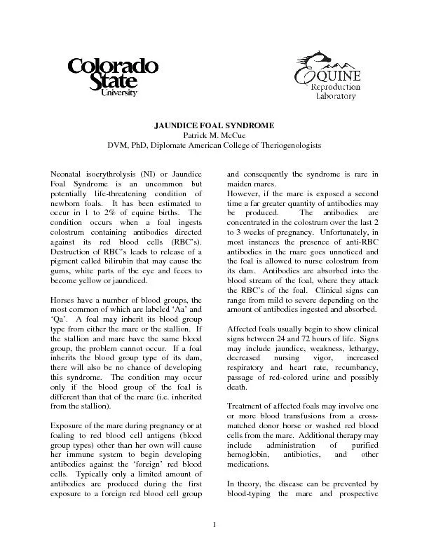
Auscultation of the lung fields can be very misleading in newborn foals Fluid sounds are normal immediately after birth as are crackles due to simple atelectasis of the dependent lung during latera ID: 502894

Auscultation the lung fields

Share:

Link:

Embed:

Download Presentation from below link

Download Pdf The PPT/PDF document "NEWBORN FOAL NOW WHAT?L. Chris Sanchez, ..." is the property of its rightful owner. Permission is granted to download and print the materials on this web site for personal, non-commercial use only, and to display it on your personal computer provided you do not modify the materials and that you retain all copyright notices contained in the materials. By downloading content from our website, you accept the terms of this agreement.


Presentation Transcript

NEWBORN FOAL NOW WHAT?L. Chris Sanchez, DVM, PhD, DACVIMand Steeve Giguère, DVM, PhD, DACVIM*College of Veterinary MedicineUniversity of Florida, Gainesville, FL, USAand *University of Georgia, Athens, GA, USAProceedings: North American Veterinary Conference, Orlando, FL, January 2012Reprinted with PermissionA complete and thorough physical examination of both the mare and the newborn foal is essential in Auscultation of the lung fields can be very misleading in newborn foals. Fluid sounds are normal immediately after birth, as are crackles due to simple atelectasis of the dependent lung during lateral recumbency. Conversely, foals with significant bronchopneumonia can have minimal abnormal findings on auscultation. Due to these limitations, thoracic ultrasound, radiography, and arterial blood gas are commonly included in the diagnostic evaluation of compromised neonates.Holosystolic murmurs over the pulmonic or aortic valve area on the left thorax are commonly heard in newborn foals and are usually innocent flow murmurs. Continuous murmurs with PMI caudal and dorsal to the aortic valve usually represent a left to right patent ductus arteriosus (PDA). PDA are considered normal up until day 4 of life and do not require further investigation unless accompanied by other clinical signs, such as cyanosis or signs of congestive heart failure. Echocardiography with Doppler evaluation is essential for complete assessment of any loud murmur (≥ III/IV)persisting after the first week of age. Although most congenital cardiac diseases are detected in foals or young horses, some defects (especially VSD) only become apparent as performance limitationsor may even be incidental findings in older horses. Ventricular septal defect is the most common congenital anomaly of the cardiovascular system reported in newborn foals and a variety of other defects have been described. Various malformations of the respiratory system such as collapsing trachea, stenoticnares, choanal atresia, subepiglotic cysts, guttural pouch tympany, diaphragmatic hernia and lung lobe agenesis have been reported rarelyGastrointestinal tract and abdomenThe value of abdominal palpation in the neonatal foal depends on the cooperation of the foal and tonus of the abdominal musculature. In a small foal with relaxed abdominal muscles, it is sometimes possible to palpate meconium impactions and the urinary bladder. It is normally easy to auscultate borborygmi bilaterally in equine neonates. Meconium may be expelled before delivery due to uterostress. Thus, the newborn foal should be closely evaluated for any signs of yellowbrown meconium staining as spiration of meconiumcontaminated fluid during parturition can result in severe pulmonary pathology. Most foals display abdominal straining within a few hours of birth and pass most of themeconium within the first 24 h of life. A change in fecalcolor and consistency from dark brown/black pasty material to a lighter brown/yellow, less tenacious material indicates that meconium has been passed. Meconium retention is the most common cause of colic in the newborn foal.Abdominal distension may result from accumulation of gas and fluid in the GI tract or accumulation of urine, blood or excessive peritoneal fluid in the abdominal cavity. Abdominal radiographs, abdominocentesis and abdominal ultrasonography can further define the cause of abdominal distension. Rarely, a severely enlarged bladder can result in abdominal distension.Congenital defects of the GI tract include cleft palate, poor jaw conformation, and atresia coli, recti or ani. Congenital aganglionosis has also been described in the Overo Lethal White Syndrome. Urogenital systemMean time to first urination is 6 h in colts and nearly 11 h in fillies. Because of the large volume of milk consumed (up to 25% BW/day), foals produce large volumes(148 ml/kg/daof dilute urineUSG 1.0011.015). Compared to that of adult horses, foal urine is more acidic (pH 5.58.0) and has increased protein for the first 36 h of life due to colostral absorption and subsequent protein excretion.During initial physical examination, the umbilicus should be palpated externally. Infection or abscessation of the internal remnantscommonly occurs in the absence of external signs of disease. Therefore, ultrasonographic examination is essential for complete evaluation. Sonographyof the umbilical remnants includes a thorough examination of the external stump, umbilical vein(from the stump to the liver), umbilical arteries(stump to lateral bladder)and urachus(stump to bladder). The external genitalia should be examined for the presence of congenital abnormalities. The penis is not easily extracted from the prepuce without sedation and colts often do not exteriorize the penis to urinate for the first few days of life.Congenital defects of the urinary system include ruptured bladder or ureter, ectopic ureter, renal agenesis and polycystic kidneys. Cryptorchism and inguinal or scrotal hernia are common congenital anomalies of the genital tract. The testes of neonates may be present in the scrotum or remain retracted in the abdomen. Definitive diagnosis of a cryptorchid should not be made until at least 1 year of age.Musculoskeletal systemThe passive range of motion of the joints, softness of the ears, the haircoatand muscle development should be assessed for evaluation of immaturity and prematurity. In the immature appearing neonate, radiographs of at least one carpus and one tarsus are recommended to assess the degree of ossification present. All four limbs should be carefully examined for angular limb deformities, contracture, laxity or other malformations. Any heat, swelling, edema, or pain around the joints and/or physes should be taken seriously and presumed infected until proven otherwise. The gait of thefoal should be observed fort lameness and weakness.The eyehe anterior and posterior Y sutures of the lens are usually visible and must not be mistaken for a cataract. The normal neonatal lens may also appear slightly cloudy for the first two days oflife. Blood may be present in the hyaloid vessels for the first few hours after birth. Empty blood vessels may be seen traversing the posterior lens capsule for the first 14 days of life. The point of attachment of the hyaloid vessels, represented by an opaque central dot just posterior to the posterior capsule, may be seen for 48 weeks. Evaluation of sight in the neonatal foal may be difficult because the menace reflex is usually absent until about 2 weeks of age. Ocular problemsoccur far more commonly than most clinicians recognize. Unlike adults, foals with corneal ulcers often fail to demonstrate the classical clinical signs of blepharospasm, epiphora and ocular pain. Some work suggestthat the neonatal cornea may be less sensitive than that ofolder foals and adults. Entropion is also seen commonly in sick foals and managed by suturing or placement of a bleb of procaine penicillin G. Rehydration alone maycorrect the problemin sick foals; surgical correction is rarely necessary. Neurological evaluationCritically ill neonates are often weak, depressed, recumbent and unable to suckle. These clinical signs may represent the presence of a primary neurological disorder or, alternatively, may only be secondary to a severe systemic disease such as sepsis. Close examination of the standing foal often requires restraint. Normal equine neonates often respond to such restraint by an initial period of struggle followed by a sudden relaxation. Equine neonates differ from adult horses during neurological examination in many regards. Head movements in response to auditory or tactile stimuli are jerky and exaggerated. The pupillary light response and dazzle response are present soon after birth. The PLR may be slightly delayed in excited foals. Thmenace reflex (eyelid closure to threatening gesture) is not consistently present in foals less than 2 weeks of age. But, by one day of age, an alert foal will withdraw its head from the menacing gesture. The slap test is inconsistently present before 1 month of age. The gait of the newborn foal is springy and dysmetric. At rest, foals often assume a widebased stance. Reflexes are increased or hyperreflexic (relative to adults) up to several weeks of age. The withdrawal reflex is accompanied by a crossed extension of the contralateral limb in normal foals up to 34 weeks of age.Premature foalsThe physical characteristics of immaturity include a low birth weight, weakness, a short, silky hair coat, an increased range of joint motion, rear limb flexural laxity, a prominent or domed forehead, and incomplete skeletal ossification (assessed radiographically). Premature foals often take longer than normal to stand (�60 min) and suckle from the mare (�120 min). The suckle reflex may lack vigor. Severely affected premature foals may demonstrate forelimb flexural laxity, “floppy” ears and a progressive decline in neurological function. Prematurity is discussed in detail later. LABORATORY FINDINGS IN NEONATESHematologyPCV and Hgbvalues increase during fetal development reaching values similar to those of adults by day 300 of gestation. PCV and Hgb increase slightly during the first 30 min after birth and then decrease slightly within the next 2 weeks. This is often referred to as anemia of newborn foals. Although most foals are born with total white blood cell count similar to that of an adult there can be considerable variation. Absolute neutrophil numbers are low (1500/µL) in the fetus prior to day 300 of gestation. Thereafter they begin to increase reaching mean values of about 5,500/µL at birth. Band neutrophils are absent or present at very low number in foals ( 150/µl). Lymphocytes greatly outnumber neutrophils in the fetus (  3000/µL between 150300 days of gestation). Lymphocyte counts decrease to mean values of about 1,400/µL a few hours after birth and then begin a gradual increase until about 5 months of age. Lymphocyte counts 1000/µL during the first few days of life may be normal or from sepsis or prematurity. Normal term foals should have N/L ratios  3. Premature foals with N/L ratios 1.5 during the first 24h may have a poorer prognosis. Total plasma protein increases from a mean of about 5 g/dL before suckling to about 6 g/dL after absorption of Igfrom colostrum. Total protein can vary widely in foals and, unlike in calves, is not a useful indicator of adequate absorption of IgG. Chemistry Interpretation of biochemical analysis is fairly similar to adults with the exception of alkaline phosphatase, which is increased for the first 3 months of life. This is primarily due to increased metabolic activity associated with bone growth. Levels approaching 3000 IU/L are common in newborns. Hepatobiliary enzymes such as GGT and, to a lesser extent, SDH may also be transiently increasedduring the first two months of life. Icterus occurs commonly in newborn foals but should be mild and associated with increasesin the indirect or unconjugated bilirubin. This transient increase may be due to a combination of accelerated breakdown of neonatal RBC and inadequate hepatic function at birth. Neonatal hyperbilirubinemia is more pronounced during the first week of life but bilirubin concentrations are slightly higher than that of adult horses until approximately 1 month of age. A total bilirubin above 4.5 mg/dl probably requires investigation; NI, liver disease or sepsis should be ruled out. Serum glucose concentrations are increasedrelative to adults due to frequent milk intake.Creatinine concentrations are sometimes increased(1.24.5 mg/dl) in normal foals in the first 36 h of life. More substantial increases can occur in clinically healthy foals; the mechanism of this increase is not completely understood but is likely related to placental dysfunction. If the increase in creatinine is due solely to placental dysfunction (and not renal disease), it should decrease significantly by day 2 and return to normal by day 5. BUN concentrations are initially within the normal adult range. A decrease in BUN is observed between 3 days and 2 months of age and may be associated with an increased demand for amino acids necessary for protein synthesis.