/
CASE REPORT: SUCCESSFUL TREATMENT OF            ADULT REFRA CASE REPORT: SUCCESSFUL TREATMENT OF            ADULT REFRA

CASE REPORT: SUCCESSFUL TREATMENT OF ADULT REFRA - PowerPoint Presentation

luanne-stotts
luanne-stotts . @luanne-stotts
Follow
406 views
Uploaded On 2017-06-04

CASE REPORT: SUCCESSFUL TREATMENT OF ADULT REFRA - PPT Presentation

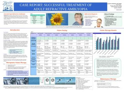
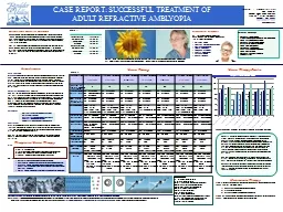
Jennifer S Simonson OD FCOVD Clinic Director Boulder Valley Vision Therapy 1790 30 th Street Suite 311 Boulder CO 80301 wwwbouldervtcom 3034432257 A 35yearold Caucasian female presented with symptoms of blurred vision in the right eye poor night vision and ID: 555558

eye vision test therapy vision eye therapy test graefe seconds visual 20cc patient amblyopia treatment pursuits acuity 12saccades ilehorizontal

Share:

Link:

Embed:

Download Presentation from below link

Download Presentation The PPT/PDF document "CASE REPORT: SUCCESSFUL TREATMENT OF ..." is the property of its rightful owner. Permission is granted to download and print the materials on this web site for personal, non-commercial use only, and to display it on your personal computer provided you do not modify the materials and that you retain all copyright notices contained in the materials. By downloading content from our website, you accept the terms of this agreement.


Presentation Transcript

Slide1

CASE REPORT: SUCCESSFUL TREATMENT OF ADULT REFRACTIVE AMBLYOPIA

Jennifer

S. Simonson, OD, FCOVD Clinic Director Boulder Valley Vision Therapy1790 30th Street, Suite #311 Boulder, CO 80301 www.bouldervt.com303-443-2257

A

35-year-old Caucasian female presented with symptoms of blurred vision in the right eye, poor night vision, and inability to see 3D. She experienced frequent headaches, halos around lights, and skipping or repeating of lines when reading. She noted blurred vision around lights and projection screens. These vision difficulties had been present her entire life. Medical history included a drug allergy to Sulfa medications and she was taking Juice Plus vitamin supplements. She had no history of eye injury or disease.

Case Summary

Examination Findings

See Table

2.

Maintenance vision therapy was prescribed for home practice, including computer and free space activities. See sample images. 1) Eccentric circles (convergence and divergence)2) Free-space fusion images3) Magic eye books and random dot stereo images The patient was referred to the co-managing optometrist for contact lens evaluations and annual comprehensive vision and ocular health examinations.

Acknowledgments: Thank you to the following colleagues for their participation in the care of this patient: Ashlee Elmont, OD, Rachel Potter, OD, and Barbara Nelson, COVTReferences: 1.) Applied Concepts in Vision Therapy by Leonard Press, OD, FCOVD 2.) Fact Sheets on Conditions of the Visual System Treated with Vision Therapy by the COVD Clinical Standards Committee 3.) Care of the Patient with Amblyopia by the American Optometric Association Clinical Practice Guidelines http://www.aoa.org/optometrists/tools-and-resources/clinical-practice-guidelines. 4. Bernell Corporation (Vision Therapy Equipment)

Anisometropic Hyperopia (367.31, 367.0)

Asthenopia (368.13)Saccadic oculomotor dysfunction (379.57)Refractive Amblyopia (368.03)Suppression of Binocular Vision (368.31)

Diagnoses

Treatment options of glasses, contact lens, patching, and active in-office vision therapy were discussed. A contact lens fitting for the right eye and vision therapy treatment were recommended with patient goals of gaining depth perception, better vision clarity, and improved reading skills. The patient was fitted with a contact lens (Air Optix) in the right eye and began weekly office therapy April 7, 2011 with assigned home therapy reinforcement activities.

Treatment

Results: The patient completed weekly in-office visits with home practice from April 2011 to February 2012. Vision Therapy was tapered through February 2013. Therapy included oculomotor, accommodative and binocularity training and vision perception (speed of information processing, perceptual span) Examination resulted in reduced fusional, accommodative and oculomotor efficiency findings. Due to clinical measurements of stable vision skills including acuity and stereopsis and the patient remaining symptom-free, in-office therapy was completed after the evaluation on February 22, 2013. A post-treatment evaluation was then completed on June 21, 2013The outcome was increased vision clarity, reading speed, stereopsis, and visual comfort. Retesting resulted in 20/20- acuity at distance and 20/32 at near OD with stable refraction, 70” stereopsis, and elimination of symptoms. Discussion: The patient achieved all therapy goals, including those beyond improved visual acuity through refractive correction and vision therapy as an adult. Treatment of amblyopia should be recommended for all patients regardless of age.

Anisometropic Refractive Amblyopia

Introduction

Vision Therapy

Results

Progress Graph (Standard Scale)

Maintenance Therapy

Diagnostic Test

Initial Examination3/16/2011Progress Examination6/22/2011Progress Examination9/14/2011Progress Examination12/23/2011Progress Examination6/4/2012Progress Examination2/22/2013Post-Therapy 6/21/2013Number of therapy sessions completed0102128405757 Distance Visual Acuity (20 ft./6 M)sc: OD 20/200+cc: OD +3.50 DS 20/70-sc: OS 20/20cc: OD 20/50+2sc: OS 20/20cc: OD 20/40-1sc: OS 20/20cc: OD 20/40-1sc: OS 20/20cc: OD 20/30+3sc: OS 20/20cc: OD 20/25 (full chart)sc: OS 20/20cc: OD 20/20-sc: OS 20/20Near Visual Acuity (16 inches/40 cm)cc: OD 20/200sc: OS 20/20cc: OD 20/50+2sc: OS 20/20cc: OD 20/40sc: OS 20/25cc: OD 20/40sc: OS 20/25cc: OD 20/40sc: OS 20/20cc: OD 20/32sc: OS 20/20cc: OD 20/32sc: OS 20/20AccommodationNRA: +0.50PRA: -1.50BXC (FCC): +1.50+/-2.00 Flipper: 4 cpmNRA: +2.00PRA: -2.00 (doubles)BXC (FCC): +1.50+/-2.00 Flipper: 7 cpmNRA: +2.25PRA: -1.50BXC (FCC): +1.50+/-1.00 Flipper: 14 cpmNRA: +1.75PRA: -1.25+/-1.00 Flipper: 14 cpmNRA: +1.25PRA: -2.25BXC (FCC): +1.00NRA: +2.00PRA: -2.00 (doubles)BXC (FCC): +1.50+/-1.00 Flipper: 18 cpmNRA: +3.00PRA: -1.50BXC (FCC): +0.75Distance Eye AlignmentVon Graefe: 4 esoCover test: flick esophoiraVon Graefe: 4 esoCover test: flick esophoiraVon Graefe: 2 exoCover test: flick esophoriaVon Graefe: 2 exoCover test: flick esophoiraVon Graefe: OrthophoriaCover test: OrthophoriaVon Graefe: OrthophoriaCover test: OrthophoriaVon Graefe: OrthophoriaCover test: OrthophoriaNear Eye AlignmentVon Graefe: 8 (variable) eso, intermittent suppressionCover test: flick esophoiraVon Graefe: 8 esoCover test: 8 esophoriaVon Graefe: 8 esoCover test: flick esophoriaVon Graefe: 8 esoCover test: flick esophoiraVon Graefe: 3 exoCover test: 4Δ esophoiraVon Graefe: 4Δ exoCover test: 4Δ esophoriaVon Graefe: 3Δ exoCover test: 4Δ esophoriaStereopsisAbsent400” arc seconds100” arc seconds100” arc seconds100” arc seconds70” arc seconds70” arc secondsNear Point of Convergence2 cm/4 cm2 cm/4 cmTo the noseTo the noseTo the noseTo the noseTo the noseDistance Fusional RangesBO: 22/24/6BI: x/8/2BO: 16/32/10BI: x/8/6BO: 16/26/16BI: x/10/6BO: 12/24/20BI: x/8/6BO: 16/24/20BI: x/8/6BO: 26/32/22BI: x/6/5BO: 26/32/26BI: x/7/5Near Fusional RangesBO: 16/20/9BI: 10/20/7Fusion facility: unableBO: x/18/11BI: x/19/14BO:22/28/16BI: x/22/18BO:12/23/18BI: x/18/17Fusion facility: 11 cpmBO: 33/33/28BI: 17/18/13Fusion facility: 13 cpmBO: 29/33/31BI: x/18/14Fusion facility: 14 cpmBO: 29/33/30BI: x/17/13Oculomotor SkillsColorado study group: Pursuits: 10/12Saccades: 10/12DEMVertical: 48 seconds <1st%ileHorizontal: 52 seconds <1st%ileColorado study group: Pursuits: 11/12Saccades: 11/12DEM:Vertical: 35 seconds26th%ileHorizontal: 40 seconds 16th%ileColorado study group: Pursuits: 11/12Saccades: 11/12DEM:Vertical: 32 seconds 42nd%ileHorizontal: 34 seconds 44th%ileColorado study group: Pursuits: 11/12Saccades: 11/12DEM:Vertical: 31 seconds 48th%ileHorizontal: 37 seconds 27th%ileColorado study group: Pursuits: 12/12Saccades: 12/12DEM:Vertical: 32 seconds 42nd%ileHorizontal: 35 seconds 38th%ileColorado study group: Pursuits: 12/12Saccades: 12/12DEM: Vertical: 30 seconds 56th%ileHorizontal: 35 seconds 38th%ileColorado study group: Pursuits: 12/12Saccades: 12/12DEMVertical: 30 seconds 56th%ileHorizontal: 35 seconds 38th%ile

Vision Testing

Table 2.

Abbreviations: sc = without corrective lensescc = with corrective lensesOD = right eye, OS = left eye, OU = both eyesNRA = negative relative accommodationPRA = positive relative accommodation BXC = binocular cross cylinderFCC = fused cross cylindercpm = cycles per minuteeso = inward, exo = outwardBO = base-out, BI= base-in, BIM = base-in prism with minus lens, BOP = base-out prism with plus lensDEM = Developmental Eye Movement Test SILO = small/in, large/out

The patient was referred by her primary care optometrist for an evaluation on March 16, 2011. At this initial appointment, refraction was OD +3.50 DS and OS -0.25 DS with acuity of 20/70- at 20 feet and 20/200 at near OD and 20/20 at all distances OS. Depth perception was absent. All focusing skills were below typical performance. Eye alignment testing showed esophoria with convergence excess. Pursuit movements were full and smooth, but saccadic eye movements required additional head movement, re-fixation movements, and more time than expected. Significant suppression of the right eye was noted.

Background: Anisometropic refractive amblyopia occurs due to a blurred optical image from one eye affecting development of the visual system. Due to the normal eye appearance of this visual disorder, early diagnosis and treatment are not always possible. Despite the delay of treatment, this case demonstrates effective amblyopia treatment in adulthood. For an accurate diagnosis, an amblyogenic refractive error must be present during development of the visual system in childhood (refer to Table 1). This defocus disrupts the normal neurophysiological development of the visual pathway and visual cortex. In this case, the patient had uncorrected anisometropic hyperopia of over 1 diopter.

Therapeutic Vision Therapy

Equalize Monocular SkillsBuild Sensory FusionImprove Motor Fusion Accuracy and Fusional Vergence RangesDevelop Accurate Stereopsis and Eye-Hand CoordinationSpeed, Accuracy, and Maintenance of all Visual Skills

Normal Vision is developed when both eyes send clear images to the brain that are blended into one perception. When one eye has poor vision, as in the case of anisometropic hyperopia, the brain suppresses the blurred information causing amblyopia.

Potentially Amblyogenic Refractive Errors

Isoametropia

Diopters

Astigmatism

>2.50 DHyperopia>5.00 DMyopia>8.00 DAnisometriopiaAstigmatism>1.50 DHyperopia>1.00 DMyopia>3.00 D

Table 1.

Differential DiagnosisAll forms of pathology or disorders of development must be ruled out before making the diagnosis of amblyopia.Retinal DefectsCentral Nervous System LesionsMetabolic DisordersToxin ExposureCongenital DefectsOptic Nerve InsultOcular TraumaBrain Trauma

Clinical Findings Reduced Visual Acuity (typically ranges from 20/25 to less than 20/200)Poor Ocular Motility AccuracyReduced Accommodation SkillsDecreased Contrast SensitivityPoor Spatial Judgment Crowding PhenomenonUnstable Binocular Alignment Poor Fusion SkillsReduced or Absent StereopsisSuppression

Accommodation

: Monocular, Bi-ocular, Binocular

Oculomotor

: Fixations, Pursuits, Saccades, Eye-Hand Coordination

Sensory

Fusion: Anti-suppression, Center/Peripheral, Monocular Fixation in a Binocular Field, Luster, Simultaneous Perception, Flat Fusion, StereopsisMotor Fusion: Convergence, Divergence, Jump Ductions, BIM/BOP

Plan:

Activities: