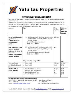
/
David SuIzer,1.21415 Ta-Kung Chen,‘j Yau Yi Lau,6 Helle Kristense David SuIzer,1.21415 Ta-Kung Chen,‘j Yau Yi Lau,6 Helle Kristense

David SuIzer,1.21415 Ta-Kung Chen,‘j Yau Yi Lau,6 Helle Kristense - PDF document

luanne-stotts
luanne-stotts . @luanne-stotts
Follow
394 views
Uploaded On 2015-10-06

David SuIzer,1.21415 Ta-Kung Chen,‘j Yau Yi Lau,6 Helle Kristense - PPT Presentation

Whether amphetamine acts principally at the plasma mem brane or at synaptic vesicles is controversial We find that Received Nov 3 1994 revised Dec 29 1994 accepted Jan 4 1994 We thank Drs ID: 152008

Whether amphetamine acts principally

Share:

Link:

Embed:

Download Presentation from below link

Download Pdf The PPT/PDF document "David SuIzer,1.21415 Ta-Kung Chen,‘..." is the property of its rightful owner. Permission is granted to download and print the materials on this web site for personal, non-commercial use only, and to display it on your personal computer provided you do not modify the materials and that you retain all copyright notices contained in the materials. By downloading content from our website, you accept the terms of this agreement.


Presentation Transcript

David SuIzer,1.21415 Ta-Kung Chen,‘j Yau Yi Lau,6 Helle Kristensen,6 Stephen Rayport,1J.415 and Andrew Ewing6 Departments of ‘Psychiatry, 2Neurology, and 3Anatomy and Cell Biology, and 4Center for Neurobiology and Behavior, Columbia University, New York, New York 10032, 5Department of Neuroscience, New York State Psychiatric Institute, New York, New York 10032, and 6Department of Chemistry, Penn State University, University Park, Pennsylvania 16802 Whether amphetamine acts principally at the plasma mem- brane or at synaptic vesicles is controversial. We find that Received Nov. 3, 1994; revised Dec. 29, 1994; accepted Jan. 4, 1994. We thank Drs. Qais Al-Awqati, Susan Amara, Robert Edwards, George Uhl, and the late Eric Holtzman for comments. This work was supported by NIDA Grant DA07418 (D.S.). NIMH MH00705 and MH44736 (S.R.). NIH \ and ONR (A.G.E.). The Parkinson’s Disease Foundation provided support in the final stages of this project (D.S.). S.R. is a Burroughs Wellcome Scholar in Experimental Therap&&s and this work was supported in part by a grant from the Burroughs Wellcome Fund. A.G.E. is a Camille and Henry Dreyfus Teacher Scholar. Correspondence should be addressed to David Sulzer, Ph.D., Black Building 307, Columbia tribute catecholamines from synaptic vesicles to the cytosol by collapsing the vesicular proton gradient that provides the free energy for neurotransmitter accumulation (Ereckinska et al., 1987; Sulzer and Rayport, 1990; Rudnick and Wall, 1992). This would increase the availability of transmitter for reverse trans- port without requiring a mobile site. In support of this model, weak bases and protonophores, which cause an AMPH-like re- lease of DA from isolated vesicles (Sulzer and Rayport, 1990) but are not substrates for the uptake transporter, induce reverse transport in cultured DA neurons (Sulzer et al., 1993). In addi- tion, low-affinity binding of AMPH to the vesicular transporter (Gonzalez et al., Materials and Methods GDC electrochemistry. The use of the Planorbis comeus giant dopa- mine cell (GDC) for electrochemical experiments has been described previously (Chien et al., 1990; Ewing et al., 1992). Briefly, adult Plan- orbis comeus were obtained from NASCO (Fort Addison, WI) and dissected to reveal the left pedal ganglion, which was immersed in 10 ml of snail saline and desheathed to expose the GDC. Snail saline con- tained 39 mM NaCl, 1.3 mM KCI, 4.5 mM The Journal of Neuroscience, May 1995, 1~75) 4103 sampled from each plotted voltammogram at 0.8 V versus a sodium- saturated calomel reference electrode and plotted with respect to time. Nomifensine was applied by bath perfusion. All experiments were con- ducted at room temperature. Single cell capillary electrophoresis. To measure the vesicular DA pool, single GDCs from juvenile snails were drawn into 25 pm i.d. X 75 cm long capillaries (dlefirowicz and Ewing, 1990; Kristensen et al., 1994). A 1 min iniection of moroholinoethanesulfonic acid buffer (pH 5.65; was timed to produce an obtimal differential lysis of the plasma but vesicular membranes. The resulting cellular-components were then separated by electrophoretic mobility in a kV field applied to the capillarv and Results Intracellular AMPH injection and DA release The left pedal ganglion of the pond snail Planorbis corneus contains a single giant DA neuron (GDC) that closely resembles mammalian DA neurons in several ways (Osborne et al., 1975). The GDC displays nomifensine-sensitive DA uptake, reserpine- sensitive vesicular DA uptake, and metabolizes DA to dihy- droxyphenylacetic acid (DOPAC). However, unlike mammalian DA neurons, the adult GDC is approximately 200 pm in di- ameter, facilitating single cell analysis; and, it lacks ascorbic acid (Barber and Kempter, 1986), facilitating the unambiguous elec- trochemical detection of DA. We measured DA release in real time with carbon fiber mi- croelectrodes (Wightman et al., 1991) touching the extracellular face of the GDC membrane (Fig. IA). Injection of 8 pl of 100 mM d-AMPH into the GDC rapidly released DA (Fig. 1B). Based on estimated cytosolic pM AMPH. Although AMPH is amphiphilic and may diffuse from B Figure 1. Intracellular injection of AMPH promotes DA release. A, Single GDCs from adult Planorbis comeus were impaled with an AMPH containing pipette. Oxidation current was measured with a pm carbon ring ultramicroelectrode touching the GDC membrane and held at a steady potential of 0.8 V. B, AMPH (8 pl of 100 mM) was injected intracellularly (arrow), thereby bypassing the uptake transport- er, and the increase in extracellular DA measured. Similar levels of DA release were found in three replications of the experiment using DC amperometry. The small, slow baseline decrease during these experi- ments results from adsorption of electrode oxidation products to the electrode surface (Lau et al., 1991). C, The shape of the voltammogram (-0.2 AMPH, which lacks hydroxyl groups, is not oxidized at these potentials. Calibration: i = 0.5 for the experiment and 1.5 for the standard. the cell, it also diffuses rapidly in aqueous medium and would reach nanomolar levels within milliseconds. Extracellular DA increased following the injection, reaching a maximum of 1.43 + 0.58 FM (mean + SEM, n = 3). The shape of a voltam- mogram taken at the peak current value closely matched the voltammogram of a DA standard (Fig. 1 C). Measurement of the vesicular DA pool To examine the effects of AMPH on the vesicular pool directly, we measured vesicular DA by capillary electrophoresis of ju- venile p,M for 15 min) reduced peak 2 to below detectable limits and correspond- ingly increased peak 1 (Fig. 2, upper trace; Table 1). I 4 PA 5 min AMPH Figure 2. Electrophoretic separation of readily oxidized components from a single Phnorhis corneus giant DA neuron. Single neurons (75 p,rn diameter) from juvenile snails were drawn into capillary tubes. Subsequent injection of morpholinoethanesulfonic acid buffer (pH 5.65) for I min produced a partial lysis of the cell membranes, presumably mainly the plasma membrane. Based on this differential lysis, peak 1 reflects free DA and DA from and KC1 (Chen et al., 1994). In each case, release events were measured for approximately 2 min following stimulation. In controls, individual exocytic events corresponded to the re- lease of 11 1,000 ? 32,000 catecholamine molecules (n = 9 cells, 92 +- 37 events per cell, mean ? SEM). AMPH (10 FM for 10 min) reduced stimulated catecholamine released per ex- ocytic event. Randomly chosen amperograms for exocytosis from control cells and those treated with AMPH are shown in Data are mean 2 SEM (n = 3) for DA (fmol) peaks in single GDCs from juvenile Planorbis ~o~ntws measured by capillary electrophoresis. Cells were exposed to IO FM AMPH for 15 min prior to sampling. The detection limit is typically l-10 amol. Figure 3, A and B. When paired with age-matched controls from sister cultures (n = 3, 139 +- 105 events per cell), the average exocytic event following incubation with AMPH (n = 7 cells, DA release due to intracellular DA injection To determine whether increased cytosolic DA is sufficient to induce release, we returned to the GDC. In the resting state, cytosolic DA is about 2.2 FM and extracellular DA below the limits of detection (Ewing et al., 1992). Intracellular injection of 4 pl of 0.5 mM DA (Fig. 4A) rapidly increased extracellular DA to.22.5 * 0.9 PM, 3.5 set postinjection (n = 3 cells); extracel- lular perfusion used uptake blockers, has no detectable effect on intracellular proton gradi- ents; Sulzer et al., 1993) attenuated the increase in extracellular DA following subsequent injections by 87 4%. After removal of nomifensine, the increase in extracellular DA due to intra- cellular DA injection returned to near control levels (20.4 ? 1.5 PM peak concentration, p = 0.265 by Student’s t test, not sig- nificantly different). Intracellular injections of control solution lacking DA induced no DA release (data not shown). Difference voltammograms taken after the first, second, and in vitro calibration. Discussion Whether AMPH acts principally at the plasma membrane uptake transporter or at the vesicular level has been controversial. We used two model systems to address this issue. Arguing for a principal mechanism of action at the vesicular level, intracellular injection of AMPH into the Planorbis GDC, which bypasses the plasma membrane uptake transporter, causes DA release. Whole- cell capillary electrophoresis of GDCs shows that AMPH pro- foundly reduces vesicular DA; and amperometric detection of DA release in PC12 cells shows that AMPH duces DA release from individual exocytic events. Taken to- gether, these results indicate that amphetamine redistributes DA from synaptic vesicles to the cytosol. Monitoring DA at the ex- tracellular face of the GDC plasma membrane shows that intra- cellular injection of DA produces rapid nomifensine-sensitive release, indicating that increased cytosolic DA is sufficient to trigger reverse transport. AMPH acts intracellularly Injection of AMPH directly into the cytosol of the GDC causes DA release even though the uptake transporter is bypassed and the AMPH concentration gradient is in the opposite direction from that required for net efflux via exchange diffusion. Togeth- 2 PA 250 ms 0 control [7 lO/.tMAMPH 30 90 K molecules Figure 3. AMPH exposure decreases the quanta1 amplitude of individual release events from PC12 pM d-AMPH for 10 min. Cells were stimulated by local perfusion (30 nl of 1 mM nicotine in 105 mM KCI saline) to induce vesicular exocytosis. C, A histogram shows the percentage of peak sizes in paired er with the uptake of AMPH by catecholamine transporters (Zac- zek et al., 1991), which would play a role in the specificity of transmitter release, and the ability of AMPH to release mono- amines from isolated vesicles PM AMPH, a level reached in the brain with a standard dose (1 mg/ kg) (Melega et al., 1992), and shows for the first time a reduction PA B ,\_-_--- ,\F-.-’ I 5 PA -2hO b 2bO 800 mV Figure 4. Intracellular injection of DA induces reverse transport. A, Intracellular injection of 4 pl of 0.5 mM DA (arrows) reliably increases extracellular DA. During extracellular perfusion with 10 pM nomifensine (dashed line), the DA release due to the same DA injections was markedly attenuated. Perfusion with nomifensine-containing medium and its replacement by control medium produce current spikes (asrerisks). These data have Elevated cytosolic DA induces reverse transport Injection of DA into the cytosol of GDCs leads to a rapid release of DA that is nomifensine sensitive. Uptake transport blockers like nomifensine are thought to block transport by binding to the extracellular face of the DA transporter; however, unlike AMPH, they are not transported (Anderson et al., 1989; Ritz et al., 1990). We have previously shown in DA neuron cultures that nomifensine blocks DA 4A). This slower action of AMPH would be consistent with a rate-limiting step of AMPH-mediated redistribution of DA from synaptic vesicles to the cytosol. Altered substrate plasma membrane concentration gradients due to vesicular AMPH sequestration could further affect DA flux Conclusions These findings demonstrate in invertebrate DA neurons and PC12 cells that AMPH decreases vesicular DA content, presum- ably resulting in DA redistribution to the cytosol. Cytosolic DA is, in turn, rapidly released by reverse transport. Since AMPH- induced DA release occurs independently of specific AMPH up- take by the plasma membrane transporter and can be induced in isolated monoamine vesicles (Slotkin and Kirshner, 197 1; Knep- per et al., 1988; Sulzer and Rayport, 1990), the present findings favor the release of DA from the vesicular pool. This mechanism has important implications for neurotrans- mission and pathology. It suggests that AMPH affects synaptic transmission both by increasing activity-independent reverse transport and by decreasing the amount of transmitter released during activity-dependent vesicular exocytosis. The result should be reduction in the signal-to-noise ratio of stimulation-depen- dent monoamine synaptic transmission. Furthermore, this redis- tribution of DA from the relatively reducing environment of the synaptic vesicle to the cytosol appears to cause local increases in intracellular oxidative stress Adam-Vizi V (1992) External Ca*+-independent release of neurotrans- mitters. J Neurochem 58:395+05. Amara SG, Kuhar MJ (1993) Neurotransmitter transporters: recent progress. Annu Rev Neurosci 16:73-93. Andersen PH (1989) The dopamine uptake inhibitor GBR 1209: se- lectivity and molecular mechanism of action. Eur J Pharmacol 166: 493-504. Attwell D, Barbour B, Szatkowski M (1993) Nonvesicular release of neurotransmitter. Neuron 11:401+07. Barber A, Kempter B (1986) Release of endogenous dopamine from nervous tissue of the pond snail Planovbis corneus: a study using high performance liquid chromatography with electrochemical detec- t&-n. ‘Camp Biochem Physiol 84CT17i. - Bernath S. Zigmond MJ (1990) Calcium-indeuendent GABA release from stiiatay slices: the ;ole of calcium chainels. Neuroscience 36: 677-682. Chen TK, Luo G, Ewing AG (1994) Amperometric monitoring of stim- ulated catecholamine release from rat pheochromocytoma (PC 12) cells at the zeptomole level. striatum. J Neurochem 55:1556-1562. Rudnick G, Wall SC (1992) The molecular mechanism of “ecstasy” [3,4-methylenedioxymethamphetamine (MDMA)]: serotonin trans- 4108 Sulzer et al. *Amphetamine-Induced Dopamine Redistribution Sulzer D, Maidment NT, Rayport S (1993) Amphetamine and other weak bases act to promote reverse transport of dopamine in ventral midbrain neurons. J Neurochem 60527-535. Szatkowski M, Barbour B, Attwell D (1990) Non-vesicular release of glutamate from glial cells by reversed electrogenic glutamate uptake. Nature 348:443446. Turner TJ, Adams ME, Dunlap K (1992) Calcium channels coupled to glutamate release identified by omega-Aga-IVA. Science 258:3 I O- 313. Wightman RM, Jankowski JA, Kennedy RT, Kawagoe KT, Schroeder TJ, Leszczyszyn DJ, Near JA, Diliberto EJ Jr, Viveros OH (1991) Temporally resolved catecholamine spikes correspond to single ves- icle release from individual chromaffin cells. Proc Nat1 Acad Sci USA 88:10754-10758. Zaczek R, Culp S, De Souza EB (1991) Interactions of [‘Hlampheta- mine with rat brain synaptosomes. II. Active transport. J Pharmacol Exp Ther 257:830-835. Zhou R, Luo G, Ewing, AG (1994) Direct observation of the effect of autoreceptors on stimulated release of catecholamines from adrenal cells. J Neurosci 14:2402-2407.