/
Journal of the Canadian Dental AssociationMay 2000 Vol 66 No 5obac Journal of the Canadian Dental AssociationMay 2000 Vol 66 No 5obac

Journal of the Canadian Dental AssociationMay 2000 Vol 66 No 5obac - PDF document

lucy
lucy . @lucy
Follow
342 views
Uploaded On 2022-10-11

Journal of the Canadian Dental AssociationMay 2000 Vol 66 No 5obac - PPT Presentation

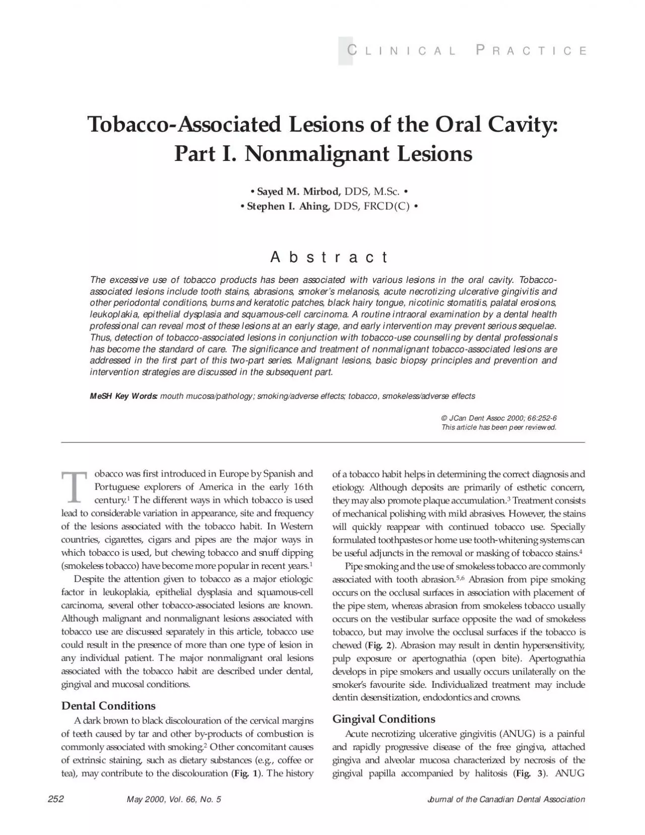
This article has been peer reviewed May 2000 Vol 66 No 5Journal of the Canadian Dental AssociationTobaccoAssociated Lesions of the Oral Cavity Part I Nonmalignant Lesions Figure 5 Smokeless ID: 958720

smoking oral cessation tobacco oral smoking tobacco cessation cigarette lesions melanosis dental dysplasia epithelial due malignant carcinoma toluidine biopsy

Share:

Link:

Embed:

Download Presentation from below link

Download Pdf The PPT/PDF document "Journal of the Canadian Dental Associati..." is the property of its rightful owner. Permission is granted to download and print the materials on this web site for personal, non-commercial use only, and to display it on your personal computer provided you do not modify the materials and that you retain all copyright notices contained in the materials. By downloading content from our website, you accept the terms of this agreement.


Presentation Transcript

Journal of the Canadian Dental AssociationMay 2000, Vol. 66, No. 5obacco was Þrst introduced in Europe by Spanish andPortuguese explorers of America in the early 16thcentury. This article has been peer reviewed. May 2000, Vol. 66, No. 5Journal of the Canadian Dental AssociationTobacco-Associated Lesions of the Oral Cavity: Part I. Nonmalignant Lesions Figure 5: Smokeless-tobacco induced lesion primarily affects young adults who smoke heavily and have poorAlthough the exact interaction between ANUGand smoking is not clear, local and systemic effects have beenThe progression of ANUG may be enhanced byischemia secondary to nicotinic vasoconstriction.Withouttreatment,ANUG may occasionally progress to involve themarginal alveolar bone. Initial therapy for cases uncomplicated by bone involvementincludes debridement under local anesthetic, instruction in oralhygiene and increasing awareness of the role of tobacco.Dramatic resolution of gingival lesions is often seen within 48 to Figure 1: Pronounced tobacco stains : Acute necrotizing ulcerative gingivitis Figure 2: Extensive abrasions : SmokerÕs melanosis Journal of the Canadian Dental AssociationMay 2000, Vol. 66, No. 572 hours with these measures alone. Antibiotics, when indicated,should be considered as adjunctive to local debridement.Penicillin and metronidazole are both effective in cases of severeANUG or where debilitation is known or suspected.Frequentfollow-up is necessary until the interdental papillary contourreturns to normal. If this does not occur, periodontal surgicalintervention may be necessary.Although gingivitis and periodontitis are caused by bacteria,smoking has been strongly implicated as a risk factor for theinitiation and propagation of periodontal diseases (Table 1Smoking has been associated with increased calculus deposition,which can accelerate plaque accumulation, recession and deep-The prevalence of periodontitis isgreater in smokers than nonsmokersand the alveolar bone ismore susceptible to resorption in smokers.Several biologicmechanisms have been suggested. Smoking has been implicatedin adverse effects on neutrophil functionreducing gingival blood ßow.Cigarette smoking has been asso-ciated with impaired healing and less improvement in pocketreduction following simple pocket-reduction surgery.SmokerÕs melanosis occurs in 25 to 31% of tobacco users andis characterized by discrete or coalescing multiple brown maculesthat usually involve the attached mandibular gingiva on theFig.4Smoking-associated melanosis is due to increased melaninproduction by melanocytes and its deposition within the basalcell layer and lamina propria. The microscopic appearance ofOther entities to considerin differential diagnosis are racial melanosis, melanosis due tomedications, Peutz-JegherÕs syndrome, AddisonÕs disease andearly mel

anoma. SmokerÕs melanosis is benign and not consid-ered to be precancerous, but a biopsy may be indicated to ruleout more serious conditions, in particular melanoma. A gradualreturn to normal pigmentation over several months to years hasbeen reported following smoking cessation.Burns and keratotic patches are common on the lips at thesite of habitual cigarette smoking, particularly where the ciga-rette or cigar is retained as a stub for lengthy periods. Thelesions characteristically appear on the mucosal surface of thelower and upper lips at the site at which the cigarette is held.They are characterized by ßat or slightly elevated whitish areaswith red striations. Cessation of smoking is the requiredtreatment.Lesions induced by smokeless tobacco characteristicallyhave a wrinkled surface that ranges from opaque white totranslucent and are located in the area where the snuff is heldFig.5). Such lesions usually resolve within a week of cessationof tobacco use. A thorough history is important in establishingthe correct diagnosis; however, a biopsy may be required torule out carcinoma or precancerous changes, especially if thelesion is associated with induration, ulceration, erythema andnonresolution within 2 weeks of stopping tobacco use.Hypertrophy of Þliform papillae and retardation of thenormal rate of desquamation on the dorsal surface of thetongue produce a hair-like appearance.A black hairy tongueis mainly seen in people who are heavy smokers. Clinically, thehairy tongue usually begins near the foramen cecum and Table 1Dental and gingival conditions associated with tobacco use MechanismSigniÞcanceManagement DiscolourationCombustion by-productsEstheticsMechanical polishing Plaque trapWhitenersAbrasion (mild)Pipe smokingDentinal/tooth Desensitization Smokeless tobaccosensitivityDirect restorationsAbrasion (severe)Pipe smokingPulp exposureEndodontic/prosthodontic Smokeless tobaccoOcclusal disharmonyoptions ANUGTar/nicotine induced plaque Severe gingival destructionUltrasonic debridementaccumulationAntibiotic therapy (rare) SmokerÕs melanosisStimulation of melanocytesMust rule out melanoma orBiopsy may be required to systemic conditionsrule out melanomaAssociation withIncreased calculusPeriodontal destructionPeriodontal therapygingivitis andand plaque deposits(debridement, excisional periodontitisIschemiaprocedures)Poor wound healingIschemiaPostsurgical careRegenerative periodontal Osteitistechniques contraindicated May 2000, Vol. 66, No. 5Journal of the Canadian Dental AssociationTobacco-Associated Lesions of the Oral Cavity: Part I. Nonmalignant Lesions spreads anteriorly and laterally with elongated Þliform papillaereaching several millimeters in length. Individuals are usuallyasymptomatic, but may have a gagging or tickling sensation ifthe papillae become particularly long. A

biopsy is usually notnecessary for diagnosis due to the typical presentation.Scraping or brushing of the tongue and smoking cessationenhance the resolution of this problem. Trimming of thepapillae may be required in extreme cases.Nicotinic stomatitis occurs almost exclusively in heavy pipesmokers and rarely in cigarette or cigar smokers. It characteristi-cally occurs posterior to the rugae as redness on the palate, whichlater assumes a grayish-white and nodularappearance due toperiductal keratinization of the minor salivary glands (Fig.6characteristic Þnding is the appearance of multiple red dots,which represent the dilated and inßamed duct openings of theminor salivary glands. Thermal and chemical agents actinglocally are responsible for the occurrence of this condition.Nicotinic stomatitis is not considered to be a precancerouslesion. The treatment of choice is smoking cessation.Painful palatal erosions due to heavy smoking may occur inaddition to nicotinic stomatitis. The erosions are due to theelevated temperature in the oral cavity for a long period.Thickening of the epithelium and white lesions may also occur.Cessation of smoking and a biopsy must be performed to ruleout epithelial dysplasia or carcinoma.The relationship between smoking and leukoplakia is notalways clear. Leukoplakia of the oral mucosa can occur inpatients who have never smoked. Although cessation of tobaccouse can sometimes result in resolution of tobacco-associatedand this may reßect malignant transformation. A large numberof oral carcinomas are associated with precursor lesions of leuko-tion, with this frequency increasing with longer follow-upThe presence or absence of malignant change in alesion of oral mucosal leukoplakia cannot be reliably determinedby visual examination. Limitations are also associated withcommercially available toluidine blue screening kits, such as highfalse-negative staining rates for carcinoma in situ, moderate tosevere epithelial dysplasia or both.Thus, screening for oralcancer by toluidine blue methods is not recommended. Lesionssuspected of being malignant or premalignant should be investi-gated by prompt incisional biopsy regardless of staining reactionto toluidine blue. However, toluidine blue may be of some use inlesions or when a lesion is larger than 3 cm. The treatment ofchoice for leukoplakia is the cessation of any tobacco habit. Inalmost always disappears when the use of snuff or chewingalways be biopsied to determine the presence of epithelialdysplasia or a carcinoma. Epithelial dysplasia is a neoplasticconnective tissue. The severity of epithelial dysplasia is deter-mined by the degree and extent of architectural and cytologicchanges within the epithelium; categories are mild, moderateor severe. Epithelial dysplasia may appear clinically white orred, due to hyperkerato

sis or epithelial atrophy, respectively.Epithelial dysplasia that involves the full thickness of epithe-lium but does not invade the connective tissue is termed carci-Patients with mild or moderate dysplasia have a signiÞcant,but not absolute, potential for reversibility and can befactors. Patients with severe dysplasia or carcinoma in situ havea low potential for reversibility and should be managed bycomplete surgical excision of the lesion(s) using a regularscalpel followed by histologic examination of the excisedtissue. Surgical ablative techniques such as electrosurgery,cryosurgery and laser surgery may be employed; however, withmargin assessment. This disadvantage can be partly overcomeby carrying out an immediate preablative biopsy of themargins but this approach does not solve the problem ofdetecting microscopic foci of early invasive carcinoma that can Table 2Mucosal conditions associated with tobacco use LesionMechanismSigniÞcanceManagementBurns and keratotic Heat from cigarette Malignant potentialBiopsy to rule out malignancy changesretained as a stubBlack hairy tongueHyperplasia of EstheticBrushing of tonguepapillae and slowGagging sensationTrimming of papillae only rarely desquamation Nicotinic stomatitisHeat irritationLow malignant potentialBiopsy if in setting of reverse smoking Palatal erosionsHeavy smokingMalignant potentialBiopsy to rule out malignancyLeukoplakiaCombustion products Malignant potentialBiopsy to rule out malignancy Snuff dippersÕlesionLeeched products ofLow malignant potentialBiopsy of residua after cessation smokeless tobacco aAll treatment should coincide with cessation of the tobacco habit. Journal of the Canadian Dental AssociationMay 2000, Vol. 66, No. 518. Hedin CA. SmokersÕ melanosis: occurrence and localization in theattached gingiva. Arch Dermatol19. Brown FH, Houston GD. SmokerÕs melanosis: a case report.JPeriodontol20. Hedin CA, Pindborg JJ, Axell T. Disappearance of smokerÕs melanosisafter reducing smoking. J Oral Pathol MedColor atlas of oral diseases. 2nd ed. Stuttgart: Thieme Medical;1994. p.64-7.22. Manabe M, Lim HW, Winzer M, Loomis CA. Architectural organiza-tion of Þliform papillae in normal and black hairy tongue epithelium: dissec-tion of differentiation pathways in a complex human epithelium accordingto their patterns of keratin expression. Arch Dermatol23. Silverman SJr, Gorsky M, Lozada F. Oral leukoplakia and malignanttransformation: a follow-up study of 257 patients. Cancer24. Silverman S Jr. Oral cancer. 4th ed. Hamilton: B.C. Decker; 1998.p.25-33.25. Martin IC, Kerawala CJ, Reed M. The application of toluidine blue asOral Surg OralMed Oral Pathol Oral Radiol Endod26. Allen CM. Toluidine blue: proceed with caution? Oral Surg Oral MedOral Pathol Oral Radiol Endod be easily found by postsurgical microscopic examination oft

he entire excised lesion. Postoperative management includeslong-term follow-up with regular monitoring for signs ofmalignant transformation and programs for cessation ofciated with tobacco use and their signiÞcance are summarized inTable 2 Acknowledgments: assistance of Dr. Shirley Gelskey and Dr. David Singer in reviewing theDr. MirbodDr. AhingDr. Sayed M. Mirbod, Faculty of Dentistry,Periodontics Clinic, University of Manitoba, 790 Bannatyne Ave., Winnipeg,MBR3E0W2. E-mail: ummirbod@cc.umanitoba.caReferences1. Routh HB, Bhowmik KR, Parish JL, Parish LC. Historical aspects ofClin Dermatol2. Regezi J, Sciubba J. Oral pathology. Clinical-pathologic correlationsed. Philadelphia: Saunders; 1998. p. 146-50.Periodontology. 2nd ed. New York: Thieme Medical; 1989. p. 49-53.4. Rosenstiel SF, Gegauff AG, Johnson WM. Randomized clinical trial ofthe efficacy and safety of a home bleaching procedure. Quintessence Int5. Bowles WH, Wilkinson MR, Wagner MJ, Woody RD. Abrasive parti-cles in tobacco products: a possible factor in dental attrition. J Am DentAssoc6. Robertson PB, Walsh MM, Greene JC. Oral effects of smokeless tobaccouse by professional baseball players. Adv Dent Res7. Wade DN, Kerns DG. Acute necrotizing ulcerative gingivitis-periodontitis: a literature review. Mil Med8. Kardachi BJ, Clarke NG. Aetiology of acute necrotizing ulcerativeJPeriodontol9. Consensus Report. Proceedings of the world workshop in clinical peri-odontics: acute necrotizing ulcerative gingivitis. The American Academy ofPeriodontology10. Salvi GE, Lawrence HP, Offenbacher S, Beck JD. Inßuence of riskPeriodontology 200011. Solomon HA, Priore R, Bross I. Cigarette smoking and periodontalJ Am Dent Assoc12. Haber J, Kent RL. Cigarette smoking in a periodontal practice.JPeriodontol13. Bergstrom J, Eliasson S. Cigarette smoking and alveolar bone height insubjects with a high standard of oral hygiene. J Clin Periodontol14. Seymour GJ. Importance of the host response in the periodontium.J Periodontol15. Bennet K, Reade PC. Salivary immunoglobulin A levels in normalOral Surg Oral Med Oral Pathol16. Clarke NG, Shephard BC, Hirsch RS. The effects of intra-arterialepinephrine and nicotine on gingival circulation. Oral Surg Oral Med OralPathol17. Preber H, Bergstrom J. Effect of cigarette smoking on periodontalhealing following surgical therapy. J Clin Periodontol ESOURCE For more information on the oral effects of tobacco andtobacco cessation, contact the CDA Resource Centre.and services by contacting us at (613) 523-6574; Memorabilia from our rich dental heritage arenow on display in the Resource Centre at theFor further information on how you may participate in contribut-ing to our rich dental history, contact Dr. Ralph Crawford,Dentistry Canada Museum, 427 Gilmour St., Ottawa, ON K2P0R5, Tel. (613) 236-4763, fax (613) 236-3