/
Oral findings in a case of Noonansyndrome in an 8yearold Japanese ma Oral findings in a case of Noonansyndrome in an 8yearold Japanese ma

Oral findings in a case of Noonansyndrome in an 8yearold Japanese ma - PDF document

lucy
lucy . @lucy
Follow
342 views
Uploaded On 2022-08-24

Oral findings in a case of Noonansyndrome in an 8yearold Japanese ma - PPT Presentation

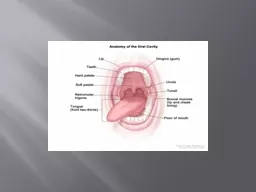
117 IntroductionNoonan syndrome was described by Noonan and Ehmkein 1963 as a multisystem disorder characterized by suchclinical features as short stature hypertelorism ptosis Journal of Oral Scie ID: 941224

syndrome fig height open fig syndrome open height bite dental arch oral angle patient noonan maxilla primary present palatal

Share:

Link:

Embed:

Download Presentation from below link

Download Pdf The PPT/PDF document "Oral findings in a case of Noonansyndrom..." is the property of its rightful owner. Permission is granted to download and print the materials on this web site for personal, non-commercial use only, and to display it on your personal computer provided you do not modify the materials and that you retain all copyright notices contained in the materials. By downloading content from our website, you accept the terms of this agreement.


Presentation Transcript

117 Oral findings in a case of Noonansyndrome in an 8-year-old Japanese male are reported.Examination of the patient revealed a narrow, high-arched palate and an anterior open bite. Cephalometricmeasurements showed a wide gonial angle, a largemandibular plane angle, a large Y-axis and long facial IntroductionNoonan syndrome was described by Noonan and Ehmkein 1963 as a multisystem disorder, characterized by suchclinical features as short stature, hypertelorism, ptosis, Journal of Oral Science, Vol. 45, No. 2, 117-121, 2003 Correspondence to Dr. Mitsugi Okada, Department of PediatricDentistry, Hiroshima University Faculty of Dentistry, 1-2-3Kasumi, Minami-ku, Hiroshima 734-8553, Japan Oral findings in Noonan syndrome: report of a caseMitsugi Okada, Naoko Sasaki, Yasutaka Kaihara, Rinzo Okada, Hideaki Amano, Kazuo Miura and Katsuyuki KozaiDepartment of Pediatric Dentistry, Hiroshima University Faculty of Dentistry, Hiroshima 734-8553Case report 118 erupted. An abnormal swallowing habit was also observed,although no nasal obstruction was present.Radiographic examinationFigure 3 shows frontal and lateral head radiographicviews of the patient. Cranial sutures were normal, as theyappeared to have closed. Digitate impressions were seenat the second, third, and fourth vertebral arch. He had anlesions seen clinically or on X-ray. All permanent teeth,xcept for the third molars, were observed to be present Fig. 1PatientÕs full face and profile at 8 years of age. Fig. 2Oral features of the maxilla, mandible and occlusion. Fig. 4PatientÕs orthopantomograph. Fig. 3Radiographs of the frontal and lateral portions of the Fig. 5Radiograph of the patientÕs hand-wrist. 119 Hand-wrist radiographs confirmed the bone age of aneight-year-old (Fig. 5). The landmarks and measurementsused, taken

according to the method of Sakamoto (9) andJapanese society of Pediatric Dentistry (10) are shown inFig. 6. Table 1 shows a summary of the lateralanalysis revealed a wide gonial angle and high total andlower facial heights (N-Me, ANS-Me). In addition, the Y-axis, mandibular plane angles and ramus were over 1 SDlarger than mean values, while the SNB and interincisalangle were over 1 SD smaller.A study model of the maxilla was prepared forof each tooth was within the normal range. The patient wasfound to have a narrow palate and a right simian creaseJapan) in which 0.5 mm interval scanning was available(Fig. 8). Table 2 shows a summary of the dental archby Ootsubo et al (11). The width of the dental arch waser 2 SD narrower at the maxilla than the standard value,and over 1SD narrower at the mandible. Further, the patienthad an over bite of -4.2 mm and an over jet of 2.8 mm.The height of the palate was measured from the maxillarycasts, using a previously established three-dimensionalhorizontal plane was defined by 4 points; P2 and P3showed the deepest point of the cervical portion on the Table 2Summary of dental arch lengh measurements Table 1Summary of cephalometric measurements Fig. 6Angular and linear measurements used in this study. Fig. 7Study model of the patientÕs maxilla. 120 palatal side of the upper first primary molars and the uppersecond primary molars on both sides, respectively (Fig.9). Four lines were drawn (3 parallel and 1 perpendicular);from the highest vertex of the papilla of the incisor (P1)portions of the upper first and second primary molars, andthe first permanent molars on each side of the palate (L1,L2, L3 and L4 in Fig. 9). The height of the palate wasdefined as the distance between the intersections and theFigure 10 shows the patientÕs palatal he

ight comparedto data from twenty normative subjects at HellmanÕs dentalstage IIIA. P1, P2, P3 and P4 show the upper palatal pointon the first permanent incisor, first primary molar, secondprimary molar, and first permanent molar, respectively. Thepalatal depth was over 1 SD higher at P2, P3 and P4 thanstandard values. The palatal depth at P3 was the highestof all measured points, with a value of 14.3 mm.The high frequency of cardiac, ophthalmic, growth, andemphasizes the need for early diagnosis. Early diagnosisof this relatively common condition would result in betterMoreover, it is important to recognize the markedphenotypic changes with age from newborn to adult. The present case was diagnosed at birth, and the resultsof procedures undertaken for pulmonary stenosis andwebbed neck have been satisfactory.The present report describes a narrow high-arched palateand an open bite with accompanying Noonan syndrome.There have been several other reports of oral findingswith this syndrome (5-8), however, few have reported anarrow high-arched palate with an open bite (7). Sugar etal. (8) described a Noonan syndrome patient who hadmarked mandibular prognathism with an anterior open bite. In the present case, the patientÕs palatal height wasmarkedly higher at the measured points of the upper firstprimary molar, second primary molar, and first permanentmolar than those of standard values, while the values fordental arch length of the maxillar and mandibular were Fig. 9Diagram of palatal height measurements used in thisincisor; L2: midpoint on the first primary molar; L3: Fig. 10Palatal height of the patient, compared with data from Fig. 8Scanning image of maxilla using PICZA. 121 normal. In contrast, the width of the dental arch wasmarkedly narrow at both the maxilla and mandibularl

andmarks. Early closure of cranial sutures may influencethe growth of the dental arch. The present case exhibitedhypertelorism, ptosis, a divergent squint and a tendencyfor exophthalmos. Noonan (14) reported that deformity ofthe sternum with an early closure of sutures was afrequently-seen feature. Unremarkable cranial sutures,along with digitate impressions were noted in this patient.Kreiborg and Pruzansky (15) suggested that, in prematurecraniosynostosis, malocclusion is best explained by a lackof maxillary sutural growth in all three planes of space.In addition, a corn-shaped tongue might also contributeto a narrow dental arch. Longitudinal data on individualThe results of cephalometric measurements showed awide gonial angle and a large mandibular plane angle. Inaddition, the facial height (N-Me and ANS-Me) was longerthan that of the mean values. It was also noted that the Y-axis was larger than that of the mean value. Nahoum (16)suggested that the severity of an open-bite malocclusionis determined by several craniofacial measurements (thegonial angle, mandibular plane and facial height) that arelarge when compared with corresponding statistical norms.Kagami (17) also showed that in Japanese open-bite cases,the gonial angle is large and the height of front lowerportion of the face (ANS-Me) is long. It is suggested thatthis case had a skeletal open-bite malocclusion, whichincluded an abnormal swallowing habit. Orthodontictreatment for the narrow dental arch and open-bite areplanned for the future, since obvious discrepancies havebeen revealed in the permanent dentition. Acknowledgmentswish to thank the Research Center for MolecularMedicine, Faculty of Medicine, Hiroshima University forthe use of their facilities.ReferencesNoonan JA, Ehmke DA (1963) Associateduner HH (19

38) A syndrome of infantilism,congenital webbed neck and cubitus valgus.3.Jamieson CR, van der Burgt I, Brady AF, van ReenM, Elsawi MM, Hol F, Jeffery S, Patton MA,4.Nora JJ, Nora AH, Sinha AK, Spangler RD, LubsHA (1974) The Ullrich-Noonan syndrome (TurnerNelson JF, Tsaknis PJ, Konzelman JL (1978)NoonanÕs syndrome: report of a case with oralBernier-Buzzanga J, Su WP (1990) NoonanÕssyndrome with extensive verrucae. Cutis 46, 242-Horowitz SL, Morishima A (1974) Palataland its variants and in NoonanÕs syndrome. Oral SurgOral Med Oral Pathol 38, 839-844Sugar AW, Ezsias A, Bloom AL, Morcos WE (1994)Orthognathic surgery in a patient with NoonanÕssyndrome. J Oral Maxillofac Surg 52, 421-4259.Sakamoto T (1959) A study on the developmentalchanges of dentofacial complex of Japanese withspecial reference to sella turcica. Nippon kyosei10.Japanese society of Pediatric Dentistry (1995) Achildren. Shouni shika gaku zasshi 33, 659-696 (inOotsubo J, Ishikawa F, Kuwahara Y (1964) Alongitudinal study of dental development between6 to 13 years of age. -Growth changes of dentition-.Nippon kyosei shika gakkai zasshi 23, 182-190 (in12.Kaihara Y, Amano H, Miura K, Nagasaka N (1998)A study of a new method for diagnosis of theLucker GP, Steijlen PM (1994) Widespreadleucokeratosis in NoonanÕs syndrome. Clin ExpNoonan JA (1968) Hypertelorism with Turnerphenotype. A new syndrome with associatedcongenital heart disease. Am J Dis Child 116, 373-Kreiborg S, Pruzansky S (1981) Craniofacial growthpremature craniofacial synostosis. Scand J PlastReconstr Surg 15, 171-186Nahoum HI (1977) Vertical proportions: a guide for17.Kagami H (1972) A morphological considerationof the open bites and over bites with reference toanalytical values of the cephalograms and casts.Nippon kyosei shika gakkai zasshi 31, 45-60