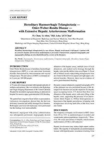
/
Original ArticlesRev Colomb Radiol 2010 214111RATIVE EVLURAL V Original ArticlesRev Colomb Radiol 2010 214111RATIVE EVLURAL V

Original ArticlesRev Colomb Radiol 2010 214111RATIVE EVLURAL V - PDF document

luna
luna . @luna
Follow
343 views
Uploaded On 2022-09-23

Original ArticlesRev Colomb Radiol 2010 214111RATIVE EVLURAL V - PPT Presentation

EYWORDS Vascular malformations ALABRASCLAVE Malformaciones vascularesImagen por resonancia magnéticaArterias cerebrales Radiologist Neuroradiology Fellow Universidad de Antioquia Medellín Colo ID: 955660

activation left functional fmri left activation fmri functional x00660069 patient language visual vascular malformations avm area lesion temporal cerebral

Share:

Link:

Embed:

Download Presentation from below link

Download Pdf The PPT/PDF document "Original ArticlesRev Colomb Radiol 2010 ..." is the property of its rightful owner. Permission is granted to download and print the materials on this web site for personal, non-commercial use only, and to display it on your personal computer provided you do not modify the materials and that you retain all copyright notices contained in the materials. By downloading content from our website, you accept the terms of this agreement.


Presentation Transcript

Original ArticlesRev Colomb Radiol. 2010; 21:(4):1-11RATIVE EVLURAL V EYWORDS Vascular malformations ALABRASCLAVE Malformaciones vascularesImagen por resonancia magnéticaArterias cerebrales Radiologist. Neuro-radiology Fellow. Universidad de Antioquia. Medellín, Colombia. Neuro-radiologist, Centro Avanzado de Diagnóstico Médico (Cedimed) andUniversidad de Antioquia. Medellín, Colombia. Functional magnetic resonance imaging in he pre-operative evaluation of cerebral vascular malformations, ontes ; errera A, argas prequirúrgico de la corteza elocuente con Rf. e usó una técnica dependiente de la concentración de oxígeno () para localizar estas zonas en relación con la malformación vascular cerebral, aplicando diferentes paradigmas. Resultados: e encontró una en el lóbulo mesotemporal derecho, con representación de la memoria visuoespacial en el hipocampo y parahipocampo contralesionales; una temporal posterior izquierda con activación contralateral exclusiva del área de Wernicke; una parietal izquierda sin afectación de la corteza sensoriomotora; una malformación cavernosa en la circunvolución angular izquierda con dominancia hemisférica del lenguaje en ese lado; una talámica derecha sin daño en la corteza elocuente; una periventricular izquierda con patrón de equidominancia del lenguaje; una pequeña occipital izquierda con activación normal en la corteza visual primaria y disminución en la activación de la corteza de asociación visual del lado izquierdo donde se encuentra la lesión, y una temporo-occipital con dominancia hemisférica izquierda y desacople neurovascular. Conclusión: a Rf puede delinear anatómicamente la relación entre la lesión y la corteza elocuente y brindar información que facilita la planeación quirúrgica, incluida la estimación del riesgo de la intervención.A case series is presented in order to illustrate the usefulness Patients and methodsWe reviewed the clinical records of patients with a diagnosis of cerebral vascular malformation referred for fMRI between March 2007 and December 2008 in order to assess the relationship between the lesion and cortical functional areas. Nine patients (�ve women and four men) ranging from 17 to 50 years of age were included in the study (Table 1), and the retrospective review was approved by the Ethics Committee of the institution. The assessment included the localization, type and morphology of the vascular malformation and its relationship (10) with the A 1.5 T Avanto (Siemens, Erlangen, Germany) magnetic resona

nce machine was used to acquire a T1 magnetization-prepared rapid gradient echo (MP-RAGE) sequence in the sagittal plane in 20 healthy volunteers as follows: FOV = 240, matrix = 192x192, resolution = 1.3x1.3x1.3 mm, RT = 1,670 ms, ET = 3.6 ms, �ip angle = 8°, IT = 1,000 ms, averages = 2, concatenations = 1, slices = 128, slice overmeasurement = 25%, distance factor = 50%, bandwidth = 180 Hz/Px, duration, 4’39”.An echoplanar T2* gradient echo (RT = 3,000 ms, ET = 50 ms; matrix = 64x64; voxel size = 3x3x3 mm) sequence with continuous acquisition was used for the functional imaging (Table 2). Functional images were acquired following a block pattern, including alternating series every 30 s between basal and active states. The paradigms (Table 3) consisted of alternating closing and opening of the hand in order to activate the sensorymotor cortex, generation of verbs to assess language function, modi�ed Roland test to assess visual and spatial memory (11), opening and closing of the eyes for visual activation, plus a IntroductionLocalization of the eloquent cortex is dif�cult even with the use of multiplanar magnetic resonance imaging (MRI). There may be distortion and displacement of this area with congenital lesions such as cerebral vascular malformations (1). Functional magnetic resonance mapping (fMRI) using a blood level oxygen-dependent technique (BOLD) has gained popularity as part of the therapeutic planning process in patients who are candidates for surgical, endovascular o radiosurgical treatment. This technique is based on the local increase in oxyhemoglobin (molecule with paramagnetic properties) concentration in the cerebral vasculature, resulting from increased blood �ow and volume in the Mapping of cerebral function in the adjacent areas may be performed using intra- or pre-operative methods, including fMRI as an alternative to positron emission tomography (3), magnetic encephalography (4,5), electrocorticography and the Wada test (1,6,7), for example. Although these procedures, in particular fMRI, are very accurate for localizing the different cerebral functions, they do not provide information about the time course of the activations and, consequently, about the organization of fMRI makes the therapeutic planning easier as it allows to de�ne the relationship between the vascular lesion and the functional cortex before proceeding to the surgical resection or endovascular exclusion, thus preventing a clinical de�cit (8). Language mapping is esse

ntial in these lesions because cerebral vascular malformations may be congenital and may give rise to the reorganization of functional areas (6,9).The purpose of this study is to describe our initial experience with the localization of the eloquent cortex using fMRI, in order to establish its relationship with cerebral vascular malformations and determine its importance in pre-operative planning. Rev Colomb Radiol. 2010; 21:(4):1-11 Original ArticlesTable 1. Patients with cerebral vascular malformations assessed with fMRI before surgery (Medellin, Colombia, 2007-2008) o.ateralityClinical anifestationalformationaradigm interpretationreatment outcome18 year-old RighteizuresRight mid-temporal AemoryOnly left hippocampal and parahippocampal activation ncomplicated embolization50 year-old femaleRightrior bleeding, eft temporal (verb generation)ixed dominance (left roca’s area and right Wernicke’s area)Resection, mild post-surgical dysphasia with full recovery39 year-old Righteft parietal Aotor (right on-displaced activation in left pre- and post-central gyri (sensorymotor cortex)ncomplicated embolization36 year-old femaleRighteizureseft temporal cavernous malformation(verb generation)eft language dominance, lesion in Wernicke’s areao intervention17 year-old femaleRightleeding, left hemiparesisRight thalamic otor (left on-displaced activation in right paracentral gyrusncomplicated embolization19 year-old female Rightleeding, right hemiparesis, dysphasiaCorona radiata and left semi-oval white matter Aand right hand motorilateral language dominance, non-displaced activation in sensorymotor cortexncomplicated embolization38 year-old Rightartial seizures componenteft occipital (opening and eyes)Reduced activation in left visual association areaWaiting for embolization12 year-old femaleRightntra-parenchymal hematoma. motor deficiteft temporoccipital (sentence completion) and breath-breathing cycle)eft hemisphere dominance and neurovascular uncouplingembolization and waiting for radiosurgery Functional magnetic resonance imaging in he pre-operative evaluation of cerebral vascular malformations, ontes ; errera A, argas Table 2. Acquisition technique arameterAnatomical equenceFunctional equence1.5 iemens Avanto magnetic resonance 1.5 iemens Avanto magnetic resonance equences-RAatrixResolutionRepetition Flip Angle (degrees)AveragesConcatenationslicesistance factorandwidthuration4 minutes 39 seconds5 minutesTable 3. Paradigms: block pattern aradigmsasal stateActive stateotor10 scan off10 scan onumber of cyclesuration3 minutes

ummyFirst cycle was discarded to eliminate 1 effecttaying stillAlternating finger movementerb generation10 scan off10 scan onumber of cyclesuration5 minutesummyFirst cycle discarded to eliminate 1 effectAlternating high-low frequency auditory inputouns every 3 s as auditory input and the patient is asked to think of a related verb (10 nouns per cycle)emory10 scan off10 scan onumber of cyclesuration5 minutesummyFirst cycle was discarded to eliminate 1 effectreath-hold15 scan off15 scan onumber of cyclesuration4 minutesummyFirst cycle was discarded to eliminate 1 effectormal breathingApnea10 scan off10 scan onumber of cyclesuration4 minutesummyFirst cycle was discarded to eliminate 1 effectyes closedyes open Rev Colomb Radiol. 2010; 21:(4):1-11 Original ArticlesThe task used for memory assessment was explained in detail before performing the scan. The patient provides information about �ve normal routes and during acquisition the patient is asked to think about the route and imagine every detail. In the basal state, the task consisted of counting odd numbers mentally (11). An of�ine process was also performed where the images acquired for the various paradigms and anatomical series were transmitted in a DICOM format. Images were spatially normalThreshold correlations for each pixel were determined with a 0.001 probability (13). Co-recording of anatomical and functional images, normalization of the data set according to the Montreal Neurological Institute template (ICBM 152 template), anatomical image segmentation and smoothing of functional images with a 6x6x6 gaussian Kernel were performed afterward. The �rst acquisition cycle for each paradigm was discarded in order to eliminate T1-dependent effects in a T2 EPI series.After preparing the EPI images (spatial smoothing and movement correction), the linear GLM model with the block pattern, test generation and the Z parameter maps were applied, followed by alignment with the anatomical images and the entire MNI Atlas. Orientation of local or regional signal variations and noise �ltering were done with spatial smoothing of the imaging data during the realignment steps, in order to prepare the Finally, the researchers selected certain regions of interest (ROI) using the WFU.PickAtlas software graphic interface, executed under SPM5 and Matlab 7.0. This software offers a means to generate ROI masks based on the Talairach Daemon database, according to the methodology described and validated by Lancaster et al. (14). The atlas include Brodma

nn’s area, lobes, hemispheres and various anatomical labels. The functional images were fused with the anatomical images for interpretation by two neuro-radiologists (DAH and SAV).Patient 1: right temporal arteriovenous malformationEighteen year-old male patient complaining of a �rst-time seizure and headache. He was taken to contrast computed tomagraphy imaging of the skull that showed a hypervascular lesion in the medial portion of the right temporal lobe. Catheter arteriography revealed a 6 cm arteriovenous malformation (AVM) in the right temporal lobe, supplied mainly by branches of the posterior communicating artery, with deep and super�cial venous drainage (Figure 1). Functional magnetic resonance was performed using a memory paradigm, with a �nding of exclusive contralateral activation in the left parahippocampal and hippocampal areas. Three embolizations with Onyx were performed, achieving 70% occlusion of the lesion. The patient Patient 2: left temporal arteriovenous malformationFifty year-old female with a history of bleeding due to a left temporal AVM and four years with headaches. Functional MRI was used for pre-operative assessment. Activation was found mainly in the left inferior frontal gyrus (Broca’s area), and exclusively in Wernicke’s area, in the right temporal lobe, when a verb-generation paradigm was used (Figure 2). This constitutes a mixed language dominance pattern. The patient was taken to surgery, but presented mild dysphasia during the immediate post-Patient 3: left parietal arteriovenous malformationThis was a 39 year-old male patient complaining of headaches, with a diagnosis of left parietal AVM. Functional magnetic resonance was performed in order to assess the motor area, using a paradigm consisting of alternating right-hand opening and closing. The test revealed activation in the left pre-central gyrus near the sigmoid portion. The AVM did not affect the sensorymotor cortex (Figure 3), but embolization was performed, nonetheless, in order to achieve 90% occlusion of the malformation, with no Patient 4: left temporal cavernous malformationThirty-six year-old female patient with convulsive syndrome since she was 20. Over the past three years, seizures became refractory to medical treatment and she was referred for brain MRI. The �ndings revealed a cavernous malformation affecting the anterior and subcortical portions of the left angular gyrus. A verb-generation paradigm was used, showing left hemisphere language dominance as a result of activatio

n in Broca’s area, mainly in the left side. When the language reception area (Wernicke’s) was assessed, there was activation of the transverse, superior temporal and supramarginal gyri, surrounding the Patient 5: right thalamic arteriovenous malformationThis is the case of a young 17 year-old female with a history of cerebral hemorrhage due to right thalamic AVM, who was left with a moderate left hemiparesis. A functional MRI was performed in order to assess the motor area (Figure 5). The AVM was causing a motor de�cit as a result of the compression caused by the residual hematoma on the corticospinal tract in the posterior branch of the left internal capsule. A motor paradigm was used for the fMRI with alternating left-hand opening and closing, giving rise to activation of the right pre-central gyrus. Embolization was performed, achieving a 70% occlusion of the Patient 6: left periventricular arteriovenous malformationNineteen year-old female with a history of bleeding from an AVM in the corona radiata and left semioval white matter, with residual moderate left hemiparesis and conductive dysphasia. The AVM was causing a motor de�cit as a result of a lesion of the corticospinal tract, while the language de�cit was due to damage of the arcuate fasciculus. A motor paradigm with alternating right-hand opening and closing was used in the fMRI. There was activation of both sensorymotor cortices, predomi Functional magnetic resonance imaging in he pre-operative evaluation of cerebral vascular malformations, ontes ; errera A, argas Figure 1. (a) Lateral view of the arteriogram prior to embolization, showing an AVM (white arrows) with a dilated posterior communicating artery supplying the lesion. (b) On a fMRI coronal section there is activation of the left hippocampus and parahippocampus (arrow heads), contralateral to the lesion (white arrows). (c) Lateral view of the arteriogram after the third embolization with 70% occlusion of the lesion. Figure 2. (a) Axial fMRI image in a patient with left temporal AVM (curved arrow) using a verb-generation paradigm. There was activation of Wernicke’s area only, in the right hemisphere (straight arrows). (b) Predominant left frontal activation (Broca’s area) (arrow heads). Rev Colomb Radiol. 2010; 21:(4):1-11 Original Articles Figure 3. Motor paradigm. (a) Axial fMRI image. (b) Sagittal fMRI image. Patient with left parietal AVM (curved arrow) with activation of the left pre-central gyrus (straight arrows), with no sensory-motor cortex involvem

ent.Figure 4. Patient with left temporal cavernous malformation (curved arrows) and ipsilateral language dominance, as shown by greater left inferior frontal activation. The activation zone appears in contact with the lesion. (a) Axial section of susceptibility image fusion (SWI) and language fMRI. (b) Sagittal section of SWI fusion and language fMRI. Functional magnetic resonance imaging in he pre-operative evaluation of cerebral vascular malformations, ontes ; errera A, argas nantly in the left side. Bilateral activation was secondary to the patient’s inability to keep the left hand still during the test, an involuntary response elicited by the effort required to move the paretic limb. The fMRI for language assessment using a verb-generation paradigm did not show a signi�cant difference in frontal activation between the two hemispheres, suggesting equal dominance (Figure 6). Embolization was performed, resulting in a 95% occlusion of the malformation, with no residual Patient 7: left occipital arteriovenous malformationThis case is of a 38 year-old male with partial seizures with visual component in the form of plain hallucinations. The MRI scan showed a small left occipital AVM (Figure 7). A visual paradigm consisting of eye closing and opening with a block pattern revealed normal activation in the primary visual cortex and reduced activation in the visual association cortex in the left side, where the lesion is located. The patient is waiting for Figure 5. Patient with right thalamic AVM (curved arrow). (a) Image with axial T2 information. (b) Motor left-hand coronal fMRI showing the relationship between the lesion and the activated cortex (straight arrows). Figure 6. Patient with AVM (curved arrow) involving the periventricular white matter and the left peri-insular region. (a) Axial section with T2 information. (b) Axial fMRI showing a bilateral dominance pattern for language, with symmetrical frontal (Broca’s area) and temporal (auditory) activation (straight arrows). Rev Colomb Radiol. 2010; 21:(4):1-11 Original ArticlesPatient 8: left temporo-occipital arteriovenous malformation The case corresponds to a 12 year-old girl with a history of intra-parenchymal hematoma due to bleeding from a left temporo-occipital AVM (Figure 8a), with no neurological de�cit. The fMRI was performed with the use of a language paradigm consisting of sentence completion, and revealed activation of Broca’s area in the left side. A paradigm for assessing cerebrovascular reactivity (breath-holding) was also us

ed with normal breathing cycles and 20-second cycles of apnea, showing neurovascular uncoupling at the malformation site (Figure 8b). This patient later underwent embolization of 50% of the lesion and is now awaiting radiosurgery.This potential change in the structure of neural networks makes it critical to determine the exact anatomical localization of the eloquent cortex before undertaking any form of intervention (15). An example of this is the representation of the visual and spatial memory exclusively in the contralateral hemisphere in one of our patients with a right mid-temporal AVM who did not It has been accepted that fMRI is no substitute for intra-operative electrical cortical stimulation. However, it is useful for pre-operative planning and mapping, because it reduces the length and extent of the craniotomy (16, 17). The determination of hemispheric dominance of language using fMRI is highly consistent with the Wada test (18), and has the added advantage of being a non-invasive technique that provides additional information about the spatial relationship between the lesion and the language area (19). In this study, a qualitative method was used to determine language lateralization and to de�ne dominance in Figure 8b. Axial and sagittal sections using a breath-holding paradigm that shows neurovascular uncoupling at the site of the malformation. Figure 7. The axial image with T2 information shows a pial AVM (curved arrow) in the left occipital lobe. Using a visual fMRI paradigm, normal activation was found in the primary visual cortex (arrows) and reduced activation of the visual association cortex on the left side, where the lesion is located. DiscussionThe eloquent cerebral tissue may be at risk during treatment of vascular malformations. It is important to know the relationship between the lesion and the eloquent cerebral territories in order to balance the bene�t from obliterating the malformation Magnetic resonance imaging provides excellent anatomical details and is a reasonable approximation to the location of the eloquent areas in the normal brain. However, conventional imaging does not provide direct information about functional areas and may lead to localization errors, especially in patients with intracranial masses that cause anatomical distortions. This problem also occurs in cases of vascular malformations that may give rise to developmental cortical reorganization (1). Figure 8a. The axial and sagittal sections show a left temporo-occipital AVM. The application of the language pa

radigm showed activation of Broca’s area on the left side. A Family Wise Error with a corrected p value of 0.05 was used. Functional magnetic resonance imaging in he pre-operative evaluation of cerebral vascular malformations, ontes ; errera A, argas the side that showed greater inferior frontal activation (Broca’s area). This interpretation modality has been described previously with good agreement among different evaluators (19). Three activation patterns were identi�ed in the patients studied: left dominance for language (patients 4 and 8), mixed dominance (patient 2) and bilateral dominance (patient 6). These types of responses with the use of language paradigms in fMRI have already been described (20). We did not �nd a fourth atypical pattern described as right language dominance where Broca’s area is localized in the right frontal lobe (19,20).Aside from language reorganization found in patients with vascular malformations, there are also blood-�ow abnormalities such as blood steal, with retrograde feedback of the distal territory that may interfere with signal intensity changes in fMRI when the BOLD technique is used (6). This fMRI method detects small changes in the magnetic properties of blood resulting from metabolic and vascular responses of neuronal activity (9). A study found that blood-�ow abnormalities associated with This effect might explain the post-operative de�cit (dysphasia) found in one of the patients in our study after the resection of a left temporal AVM, with exclusive representation of Wernicke’s area in the right side, according to the fMRI �ndings (Figure 2). In order to avoid mistakes in the interpretation of the activation areas, it is important to determine whether there is neurovascular uncoupling when breath-holding or apnea Vates et al. (15) conducted a retrospective analysis of 30 patients with vascular malformations in order to study the effect on the primary motor or sensory cortex, and identi�ed changes in cortical function topography in more than one third of the malformations (15). In the patients in our study we did not �nd displacement in the representation of the motor area; however, in all instances, there was a separation plane between the lesion Regarding visual cortex assessment, the screening test most commonly used for assessing neurological de�cit is the Hooper visual organization test, with good sensitivity for visual and visual-spatial organization, a multi-factoria

l cognitive process that includes mental rotation, visual memory, object identi�cation and name recall. This test has been adapted to fMRI paradigms where the patient is asked to answer right or wrong when presented with a linear drawing of a simple object cut into two or three pieces, followed by a single word designating the object.Due to the complex and multi-factorial nature of the task, multiple regions are involved and there are responses in both lobes, with occipital and right parietal predominance (22). Noet al. (23) studied 40 children under sedation using fMRI to map activation areas when passive visual and auditory tasks (8 Hz �ash of light and a recording of the mother’s voice) were presented. The researchers assessed activation of the auditory and visual cortices and found temporal and frontal activation and negative values in the rostral region in the �rst and second instance, respectively (23). In our patient with a left occipital AVM we applied a visual paradigm with a block pattern, con sisting of opening and closing the eyes, and we found normal activation in the primary visual cortex, and reduced activation of the visual association cortex on the left side, where the lesion ConclusionWe described the use of fMRI in patients with vascular malformations, showing complex activities of the cerebral cortex in one or both hemispheres, depending on the paradigm employed and the relationship with the lesion, as a way to provide useful information for treatment planning. One of the limitations of the study was that fMRI �ndings were not validated by a different invasive or non-invasive technique. This is a preliminary report describing the use of this method in patients with this type of abnormality. A correlation of fMRI with other functional mapping techniques is required in order to determine its role in the ReferencesLatchaw RE, Hu X, Ugurbil K, Hall WA, Madison MT, Heros tool for cerebral arteriovenous malformations. Neurosurgery. Thickbroom GW, Byrnes ML, Morris IT, Fallon MJ, Knuckey NW, Mastaglia FL. Functional MRI near vascular anomalies: Clin Neurosci. 2004;11(8):845-8.González J, Felipe-Morán A, Benavides-Barbosa J, et al. DiagDel Río D, Santiuste M, Capilla A, Maestú F, Campo P, Fernández-Lucas A, et al. Bases neurológicas del Lenguaje. AportacioOrtiz T, Maestú F, Fernández-Lucas A, Amo C, Campo P, Capilla A. Correlatos neuromagnéticos del lenguaje. RevLehéricy S, Biondi A, Sourour N, Vlaicu M, du Montcel ST, Cohen L, et al. Arteriovenous brain malf

ormations: is functional MR imaging reliable for studying language reorganization in patients? Initial observations. Radiology. 2002;223(3):672-82.Byrne JV. Cerebrovascular malformations. Eur Radiol. Lee CZ, Young WL. Management of brain arteriovenous malformations. Curr Opin Anaesthesiol. 2005;18(5):484-9.clinical applications. New York: Springer; 2006.Joint Writing Group of the Technology Assessment Committee American Society of Interventional and Therapeutic Neuroradiology, Joint Section on Cerebrovascular Neurosurgery a Section of the American Association of Neurological Surgeons and Congress of Neurological Surgeons, Section of Stroke and the Rev Colomb Radiol. 2010; 21:(4):1-11 Original ArticlesSection of Interventional Neurology of the American Academy of Neurology,Atkinson RP,Awad IA,11.Avila C, Barrós-Loscertales A, Forn C, Mallo R, Parcet MA, Belloch V, et al. Memory lateralization with 2 functional MR imaging tasks in patients with lesions in the temporal lobe. AJNR functional imaging data. En: Toga AW, Mazziotta JC (editores). Brain mapping: the methods. New York: Academic Press; 1996. Komisaruk BR, Mosier KM, Liu WC, Criminale C, Zaborszky L, Whippie B, et al. Functional localization of brainstem and cervical spinal cord nuclei in humans with fMRI. AJNR Am J Lancaster JL, Woldorff MG, Parsons LM, Liotti M, Freitas CS, Rainey L, et al. Automated Talairach atlas labels for functional Vates GE, Lawton MT, Wilson CB, McDermott MW, Halbach W, Roberts TP, et al. Magnetic source imaging demonstrates nous malformations. Neurosurgery. 2002;51(3):614-27.Giussani C, Roux FE, Ojemann J, Sganzerla EP, Pirillo D, Papreliable for language areas mapping in brain tumor surgery? direct cortical stimulation correlation studies. Neurosurgery. 2010;66(1):113-20.rect cortical stimulation. Neurosurgery. 2003;52(6):1335-47Álvarez-Linera J, Martín-Plasencia P, Maestú F, García de Sola tareas. Rev Neurol. 2002;35(2):115-8.Smits M, Visch-Brink E, Schraa-Tam CK, Koudstaal PJ, van der Lugt A. Functional MR imaging of language processing: an Stippich C. Clinical functional MRI: Presurgical functional neuMoritz CH. Technical procedures for quality control assessment sented at: ASFNR. 4th Annual Meeting of the American Society of Functional Neuroradiology; 2010 February 24-26; Las Vegas, Moritz CH, Johnson SC, McMillan KM, Haughton VM, the Hooper Visual Organization Test.tory and visual functional MR imaging. Radiology. Correspondence Diego Alberto Herrera Received for evaluation: eptember 2, 2010 Approved for publication: October 25, 20