/
wwwcdaadccajcda  May 2006 Vol 72 No 4 llowing thisthe maxil wwwcdaadccajcda  May 2006 Vol 72 No 4 llowing thisthe maxil

wwwcdaadccajcda May 2006 Vol 72 No 4 llowing thisthe maxil - PDF document

lydia
lydia . @lydia
Follow
342 views
Uploaded On 2022-08-21

wwwcdaadccajcda May 2006 Vol 72 No 4 llowing thisthe maxil - PPT Presentation

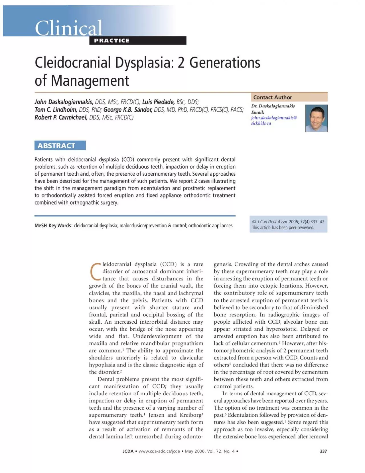
Figure 2f Presurgical panoramic radiographFigure 2ePresurgical occlusal relationship Figure 2h Intraoral views after removal of orthodontic appliances and completion of the mandibular implantsup ID: 939457

figure teeth molars ofthe teeth figure ofthe molars mandibular eruption denture maxillary surgery impacted prosthesis bone arch intraoral radiograph

Share:

Link:

Embed:

Download Presentation from below link

Download Pdf The PPT/PDF document "wwwcdaadccajcda May 2006 Vol 72 No 4 ll..." is the property of its rightful owner. Permission is granted to download and print the materials on this web site for personal, non-commercial use only, and to display it on your personal computer provided you do not modify the materials and that you retain all copyright notices contained in the materials. By downloading content from our website, you accept the terms of this agreement.


Presentation Transcript

www.cda-adc.ca/jcda ¥ May 2006, Vol. 72, No. 4 ¥llowing this,the maxillary lateral incisors and Þrstpremolars were exposed and traction hooks were placed onthem (ig.2c).At the same time,forced eruption ofthedeeply impacted mandibular second molars wasattempted by bonding parylene-coated neodymiumÐironÐboron magnets to them.A mandibular Hawley appliance was fabricated with2 larger magnets in direct juxtaposition with the magnetson the teeth to attempt their disimpaction by making useofthe attractive magnetic forces through the tissue.Thisapproach proved unsuccessful and,despite a number ofmodifications to achieve the best position for the largermagnets on the appliance,it was eventually aborted.The maxillary arch was

bonded from first molar to firstmolar and the traction hooks were tied to a stiffstainlesssteel archwire with elastic thread (ig.2dSequential extractions and exposures followed byforced orthodontic eruption continued over several yearsas the patientÕs compliance with appointments dwindledunder the taxing burden ofcare.The limited mouthopening (16 mm) made access to the posterior teeth verydifficult.The maxillary first molars were lost to caries and,later,the maxillary second molars and themandibular left second molar were deemed to be anky-losed.They were subsequently removed along with thethird molars.entually,good arch alignment ofthe remaining teethwas achieved,although a Class III interarch relation andan anter

ior crossbite remained due to the maxillarypoplasia.The plan was to address this through orthog-nathic surgery.To avoid over-retraction ofthe mandibularincisors,which would compromise the skeletal correction, Figure 2f: Presurgical panoramic radiograph.Figure 2e:Presurgical occlusal relationship. Figure 2h: Intraoral views after removal of orthodontic appliances and completion of the mandibular implant-supported crowns. Figure 2g: ofteeth in a patient already deÞcient in alveolar bone.Puseyand Duriesuggested removal ofonly the erupted teeth anduse ofa removable prosthesis to minimize alveolar bone loss.subsequent eruption ofretained teeth can requirefurther surgery and modiÞcation ofthe prosthesis.The current Òstat

e-of-the-artÓtreat-ment involves a combination ofortho-dontics and maxillofacial surgery.Our protocol involves timely extractionofdeciduous teeth,staged surgicalremoval ofsupernumerary teeth,expo-sure ofselected unerupted permanenteth and orthodontic forced eruption.The process is usually carried out instages,as teeth that are guided into theirideal position in the arch can subse-quently serve as vertical stops to main-tain the vertical dimension while thenext group ofunerupted teeth is exposedand bonded.Following alignment ofallpermanent teeth,any underlying skeletaldiscrepancy (most commonly a Class IIIskeletal malocclusion) can be correctedthrough orthognathic surgery after com-pletion ofgrowth.hat follows is a r

eport ofthe treat-ment of2 patients with CCD,a motherand her son.The contrast between treat-ments ofthe 2 patients reflects the shiftin the management paradigm over thespan ofa generation.A 39-year-old woman with a historyofCCD originally presented with thehiefcomplaint ofan ill-Þtting mandi-bular complete denture.Most ofhermandibular teeth had been removed at aoung age and she had not been able toolerate a lower denture since her teenageears.In the maxilla,however,the patientwore a denture comfortably.The onlyocclusal contact ofthe upper denture wasith tooth 46.Multiple impacted teethpresent in the maxilla,whereas in themandible,both third molars were hori-ntally impacted and tooth 47 was verti-cally impacted (ig.1

aminimize the risk ofa pathologicfracture ofthe mandible,the deeplyimpacted mandibular molars weretained.Dental implants (solid screw,4.1-mm diameter,SLA;Straumann,aldenburg,Switzerland) were placed at sites 33 and 43 and a bar-retained erdenture was provided for the mandibular archThe patient has returned annually for 4 years.Herimplants remain stable,there is no radiographic evidence ofany marginal bone loss and the prostheses remain well Þtting. www.cda-adc.ca/jcda ¥ May 2006, Vol. 72, No. 4 ¥ Figure 1a:Pretreatment panoramic radiograph. Figure 1b:Post-treatment panoramic radiograph.Figure 1d:Post-treatment frontal intraoral view of the prosthesis and Òtooth-to-lipÓ Figure 1c: Post-treatment intraoral views. Case