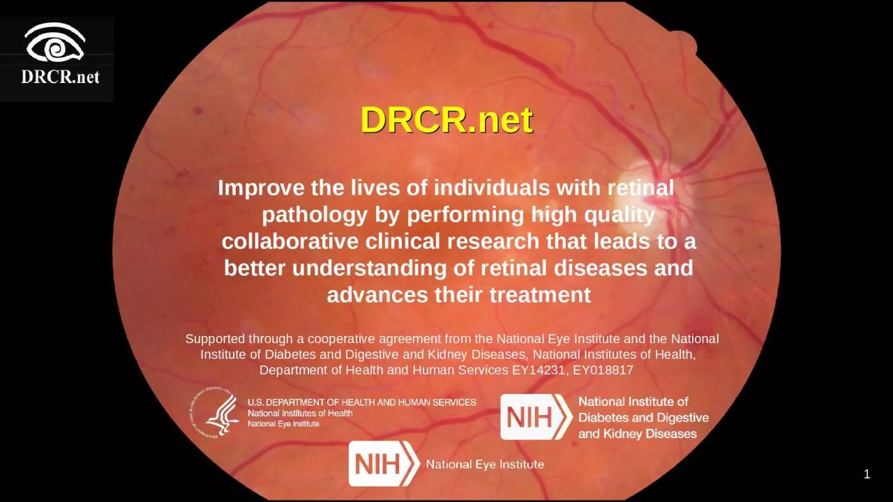
/
DRCR.net Improve the lives of individuals with retinal pathology by performing high quality DRCR.net Improve the lives of individuals with retinal pathology by performing high quality

DRCR.net Improve the lives of individuals with retinal pathology by performing high quality - PowerPoint Presentation

madison
madison . @madison
Follow
0 views
Uploaded On 2024-03-13

DRCR.net Improve the lives of individuals with retinal pathology by performing high quality - PPT Presentation

that leads to a better understanding of retinal diseases and advances their treatment Supported through a cooperative agreement from the National Eye Institute and the National Institute of Diabetes and Digestive and Kidney Diseases National Institutes of Health Department of Health a ID: 1047381

study dme eyes treatment dme study treatment eyes protocol eye acuity visual criteria clinical anti oct vegf prior injections

Share:

Link:

Embed:

Download Presentation from below link

Download Presentation The PPT/PDF document "DRCR.net Improve the lives of individual..." is the property of its rightful owner. Permission is granted to download and print the materials on this web site for personal, non-commercial use only, and to display it on your personal computer provided you do not modify the materials and that you retain all copyright notices contained in the materials. By downloading content from our website, you accept the terms of this agreement.


Presentation Transcript

1. DRCR.netImprove the lives of individuals with retinal pathology by performing high quality collaborative clinical research that leads to a better understanding of retinal diseases and advances their treatment Supported through a cooperative agreement from the National Eye Institute and the National Institute of Diabetes and Digestive and Kidney Diseases, National Institutes of Health, Department of Health and Human Services EY14231, EY018817 1

2. DRCR.net OverviewThe DRCR.net supports the identification, design, and implementation of multicenter clinical research initiatives focused on retinal disorders. Principal emphasis is placed on clinical trials, but epidemiologic outcomes and other research may be supported as well.Funding:National Eye Institute (NEI) and The National Institute of Diabetes and Digestive and Kidney Diseases (NIDDK)-sponsored cooperative agreement initiated September 2002.Current award 2014-20182

3. Priority InitiativesInvolvement of community-based practices, as well as “academic” or university-based centers. Collaborate with industry to facilitate investigations and pursue opportunities otherwise not possible and to do so in a manner consistent with the Network’s dedication to academic integrity and optimal clinical trial performance.3

4. DRCR.net Status(as of 10/18/18)4ActiveTotalSites (Community & Academic Centers)155362 Community Sites106 (68%)234 (65%)Investigators5091,343Other Personnel1,2256,465States3749Provinces in Canada811

5. How to Join the NetworkAll retina specialists in the U.S. and Canada are welcome to applyE-mail drcrnet@jaeb.orgYour request will be reviewed and if approved the necessary paperwork will be sent to you5

6. How to Submit a Protocol IdeaGo to the public* website: drcr.net Click on Information for Investigators. Scroll down to Protocol Idea Form.E-mail form to drcrnet@jaeb.orgIt will be reviewed by the Operations Group every six months.* Forms also available on the study website6

7. 7Designated ideas presented to investigators at semiannual meetingBased on Investigator feedback, ideas are prioritized by EC.Protocol Development Committee is formed.Protocol Idea Review Process

8. Completed DRCR.net Protocols8

9. DRCR.net Protocols Enrolling or in Follow-up9

10. What Has Been Learned?Diabetic Macular Edema TreatmentProtocol A: Although some ophthalmologists considered using a modified macular grid (MMG) photocoagulation technique over the focal photocoagulation technique modified from the ETDRS, this trial showed that at 12 months after treatment, the MMG technique was less effective at reducing OCT measured retinal thickening. Protocol B: Over 2 years, focal/grid photocoagulation is more effective and has fewer side effects than 1 mg or 4 mg doses of preservative-free intravitreal triamcinolone. Protocol E: In cases of DME with good visual acuity, peribulbar triamcinolone, with or without focal photocoagulation, is unlikely to be of substantial benefit. Protocol H: Intravitreal bevacizumab can reduce DME in some eyes, but the study was not designed to determine whether the treatment was beneficial.10

11. What Has Been Learned?Diabetic Macular Edema TreatmentProtocol I: Intravitreal ranibizumab with prompt or deferred (≥24 weeks) focal/grid laser is more effective through 2 years in increasing visual acuity compared with focal/grid laser treatment alone for the treatment of DME involving the central macula. Focal/grid laser treatment at the initiation of intravitreal ranibizumab is no better, and possibly worse, for vision outcomes than deferring laser treatment for 24 weeks or more in eyes with DME involving the fovea and with vision impairment.Protocol K: Sixteen weeks after focal/grid laser for DME in eyes with a definite reduction, but not resolution, of central edema, 23% to 63% likely will continue to improve without additional treatment.11

12. What Has Been Learned?Diabetic Macular Edema TreatmentProtocol R: At 1 year in eyes with non-central DME, this study could not identify a difference between topical nepafenac 0.1% and placebo on OCT parameters or visual acuity.Protocol T*: The 2-year clinical trial compared 3 drugs for diabetic macular edema (DME) and found that gains in vision were greater for participants receiving the drug aflibercept than for those receiving bevacizumab, but only among participants starting treatment with 20/50 or worse vision. At one year aflibercept had superior gains to ranibizumab in this vision subgroup, however a difference could not be identified at 2 years. The 3 drugs yielded similar gains in vision for patients with 20/32 or 20/40 vision at the start of treatment. 12

13. What Has Been Learned?Diabetic Macular Edema TreatmentProtocol U: Although its use is more likely to reduce retinal thickness and increase intraocular pressure, the addition of intravitreous dexamethasone to continued ranibizumab therapy does not improve visual acuity at 24 weeks more than continued ranibizumab therapy alone among eyes with persistent DME following anti-VEGF therapy.13

14. What Has Been Learned?Diabetic Retinopathy TreatmentProtocol F: Results suggest clinically meaningful differences are unlikely in OCT thickness or visual acuity following application of PRP in 1 sitting compared with 4 sittings. Protocol J: The addition of 1 intravitreal triamcinolone injection or 2 intravitreal ranibizumab injections in eyes receiving focal/grid laser for DME and PRP is associated with better visual acuity and decreased macular edema by 14 weeks. Whether continued long-term intravitreal treatment is beneficial could not be determined from this study. 14

15. What Has Been Learned?Diabetic Retinopathy TreatmentProtocol N: The study suggested little likelihood of a clinically important difference between ranibizumab and saline on the rate of vitrectomy by 16 weeks in eyes with vitreous hemorrhage from PDR. Short-term secondary outcomes including visual acuity improvement, increased panretinal photocoagulation completion rates, and reduced recurrent vitreous hemorrhage rates suggest biologic activity of ranibizumab. Long-term benefits remain unknown. Protocol S: This study showed that ranibizumab injections are effective in treating proliferative diabetic retinopathy. At two years, vision of the ranibizumab group on average improved by half a line on an eye chart. Vision of the laser group remained unchanged. 15

16. What Has Been Learned?OCT and Retinal ThickeningProtocol C: Although on average there are slight decreases in retinal thickening during the day, most eyes with diabetic macular edema have little meaningful change in OCT central subfield thickening or visual acuity between 8 AM and 4 PM. Protocol C: Reproducibility of retinal thickness in DME was better for central subfield thickness measurements than for center point measurements. A change in central subfield thickness exceeding 11% is likely to be real. Protocol G: While subclinical DME may be uncommon, this study suggests that between approximately one-quarter and one-half of eyes with subclinical DME will progress to more definite thickening or be judged to need treatment for DME within 2 years after its identification.16

17. What Has Been Learned?Optical Coherence TomographyProtocol G: CST (central subfield thickness) on Stratus OCT™ in people with diabetes and minimal or no retinopathy are similar to thicknesses reported from a normative database of people without diabetes. CST is greater in men than in women. Studies involving comparisons of retinal thickness to expected norms should consider different mean values for women and men.Protocol O: Mean CST is ~70 µm thicker when measured with Heidelberg Spectralis OCT as compared with Stratus OCT among individuals with diabetes in the absence of retinopathy or with minimal non-proliferative retinopathy and a normal macular architecture.  CST values ≥320 µm for men and 305 µm for women are proposed as gender-specific thickness levels.17

18. What Has Been Learned?Optical Coherence TomographyProtocol O: Conversion equations may be used to transform CSF values obtained on a SD-OCT to a TD scale for group comparisons. However, the CST conversion equations do not appear to predict TD values for an individual accurately enough to warrant use of these conversion equations confidently in clinical decision-making at the patient level. 18

19. Access to Publications and PresentationsPublications and Presentations can be found on the public website or on the study website (log in required)19

20. Recently Published Results20

21. Protocol SFive-Year Outcomes of Panretinal Photocoagulation vs Intravitreous Ranibizumab for Proliferative Diabetic Retinopathy:Randomized Clinical Trial21

22. Study DesignStudy eye(s) meeting all of the following criteria (a participant can have 2 study eyes):PDRNo history of PRPBest corrected visual acuity letter score ≥24 (~Snellen equivalent 20/320 or better) Eyes with or without central-involved DME were eligible Randomized, multi-center clinical trial (55 Sites)Primary Objective: Compare the efficacy and safety of PRP with that of intravitreous ranibizumab (0.5-mg in 0.05 mL) for proliferative diabetic retinopathy (PDR)

23. Conclusions: Five-Year OutcomesMean change in VA with ranibizumab similar to PRP at 5 yearsLoss to follow-up was relatively high in both groupsOther outcomes:Favored ranibizumab:Decreased development of central-involved DME with vision impairmentDecreased development of retinal detachmentsFavored PRP:Fewer visitsFewer injectionsBoth Groups:Substantial VA loss rare (6% in each group)Visual field loss progressed in both groups in years 2-5; difference between groups diminishedVitreous hemorrhage in almost 50% of both groups23

24. 24Active StudiesImage: National Eye Institute, National Institutes of Health

25. Protocols Currently Enrolling25

26. Protocol ACRandomized Trial of Intravitreous Aflibercept versus Intravitreous Bevacizumab + Deferred Aflibercept for Treatment of Central-Involved Diabetic Macular Edema (DME)26

27. BackgroundAflibercept treatment in Protocol T resulted in better VA, on average, for eyes with worse baseline VA than bevacizumab However, bevacizumab was effective for many eyes with worse VA at baseline.Approximately 2/3 of bevacizumab-treated eyes had >10 letter improvement at 2 yearsAlmost half had resolution of DME at 2 yearsCost is an issue 27

28. BackgroundReal Life Application: Compare starting with bevacizumab and switching to aflibercept if needed vs. starting with aflibercept?What are the implications of insurance companies mandating this approach or patients choosing this approach?Cost savingsVisual outcomes 28

29. Study ObjectiveTo compare the efficacy of intravitreous aflibercept with intravitreous bevacizumab + deferred aflibercept if needed in eyes with CI DME and moderate vision loss. 29

30. Study DesignAt least 1 eye that meets all of the following criteria:VA letter score ≤ 68 and ≥ 24 (≈20/50 to 20/320)Ophthalmoscopic evidence of CI-DME Central-involved thickening on OCTCirrus: ≥ 290 µm for women; ≥ 305 µm for menSpectralis: ≥ 305 µm for women; ≥ 320 µm for menNo history of anti-VEGF treatment for DME in past 12 months and no history of any other treatment for DME in past 4 months No history of major ocular surgery within prior 4 months or anticipated within next 6 monthsMulti-Center Randomized Clinical Trial (312 Eyes, 260 Participants)Primary Outcome: Mean change in VA over 2 years (AUC)AfliberceptBevacizumab (Aflibercept if needed)30

31. Treatment Groups31Centrally repackaged 1.25-mg bevacizumab Switched to intravitreous aflibercept if eye is not “successful”2.0-mg intravitreous afliberceptAfliberceptBevacizumab (Aflibercept if needed)

32. Study Treatment OverviewAt randomization, study eyes will receive an intravitreous injection according to their assigned treatment groupAfter the initial injection, each eye will be treated according to the retreatment protocol (Protocol T retreatment criteria)Bevacizumab will be provided by DRCRnet and will have a study drug numberAflibercept will be from your clinic stock32

33. Early Switching CriteriaConcept: 3-5 bevacizumab injections and eye has not improved and vision is still poor Details: At the 3, 4, and 5 month visit, study eyes assigned to the bevacizumab (aflibercept if needed) group will switch from bevacizumab injections to aflibercept injections the first time all of the following criteria are met at 2 consecutive visits:OCT CST is above the following cutoffs:Cirrus: ≥290 µm in women or ≥305 µm in menSpectralis: ≥ 305 µm in women or ≥320 µm in menOCT CST has not improved at least 10% from prior visitVA has not improved 5 letters from prior visitVA is 20/50 or worse 33

34. Additional Switching CriteriaConcept: At least 6 bevacizumab injections, eye is no longer improving and VA is at least mildly reducedDetails: Beginning at the 6 month visit, study eyes that have not already switched to aflibercept will switch the first time all of the following criteria are met at 2 consecutive visits:OCT CST is above the following cutoffs:Cirrus: ≥290 µm in women or ≥ 305 µm in menSpectralis: ≥ 305 µm in women or ≥320 µm in menOCT CST has not improved at least 10% from prior visitVA has not improved at least 5 letters from prior visitVA is 20/32 or worse34

35. The SwitchEyes assigned to the bevacizumab group that meet the switch criteria will receive 2 initial monthly injections of aflibercept, then will continue with aflibercept injections throughout study according to the Protocol T retreatment regimen35

36. The SwitchIf bevacizumab injections are deferred because of success according to the initial retreatment protocol and then the eye worsens, injections will resume using bevacizumab. Then the eye will be switched to aflibercept using the same criteria listed previously, following two consecutive bevacizumab injections.36

37. Failure CriteriaFor study eyes in both treatment groups, when failure criteria is met, treatment is up to investigator discretionTreatment with focal/grid laser will not be permitted in the study eye(s) during the study. If the failure criteria are met, treatment (including laser) is up to investigator discretion.37

38. Protocol AGRandomized Clinical Trial Assessing the Effects of Pneumatic Vitreolysis on Vitreomacular Traction38

39. Study ObjectivePrimaryTo compare the proportion of eyes with foveal VMT (symptomatic VMA) release on OCT after PVL with gas injection vs. observation (sham injection) in eyes with VMT without an associated macular holeSecondaryTo evaluate visual function outcomes at 24 weeks after gas injection is performed compared with observation. 39

40. Study DesignAt least 1 eye that meets all of the following criteria:Vitreomacular adhesion on OCT ≤3,000 µmReading center confirmation required for eligibilityBest corrected ETDRS visual acuity equivalent of 20/32 to 20/400Decreased visual function attributed to VMTNo macular or lamellar holePrompt Vitrectomy not required Multi-Center Randomized Clinical Trial (124 Eyes)Primary Outcome: Proportion of eyes with VMT release on OCT without rescue vitrectomy at 24 weeksPVL (Injection of C3F8 gas)Observation (Sham injection)40

41. Dropped IneligibleDropped IneligibleReading Center ReviewRandomizationObservation (Sham Injection)Injection of C3F8 Gas (0.3 mL)1 week1 week4 weeks4 weeks12 weeks12 weeks24 weeks24 weeksInformed Consent, Baseline TestingStudy Design41Primary outcome visit

42. Protocol AHSingle-Arm Study Assessing the Effects of Pneumatic Vitreolysis on Macular Hole 42

43. Rationale for Single-Arm StudyEyes with MH need treatment, therefore randomization to a sham arm would be inappropriateVitrectomy results in nearly 100% hole closure making it an unnecessary (and expensive) control group choiceEven if this proposed study finds that PVL is only moderately successful, physicians and patients may decide to attempt PVL in the office first, before proceeding with more costly, invasive surgeryThus, even without a control group, the results from this study will provide data of value for physicians and patients to make informed decisions about treatment course43

44. Study ObjectiveTo obtain estimates for the proportion of eyes with macular hole closure of the inner retinal layers for eyes with VMT and full-thickness macular holes treated with PVL 44

45. Study DesignAt least 1 eye that meets all of the following criteria:Full-thickness macular hole ≤250 microns at the narrowest point, confirmed by central reading centerVitreomacular adhesion on OCT ≤3,000 microns, confirmed by central reading center Best corrected ETDRS visual acuity equivalent of 20/25 to 20/400Multi-Center Single-Arm Study(50 Eyes)Primary Outcome: Proportion of eyes with macular hole closure of the inner retinal layers at 8 weeks without rescue treatmentPVL (Injection of C3F8 gas)45

46. Informed Consent, Baseline TestingDropped IneligibleDropped IneligibleReading Center ReviewInjection of C3F8 Gas (0.3 mL)1 week4 weeks8 weeks24 weeksStudy Design46Primary outcome visit

47. GeneticsGenes in Diabetic Retinopathy Project47

48. Genes in Diabetic Retinopathy ProjectObjectiveTo create a repository of genetic material and clinical phenotype information as a resource for the research community The database may provide the opportunity to assess genetic susceptibility and resistance to DR and also variants impacting visually-important biomarkers for ME and neovascularization. Major Eligibility CriteriaPrevious/current participant in a DRCR.net studyEnrollment (Ongoing)Total enrolled: 2,350 subjects (as of 10/18/18)48

49. ProtocolsCurrently in Follow-up49

50. Protocol AB50Intravitreous Anti-VEGF vs. Vitrectomy for Vitreous Hemorrhage from PDR

51. Study ObjectivesCompare visual acuity outcomes over time for the following two treatment regimens: Prompt Vitrectomy + PRPIntravitreous Anti-VEGF injectionsin eyes presenting with vitreous hemorrhage from PDR causing vision impairment for which intervention is deemed necessary. 51

52. Study DesignAt least one eye that meets the following criteria:Vitreous hemorrhagecausing vision impairment,presumed to be from PDR, andrequiring intervention (vitrectomy or anti-VEGF)Multi-Center Randomized Clinical TrialPrimary Outcome: Visual Acuity AUC over 6 monthsVitrectomy+PRPAnti-VEGFSample Size: 200 eyes52

53. Additional Eligibility CriteriaVisual acuity 20/32 or worse and at least light perceptionInvestigators should use particular caution when considering an eye with visual acuity 20/32 to 20/40 to ensure that the need for vitrectomy and its associated potential benefits outweighs the potential risksNo anti-VEGF treatment within 2 months prior to vitreous hemorrhage onsetNo prior vitrectomyNote: Prior PRP is not a requirement nor an exclusion No RRD, no TRD involving or threatening the fovea53

54. Secondary Outcomes54Treatment Group ComparisonsMean visual acuity at annual visitsVisual acuity Area Under the Curve (AUC) at 12 and 24 monthsPercent 20/20 and 20/40 or better at annual visitsPercent 20/200 or worse at annual visitsRates of recurrent VH on clinical examDifference in productivity from WPAI questionnaireTreatment and follow-up costs

55. Additional Outcomes of InterestVitrectomy Group OutcomesPercent requiring repeat vitrectomyRates of post-surgical complicationsRetinal detachment/tearCataract/cataract surgeryAnti-VEGF Group OutcomesNumber of injections performedPercent requiring vitrectomyPercent requiring PRP55

56. Study Treatment OverviewAnti-VEGF Group Follow-up injections every 4 weeks according to protocol criteria and no PRP unless failure criteria are metVitrectomy only permitted prior to 16 weeks if criteria for rescue treatment met:TRD threatening or involving the maculaRhegmatogenous retinal detachment NVG, NVA, progressive NVI, or ghost cell glaucomaAfter 16 weeks, vitrectomy may be performed if persistent VH after 2 consecutive 4-week injections56

57. Anti-VEGF Group57

58. Study Treatment OverviewVitrectomy GroupVitrectomy scheduled within 2 weeks of randomization Performed according to surgeon’s usual routine including +/-:Pre-op intravitreous anti-VEGF Removal of the internal limiting membraneUse of agents to improve visualization of membranesUse of intraoperative corticosteroids Cataract extractionPRP during surgery is required, unless it is determined to already be “complete”Anti-VEGF will be permitted during follow-up in certain cases for recurrent hemorrhage58

59. Vitrectomy Group59Injections during follow-up if needed for recurrent hemorrhageIf cataract becomes visually significant

60. Protocol W 60Intravitreous Anti-VEGF Treatment for Prevention of Vision Threatening Diabetic Retinopathy in Eyes at High Risk

61. Primary (Short-term) ObjectiveObservation (sham injections)To determine safety and efficacy of anti-VEGF versus observation in eyes presenting with severe NPDR and no CI-DME for prevention of vision threatening outcomes (DME or PDR)Intravitreous anti-VEGF Primary outcome: Proportion of eyes that develop PDR/PDR-related outcomes or center-involved DME causing visual acuity loss by 2 years61

62. RationaleThe application of anti-VEGF therapy earlier in the course of disease could help to reduce future potential treatment burden in patients, at the same time resulting in similar or better long-term visual acuity outcomes.If this study demonstrates that anti-VEGF treatment is effective and safe in the setting of severe NPDR, a new strategy to prevent vision-threatening complications will be available for patients. 62

63. Composite Primary Outcome: Part 1 – PDRDevelopment of NV within the 7 modified ETDRS fields on fundus photography or FA, confirmed by Reading Center, or…NVI, NVA, or NVG on clinical exam, or…Traction retinal detachment, vitreous hemorrhage, or pre-retinal hemorrhage presumed to be from PDR, documented on fundus photography or FA, or…PRP, anti-VEGF, or vitrectomy for PDR, images should be obtained prior to treatment63

64. Composite Primary Outcome: Part 2 – DMECenter-involved DME on clinical exam with ≥10% increase in central subfield thickness from baseline and visual acuity loss from DMETreatment for DME performed without meeting above criteria (protocol deviation)Note: An eye will be considered to have met the primary outcome if any one of the above PDR or DME criteria are met64

65. Major Inclusion/Exclusion Criteria in Study EyeSevere NPDR (ETDRS level 53) according to investigator4-2-1 ruleSevere hemorrhages in at least 4 midperipheral quadrants, orDefinite venous beading in at least 2 quadrants, orModerate IRMA in at least 1 quadrantVA letter score 20/25 or betterNo CI-DME using standard OCT thresholdsNo history of DME/DR treatment in prior 12 months and <4 prior injections at any timeNo prior PRP65

66. Follow-Up and Treatment OverviewTotal duration: 4 years Primary outcome: 2 years for anatomic outcome – if anatomic benefit through 2 years, continue follow-up through 4 years to determine if VA benefit tooVisits at 1, 2, and 4 months; every 4 months thereafterInjections (intravitreous or sham) required at each of the above visits through 2 yearsThereafter, evaluate at each visit for retreatment:If eye is “Mild NPDR” or better, defer injectionIf “Moderate NPDR” or worse, injection is required66

67. Protocol T – Follow-up Extension Study67A Comparative Effectiveness Study of Intravitreal Aflibercept, Bevacizumab and Ranibizumab for Diabetic Macular Edema

68. 68Eligibility Criteria:Any participant, not known to be deceased, who was randomized in the Protocol T main study at clinical sites that are currently active in the DRCR.net. One follow-up visit approximately 5 years after randomization in TStudy DesignObservational cohort study

69. Primary ObjectiveDescriptive analyses for the following:Types of DME treatments since last T visitFrequency of DME treatments since last T visitTreatments for diabetic retinopathy since last T visitVisual acuity outcomes at 5 years Mean change in VA, proportion of eyes with 2 or 3 or more letter gain/loss, distribution of VA at 5 yrsDME outcomes at 5 years Mean change in OCT, proportion of eyes with OCT < gender and machine specific cutoffsDiabetic retinopathy outcomes at 5 years APTC events occurring in participants since last T visit69

70. 5 Year Visit Examination ProceduresBest Corrected Visual Acuity, including refractionIOP measurementEye ExamHbA1cOCTFundus PhotosVisit and Treatment Log70

71. Protocol AA71Peripheral Diabetic Retinopathy (DR) Lesions on Ultrawide-field Fundus Images and Risk of DR Worsening Over Time

72. ObjectivesPrimary objective To assess whether evaluation of the retinal far periphery on UWF images improves our ability to assess DR and predict rates of DR worsening over time as compared with evaluation only of the area within the 7 standard ETDRS fields. 72

73. 73At least one eye meeting all of the following criteria:NPDR based on clinical exam (Confirmed ETDRS level 35 - 57 on 7-field photos) No CI-DME on clinical exam or OCTNo history of PRP or vitrectomyNo intravitreal Tx over prior 12 mos. and not anticipated for next 6 mos.Annual Visits for 4 yearsStudy DesignProspective, observational longitudinal studyPrimary outcome: Relative risk of 2 or more step worsening of DR severity over 4 years in groups with and without any predominantly peripheral lesions on UWF images at baseline.

74. Major Eligibility CriteriaEnrollment Criteria (one or two study eyes) Adults with Type 1 or type 2 diabetesNPDR based on clinical examConfirmed ETDRS level 35 - 57 on 7-field photos No history of PRP or VitrectomyNo intravitreal treatment over prior 12 months and not anticipated for next 6 monthsEnrollment will be limited to only 50% of the cohort with any prior intravitreal anti-VEGF or steroid for DME. No DME in the central subfield on clinical exam or OCTCirrus: < 290 µm for women; < 305 µm for menSpectralis: < 305µm for women; < 320 µm for men74

75. Major Eligibility Criteria Cont.No substantial non-diabetic intraocular pathology including AMD or other conditions that could lead to ocular neovascularizationPupillary dilation is adequate for DRCR.net protocol 7 std fld acquisition (at least 4mm or wider)No known substantial media opacities that would preclude successful imagingPrimary intraocular pathology is DRNo Hx of major ocular surgery within prior 4 months or anticipated within the next 6 months following study enrollment. 75

76. OutcomesLongitudinal AnalysisRelative risk of 2 or more step worsening of DR severity over 4 years in the groups with and without predominantly peripheral lesions on UWF images at baseline. Secondary analysis - additional risk factors including: Type of peripheral lesionsLocation of peripheral lesions Extent of peripheral or posterior non-perfusion on FAPresence or absence of peripheral lesions Whether DR severity level is different within 7-modified fields compared with UWF images76

77. Outcomes Cont.Secondary outcomes include Evaluation of risk factors for progression to PDR, improvement of DR, improvement, worsening, or development of DME, and development of VH Assess if characteristics of DR on UWF photos and UWF FA are associated with evidence of end organ damage in the kidney or cardiovascular system.Cross Sectional Analysis at BaselineLevel of agreement between DR or DME severity as graded on UWF vs DRCR.net protocol images% and type of peripheral lesions identified on UWF images not seen on DRCR.net protocol images% of time peripheral lesions seen on UWF images outside the 7 std flds could change level of ETDRS DR severity77

78. Recently Completed 78

79. Protocol V79Treatment for Central-involved DME in Eyes with Very Good Visual Acuity

80. 80Prompt laser + deferred anti-VEGFObservation + deferred anti-VEGFAt least one eye meeting all of the following criteria:Central-involved DME on OCT (Cirrus/Spectralis only)* VA letter score 20/25 (Snellen equivalent) or better*Minimal prior treatment for DME **Prompt anti-VEGFStudy DesignRandomized, multi-center clinical trialPrimary outcome: Proportion of eyes that have lost ≥5 letters of VA at 2 years*Confirmed at 2 visits (screening and randomization 1-28 days apart)**No more than 1 laser and/or 4 injections, at least 12 months ago

81. Outcome MeasuresPrimary Outcome% with VA loss of ≥ 5 letters at 2 yearsSecondary Outcomes Mean change in VA letter score% with at least 10 and 15 letter VA gain/lossVisual acuity area under the curveMean change in OCT CSF thickness% with 1 or 2 log step gain or loss on OCTNumber of injections/lasers performedWorsening/improvement of DR severity levelLow contrast visual acuitySafety outcomes81

82. Major Eligibility CriteriaType 1 or 2 diabetesStudy Eye: Central-involved DME on clinical exam, confirmed on OCT at two consecutive visits (1-28 days apart)VA letter score >79 (~20/25 or better) at two consecutive visits (1-28 days apart)The investigator is comfortable with the eye being randomized to any of the three treatment groupsMinimal history of prior DME treatmentNo more than 1 laser, 4 injections at least 12 months agoNon-study eye:Investigator must be willing to use (or switch to using) study aflibercept on the non-study eye if needed82

83. Major Exclusion CriteriaSystemicHistory of chronic renal failure requiring dialysis or kidney transplantInitiation of intensive insulin treatment (a pump or multiple daily injections) within 4 months prior to randomization or plans to do so in the next 4 monthsBP > 180/110Study eyeMacular edema not due to DME (eyes with thickening due to ERM, prior cataract surgery or other non-DME reason should not be enrolled)PRP in last 4 months or anticipated in next 6 monthsHistory of intravitreal anti-VEGF for an ocular condition other than DME in last 6 months or anticipated in next 6 months83

84. Follow-Up ScheduleTotal follow-up through 2 yearsVisit schedule will vary by treatment group and disease progressionPrompt anti-VEGF group: visits every 4 weeks through 24 weeks, then every 4 - 16 weeks depending on whether injections are being givenDeferred groups (observation and laser groups): visits at 8 and 16 weeks, then every 16 weeks unless vision and/or OCT are worsening or anti-VEGF is initiated All participants will have visits at 1 and 2 years84

85. Other Upcoming Studies85

86. DRCR.netThank you86