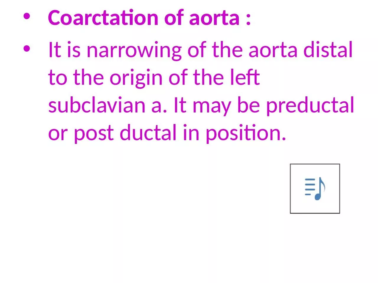
/
Coarctation of aorta : It is narrowing of the aorta distal to the origin of the left subclavian Coarctation of aorta : It is narrowing of the aorta distal to the origin of the left subclavian

Coarctation of aorta : It is narrowing of the aorta distal to the origin of the left subclavian - PowerPoint Presentation

margaret
margaret . @margaret
Follow
66 views
Uploaded On 2023-07-07

Coarctation of aorta : It is narrowing of the aorta distal to the origin of the left subclavian - PPT Presentation

Abnormal origin of Rt Subclavian a It may originate from the distal part of the Rt Dorsal aorta amp the 7th intersegmental a ampso the Rt Subclavian a is found as a branch from the arch of the aorta next to the left ID: 1006588

left amp aorta veins amp left veins aorta cardinal arch aortic umbilical subclavian liver drains vitelline forms eosophagus trachea

Share:

Link:

Embed:

Download Presentation from below link

Download Presentation The PPT/PDF document "Coarctation of aorta : It is narrowing o..." is the property of its rightful owner. Permission is granted to download and print the materials on this web site for personal, non-commercial use only, and to display it on your personal computer provided you do not modify the materials and that you retain all copyright notices contained in the materials. By downloading content from our website, you accept the terms of this agreement.


Presentation Transcript

1. Coarctation of aorta :It is narrowing of the aorta distal to the origin of the left subclavian a. It may be preductal or post ductal in position.

2. Abnormal origin of Rt. Subclavian a. It may originate from the distal part of the Rt. Dorsal aorta, & the 7th intersegmental a., &so the Rt. Subclavian a. is found as a branch from the arch of the aorta next to the left subclavian a. & it passes to the Rt. Side behind the trachea & the eosophagus, thus there may be a difficulty in breathing & swallowing , as the trachea & eosophagus may be compressed between the aortic arch & the Rt. Subclavian a.

3. Double aortic arch :The Rt. Dorsal aorta between the origin of the Rt. 7th intersegmental a. & the junction with the left dorsal aorta, will not disappear & so 2 aortic arches will be formed as a vascular ring surrounding the trachea & the eosophagus , thus there may be a difficulty in breathing & swallowing , as the trachea & eosophagus may be compressed.

4. 5. Rt. Aortic arch: It occurs by a mechanism which is the reverse of the process of forming the left arch of aorta. 6. Interrupted aortic arch: Disappearance of the Lt 4th aortic arch, thus the descending aorta becomes connected with pulmonary artery by the ductus arteriosus.

5. The venous system:During the 5th wk, each of the Rt. & left sinus horns drains 3 veins: the vitelline , umbilical, & the common cardinal v.

6. (Easy Go) The vitelline v. :The Rt. & left vitelline veins form anastomosing plexus of veins around duodenum, & another plexus inside septum transversarium. Anastomoses around duodenum forms portal vein. Anastomosis in septum transversarium forms sinusoidal capillaries of liver, (liver grows inside septum transversarium).left vitelline v. disappears later on , Rt. vitelline v. is enlarged forming Rt. Hepatocardiac channel that forms hepatic part of inf. vena cava.

7.

8. (Easy Go)The umbilical v. :Rt. & left umbilical veins present on each side of liver, then these veins will gain connection with liver substance, Rt. umbilical v. disappear, left umbilical v. remains & gain connection with hepatic part of inf. vena cava by a venous duct inside liver which is called ductus venosus. After birth, Lt, umbilical v. is closed forming ligamentum teres. Ductus venosus forms ligamentum venosum.

9. (Easy Go)The cardinal v. :During 4th wk, Lt. & Rt. common cardinal veins drain embryonic body Each of common cardinal v. drains anterior & post. cardinal v. Then after more paired cardinal veins develop during 5th -7th wk to drain venous blood , including:Subcardinal v. drains kidneys. Sacrocardinal v. drains lower limbs.Supra cardinal v. drains intercostals veins.Anastomosing channels develop between cardinal v.

10.