/
ANATOMY OF  THE DIGESTIVE SYSTEM ANATOMY OF  THE DIGESTIVE SYSTEM

ANATOMY OF THE DIGESTIVE SYSTEM - PowerPoint Presentation

marina-yarberry
marina-yarberry . @marina-yarberry
Follow
353 views
Uploaded On 2019-11-18

ANATOMY OF THE DIGESTIVE SYSTEM - PPT Presentation

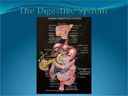
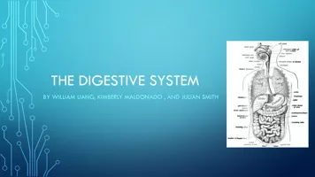
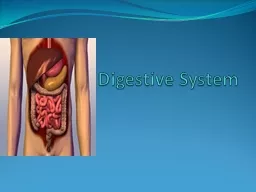
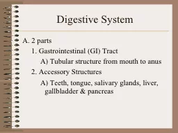
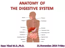
ANATOMY OF THE DIGESTIVE SYSTEM Kaan Yücel MD PhD 21November2014 Friday The oral region includes the oral cavity teeth gingivae tongue palate and the region of the palatine tonsils ID: 765301

part oral liver cavity oral part cavity liver superior duct tongue bile system stomach posterior duodenum mouth surface lies

Share:

Link:

Embed:

Download Presentation from below link

Download Presentation The PPT/PDF document "ANATOMY OF THE DIGESTIVE SYSTEM" is the property of its rightful owner. Permission is granted to download and print the materials on this web site for personal, non-commercial use only, and to display it on your personal computer provided you do not modify the materials and that you retain all copyright notices contained in the materials. By downloading content from our website, you accept the terms of this agreement.


Presentation Transcript

ANATOMY OF THE DIGESTIVE SYSTEM Kaan Yücel M.D.,Ph.D. 21.November.2014 Friday

The oral region includes the oral cavity, teeth, gingivae, tongue, palate, and the region of the palatine tonsils. The digestion starts here in the oral cavity. It is the place where the food is ingested and prepared for digestion in the stomach and small intestine. Oral Region

Food is chewed by the teeth, and saliva from the salivary glands facilitates the formation of a manageable food bolus (L. lump). Swallowing is voluntarily initiated in the oral cavity. The voluntary phase of the process pushes the bolus from the oral cavity into the pharynx, the expanded part of the alimentary (digestive) system, where the involuntary (automatic) phase of swallowing occurs.

Oral Cavity (Mouth ) Inferior to the nasal cavities . Extends from the lips to the pharynx.

Oral Cavity (Mouth ) Has a roof and floor , and lateral walls. Opens onto the face through the oral fissure. Continuous with the cavity of the pharynx at the oropharyngeal isthmus.

The oral cavity has multiple functions: Inlet for the digestive system involved with the initial processing of food, which is aided by secretions from salivary glands. Manipulates sounds produced by the larynx. Can be used for breathing because it opens into the pharynx, which is a common pathway for food and air . For this reason, the oral cavity can be used by physicians to access the lower airway .

Oral cavity (mouth ) consists of two parts: Oral vestibule Oral cavity proper

Slit-like space between the teeth and gingivae (gums) internally and the lips and cheeks externally. Enclosed by dental arches. Communicates with the exterior through the oral fissure (opening). Oral vestibule

The duct of the parotid salivary gland (Stensen’s duct) opens on a small papilla into the vestibule opposite the upper second molar tooth.

The space between the upper and the lower dental arches. Has a roof and a floor. The roof of the mouth is formed by the hard palate in front and the soft palate behind . Oral cavity proper

The floor is formedlargely by the anterior 2/3 of the tongue and by the reflection of the mucous membrane from the sides of the tongue to the gum of the mandible.

Lips Mobile, musculofibrous folds surrounding the mouth Covered externally by skin and internally by mucous membrane. Function as the valves of the oral fissure, containing the sphincter (orbicularis oris) that controls entry and exit from the mouth and upper alimentary and respiratory tracts. .

F orm the movable walls of the oral cavity. The prominence of the cheek occurs at the junction of the zygomatic and buccal regions. Cheeks (Buccae ) External aspect- Buccal region Anteriorly by lips and chin Superiorly by zygomatic region Posteriorly parotid region Inferiorly by inferior border of mandible

Teeth The chief functions of the teeth are to:Incise, reduce, and mix food material with saliva during mastication.Help sustain themselves in the tooth sockets by assisting the development and protection of the tissues that support them.Participate in articulation (distinct connected speech).

The teeth are set in the tooth sockets. There are 20 deciduous teeth and 32 permanent teeth: four incisors, two canines, four premolars, and six molars in each jaw. (4-2-4-6)

Gingivae ( Gums)Composed of fibrous tissue covered with mucous membrane. The gingiva proper (attached gingiva) is firmly attached to the alveolar processes of the mandible and maxilla and the necks of the teeth.

Forms part of the floor of the oral cavity and part of the anterior wall of the oropharynx. Tongue A mass of striated muscle covered th mucous membrane

Its anterior part is in the oral cavity and is somewhat triangular in shape with a blunt apex of tongue. The apex is directed anteriorly .The root of tongue is attached to the mandible and the hyoid bone.

Papillae The superior surface of the oral part of the tongue is covered by hundreds of papillae. 4 types of papillae in the tongue:

The superior surface of the oral part of the tongue is covered by hundreds of papillae. 4 types of papillae in the tongue. The papillae in general increase the area of contact between the surface of the tongue and the contents of the oral cavity.

Muscles of the Tongue Intrinsic and Extrinsic MusclesIntrinsic muscles: confined to the tongue, are not attached to bone. Extrinsic muscles : attached to bones and the soft palate.

Innervation of the tongue is complex and involves a number of nerves.Trigeminal nerve: sensation from 2/3 anterior tongue Glossopharyngeal nerve: sensation from 1/3 posterior tongueTaste from oral part: by the facial nerveTaste from pharyngeal part: by the glossopharyngeal nerve

Movements of the TongueProtrusion:Retraction: Depression: Shape changes: Intrinsic muscles

Palate Forms the arched roof of the mouth and the floor of the nasal cavities.Separates the oral cavity from the nasal cavities and the nasopharynx, the part of the pharynx superior to the soft palate. Consists of 2 regions: Hard palate anteriorly Ssoft palate posteriorly

Posteroinferiorly, the soft palate has a curved free margin from which hangs a conical process; uvula.

The space between the cavity of the mouth and the pharynx. Bounded Superiorly by the soft palateInferiorly by the root of the tongue Fauces ( Thorat )

Oropharyngeal isthmus ( isthmus of the fauces) is the short constricted space that establishes the connection between the oral cavity proper and the oropharynx. By closing the oropharyngeal isthmus, food or liquid can be held in the oral cavity while breathing.

SalIvary glands

Parotid Gland The largest salivary gland Lies in a deep hollow below the external auditory meatus, behind the ramus of the mandible, and in front of the SCM. The facial nerve divides the gland into superficial and deep lobes .

The parotid duct passes forward over the lateral surface of the masseter. It enters the vestibule of the mouth upon a small papilla opposite the upper second molar tooth .

Nerve SupplyParasympathetic secretomotor supply arises from the glossopharyngeal nerve. Parasympathetic stimulation of the parotid gland produces a thin watery saliva.

Lies beneath the lower border of the body of the mandible Submandibular Gland

Submandibular duct runs medially to open at the side of lingual frenulum. Parasympathetic secretomotor supply is from the facial nerve.

SMG = submandibular gland, ABD = anterior belly of digastric muscle, LN = submandibular lymph node, FV = facial vein, FA = facial artery, MH = mylohyoid muscle.

Lies beneath the floor of the mouth. The sublingual ducts (8 to 20 in number) open into the mouth.Sublingual Gland

M usculofascial half-cylinder Links oral and nasal cavities in the head to the larynx & esophagus in the neck. Superior expanded part of the alimentary system posterior to the nasal and oral cavities, extending inferiorly past the larynx. Extends from the cranial base to and is continuous with the top of the esophagus. PHARYNX

Based on these anterior relationships the pharynx is subdivided into 3 regions:Posterior apertures (choanae) of the nasal cavities open into the Nasopharynx Posterior opening of the oral cavity opens into Oropharynx 3) A perture of the larynx (laryngeal inlet) opens into the L aryngopharynx

Nasopharynx has a respiratory function; posterior extension of the nasal cavities. Oropharynx is posterior to the oral cavity, inferior to the level of the soft palate, and superior to the upper margin of the epiglottis. It opens anteriorly, through the isthmus faucium, into the mouth. Laryngopharynx lies posterior to the larynx and anterior to the vertebral column.

Waldeyer's Ring of Lymphoid Tissue 1- Pharyngeal tonsil-Adenoid2- Tubal tonsil 3- Palatine tonsil 4- Lingual tonsil

M uscular tube about 10 in. (25 cm) longExtends from the pharynx to the stomach. Begins in the neck where it is continuous with the laryngopharynx .Consists of striated (voluntary) muscle in its upper 1/3, smooth (involuntary) muscle in its lower 1/3, and a mixture of striated and smooth muscle in between. OESOPHAGUS

E xpanded part of the digestive tract between the esophagus and small intestine.Specialized for the accumulation of ingested food, chemically and mechanically prepares for digestion and passage into the duodenum . Acts as a food blender and reservoir; its chief function is enzymatic digestion. STOMACH

The size, shape, and position of the stomach can vary markedly in persons of different body types (bodily habitus) May change even in the same individual as a result of Diaphragmatic movements during respirationStomach's contents (empty vs. after a heavy meal) Position of the person.

The stomach has four parts: Cardia: part surrounding the cardial orifice (opening), the superior opening or inlet of the stomach. Fundus: dilated superior part related to the left dome of the diaphragm and is limited inferiorly by the horizontal plane of the cardial orifice. Body: major part of the stomach between the fundus and pyloric part.Pyloric part: funnel-shaped outflow region of the stomach.

Primary site for absorption of nutrients from ingested materials. Extends from the pylorus to the ileocecal junction where the ileum joins the cecum (the first part of the large intestine). SMALL INTESTINE

D uodenum first part of the small intestineShortest, widest and most fixed part. Jejunum begins at the duodenojejunal flexure where the digestive tract resumes an intraperitoneal course. Ileum ends at ileocecal junction, union of the terminal ileum & cecum. Together , jejunum and ileum are 6-7 m long.Jejunum 2/5 , Ileum 3/5 intraperitoneal section of the small intestine.

Most of the jejunum lies in the left upper quadrant (LUQ), whereas most of the ileum lies in the right lower quadrant (RLQ). The terminal ileum usually lies in the pelvis from which it ascends, ending in the medial aspect of the cecum.

Where water is absorbed from the indigestible residues of the liquid chyme, converting it into semi-solid stool or feces that is stored temporarily and allowed to accumulate until defecation occurs.Cecum AppendixAscending colonTransverse colon D escending colon S igmoid colon R ectumAnal canal LARGE INTESTINE

The large intestine can be distinguished from the small intestine by: Omental appendices: small, fatty, omentum-like projections.Teniae coli: three distinct longitudinal bands. Haustra: sacculations of the wall of the colon between the teniaeA much greater caliber (internal diameter).

L argest gland in the body After the skin, the largest single organ2.5% of adult body weightExcept for fat, all nutrients absorbed from the digestive tract are initially conveyed to the liver by the portal venous system .In addition to its many metabolic activities, the liver stores glycogen and secretes bile, a yellow-brown or green fluid that aids in the emulsification of fat.LIVER

Bile passes from the liver via biliary ducts—right and left hepatic ducts—join to form common hepatic duct, which unites with the cystic duct to form the (common) bile duct.

The liver produces bile continuously; however, between meals it accumulates and is stored in the gallbladder, which also concentrates the bile by absorbing water and salts. When food arrives in the duodenum, the gallbladder sends concentrated bile through the biliary ducts to the duodenum.

The normal liver lies on the right side and crosses the midline toward the left nipple. Liver occupies most of the right hypochondrium and upper epigastrium and extends into the left hypochondrium.

Liver hasA convex diaphragmatic surface (anterior, superior, and some posterior) A relatively flat or even concave visceral surface (posteroinferior), separated anteriorly by its sharp inferior border that follows the right costal margin.

Externally, liver is divided into 2 anatomical lobes & 2 accessory lobes by the reflections of peritoneum from its surface, the fissures formed in relation to those reflections and the vessels serving the liver and the gallbladder.

The essentially midline plane defined by the attachment of the falciform ligament and the left sagittal fissure separates a large right lobe from a much smaller left. On the visceral surface, the right and left sagittal fissures course on each side of—and the transverse porta hepatis separates:2 accessory lobes (parts of the anatomic right lobe): Quadrate lobe anteriorly and inferiorlyCaudate lobe posteriorly and superiorly.

L ies in fossa for the gallbladder on the visceral surface of the liver. This shallow fossa @ junction of right & left liver . Gallbladder

R elationship of gallbladder to duodenum is so intimate that the superior part of the duodenum is usually stained with bile in the cadaver. In its natural position the body of the gallbladder lies anterior to the superior part of the duodenum, its neck and cystic duct are immediately superior to the duodenum.

C onvey bile from the liver to the duodenum. Bile is produced continuously by the liver and stored, concentrated in the gallbladder, which releases it intermittently when fat enters the duodenum. Bile emulsifies the fat so that it can be absorbed in the distal intestine.Biliary ducts

The bile duct (formerly called the common bile duct) forms by the union of the cystic duct and common hepatic duct. The bile duct descends posterior to the superior part of the duodenum and lies in a groove on the posterior surface of the head of the pancreas.

E longated, accessory digestive gland that lies retroperitoneally, overlying and transversely on the posterior abdominal wall. Lies posterior to the stomach between the duodenum on the right and the spleen on the left. PANCREAS

The pancreas produces: Exocrine secretion (pancreatic juice from the acinar cells) enters the duodenum through the main and accessory pancreatic ducts.Endocrine secretions (glucagon and insulin from the pancreatic islets [of Langerhans]) enter the blood.Pancreas is divided into 4 parts: Head N eck Body T ail

The main pancreatic duct begins in the tail of the pancreas .Main pancreatic duct+ bile duct= hepatopancreatic ampulla (of Vater) opens into the duodenum at the summit of Major duodenal papilla

O void, usually purplish, pulpy mass about the size and shape of one's fist. Relatively delicate and considered the most vulnerable abdominal organ. Located in the superolateral part of the left upper quadrant (LUQ) or hypochondrium of the abdomen where it enjoys protection of the inferior thoracic cage. SPLEEN

As the largest of the lymphatic organs, it participates in the body's defense system as a site of lymphocyte (white blood cell) proliferation and of immune surveillance and response. To accommodate these functions, the spleen is a soft, vascular (sinusoidal) mass with a relatively delicate fibroelastic capsule. The diaphragmatic surface of the spleen is convexly curved to fit the concavity of the diaphragm and curved bodies of the adjacent ribs.

Continuous, slippery transparent serous membrane.Lines the abdominopelvic cavity and invests the viscera. Consists of two continuous layers: Parietal peritoneumlines the internal surface of the abdominopelvic wallVisceral peritoneuminvests viscera such as the stomach and intestines. PERITONEUM

A potential space of capillary thinness between the parietal and visceral layers of peritoneumWithin the abdominal cavity, and continues inferiorly into the pelvic cavity. Contains a thin film of peritoneal fluid, which is composed of water, electrolytes, and other substances derived from interstitial fluid in adjacent tissues. P eritoneal cavity

F ormed in relation to the relocation of the testis during fetal development. An oblique passage approximately 4 cm long directed inferomedially through the inferior part of the anterolateral abdominal wallL ies parallel and superior to the medial half of the inguinal ligament. INGUINAL CANAL

M ain occupant of the inguinal canal Spermatic cord in males Round ligament of the uterus in females

P ortal vein F inal common pathway for the transport of venous blood from the spleen, pancreas, gallbladder, and the abdominal part of the gastrointestinal tract. Formed by the union of splenic vein & superior mesenteric vein posterior to the neck of the pancreas . PORTAL SYSTEM

Venous drainage of the spleen, pancreas, gallbladder, and the abdominal part of the gastrointestinal tract, except for the inferior part of the rectum, is through the portal system of veins, which deliver blood from these structures to the liver. Once blood passes through the hepatic sinusoids, it passes through progressively larger veins until it enters the hepatic veins, which return the venous blood to the inferior vena cava just inferior to the diaphragm.

A rich blood supply to support its digestive activities. Arterial blood supplied mainly by Coeliac artery to the stomach, pancreas, spleen and liver Mesenteric arteries to the intestines. VESSELS & NERVES OF THE GASTROINTESTINAL SYSTEM

Venous blood drains from the stomach, pancreas and spleen via the hepatic portalvein into the liver, where the products of digestion undergo further processing and detoxification. Blood from oesophagus and rectum does not go through the liver but drains directly into the venous system.

There are two types of nerve supply to the GI tract. The enteric system, found within the walls of the GI tract, is sometimes known as the 'gut brain' and controls movement and secretion within the gut. Nerves from the autonomic nervous system also supply the GI tract.

S ympathetic systemreduce blood flow to the gut decrease secretions, motility and contractions, Parasympathetic system Increase in motility and secretion within the tract and relaxation of the gut sphincters. The vagus nerve (Xth cranial) supplies the oesophagus, stomach, pancreas, bile duct, small intestine and upper colon.

A bdominal cavityForms the superior and major part of the abdominopelvic cavity.Has no floor of its own because it is continuous with the pelvic cavity.Plane of the pelvic inlet (superior pelvic aperture) arbitrarily, but not physically, separates the abdominal and the pelvic cavities.is the location of most digestive organs, parts of the urogenital system (kidneys and most of the ureters), and the spleen.

More superiorly placed abdominal organs (spleen, liver, part of the kidneys, and stomach) are protected by the thoracic cage.

9 regions of the abdominal cavityRegions are delineated by 4 planes: 2 sagittal (vertical) 2 transverse (horizontal) planes

2 sagittal planes M idclavicular (approximately 9 cm from the midline) Midinguinal pointsmidpoints of the lines joining the anterior superior iliac spine (ASIS) and the superior edge of the pubic symphysis on each side.

Two vertical lines S ubcostal planeT ranstubercular plane inferior border of the 10th costal cartilage ilia c tubercles (5 cm posterior to ASIS on each side) and body of L5)