/
Neuroglia Neuroglia

Neuroglia - PowerPoint Presentation

marina-yarberry
marina-yarberry . @marina-yarberry
Follow
382 views
Uploaded On 2016-07-04

Neuroglia - PPT Presentation

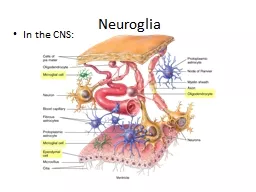
In the CNS Neuroglia In the PNS Neuroglia Myelination is the process of forming a myelin sheath which insulates and increases nerve impulse speed It is formed by Oligodendrocytes in the CNS and by ID: 390165

cell potentials channels neurons potentials cell neurons channels potential action animation postsynaptic graded membrane gated electrical neuron neurotransmitter axon

Share:

Link:

Embed:

Download Presentation from below link

Download Presentation The PPT/PDF document "Neuroglia" is the property of its rightful owner. Permission is granted to download and print the materials on this web site for personal, non-commercial use only, and to display it on your personal computer provided you do not modify the materials and that you retain all copyright notices contained in the materials. By downloading content from our website, you accept the terms of this agreement.


Presentation Transcript

Slide1

Neuroglia

In the CNS:Slide2

Neuroglia

In the PNS:Slide3

Neuroglia

Myelination is the process of forming a myelin sheathwhich insulates and increases nerve impulse speed.

It is formed byOligodendrocytes in the CNS and by Schwann cells in

the PNS.Slide4

Nodes of Ranvier

are the gaps in the myelin sheath.Each Schwann cell wraps one axon segment between two nodes of Ranvier. Myelinated nodes are about 1 mm in length and have up to 100 layers.The amount of myelin increases from birth

to maturity, and its presence greatly increases the speed of nerve conduction.Diseases like Multiple Sclerosis

result from autoimmune destruction of myelin. NeurogliaSlide5

Neuronal Regeneration

The cell bodies of neurons lose their mitotic features at birth and can only be repaired through regeneration after an injury (they are never replaced by daughter cells as occurs with epithelial tissues.)

Nerve tissue regeneration is largely dependent on the Schwann cells in the PNS and essentially doesn’t occur at all in the CNS where

astrocytes just form scar tissue.Slide6

The outer nucleated cytoplasmic layer of the Schwann cell, which encloses the myelin sheath, is the neurolemma (sheath of Schwann).When an axon is injured, the neurolemma aids regeneration by forming a regeneration tube

that guides and stimulates regrowth of the axon.Neuronal RegenerationSlide7

Neuronal RegenerationTo do any regeneration, neurons must be located in the PNS, have an intact cell body, and be myelinated by functional Schwann cells having a neurolemma.

Demyelination refers to the loss or destruction of myelin sheaths around axons. It may result from disease, or from medical treatments such as radiation therapy and chemotherapy.Any single episode of demyelination may cause deterioration of affected nerves.Slide8

Gray and White Matter

White matter of the brain and spinal cord is formed fromaggregations of myelinated axons from many neurons.

The lipid part of myelin imparts the white appearance.Gray matter (gray because it lacks myelin) of the brain and spinal cord is formed from neuronal cell bodies

and dendrites.Slide9

Electrical Signals in Neurons

Like muscle fibers, neurons are electrically excitable. They communicate with one another using two types of electrical signals:Graded potentials are used

for short-distance communication only.Action potentials allow communication over long distances within the body.Slide10

Electrical Signals in NeuronsProducing electrical signals in neurons depends on the existence of a resting membrane

potential (RMP) - similar to the electrical potential of this 9 v battery which has a gradient of 9 volts from one terminal to another.A cell’s RMP is created using ion gradients and a variety of ion channels that open or close in response to specific stimuli.

Because the lipid bilayer of the plasma membrane is a good insulator, ions must flow through these channels.Slide11

A cell’s RMP is created using ion channels to set-up transmembrane ion gradients.

Electrical Signals in Neurons

Ion channels are present in the plasma membrane of all cells in the body, but they are an especially prominent component of the nervous system.

Much of the energy expended by neurons, and really all cells of the body, is used to create a net negative charge in the inside of the cell as compared to the outside of the cell.Slide12

Electrical Signals in NeuronsWhen ion channels are open, they allow specific ions to move across the plasma membrane, down their electrochemical gradient

. Ions move from areas of higher concentration to areas of lower concentration - the “chemical” (concentration) part of the gradient.Positively charged cations move toward a negatively charged area, and negatively charged anions move toward a positively charged area - the electrical aspect of the gradient.Slide13

Electrical Signals in NeuronsActive channels open in response to a stimulus (they are “gated”). There are

3 types of active, gated channels:Ligand-gated channels respond to a neurotransmitter and are mainly concentrated at the synapse.Voltage-gated channels respond to changes in the transmembrane electrical potential and are mainly located along the neuronal axon.

Mechanically-gated channels respond to mechanical deformation (applying pressure to a receptor).“Leakage” channels are also gated but they are not active, and they open and close randomly. Slide14

Electrical Signals in NeuronsInteractions Animation

Ion Channels Animation

You must be connected to the internet to run this animationSlide15

Maintaining the RMPA neuron’s RMP is measured at rest, when it is not conducting a nerve impulse.The resting membrane potential exists because of a small buildup of negative ions in the cytosol along the inside of the membrane, and an equal buildup of positive ions in the extracellular fluid along the outside surface of the membrane. The buildup of charge occurs only very close to the membrane – the cytosol elsewhere in the cell is electrically neutral.Slide16

Maintaining the RMPThe RMP is slightly negative because leakage channels

favor a gradient where more K+ leaks out, than Na+ leaks in (there are more K+ channels than Na+ channels.)There are also large negatively charged proteins

that always remain in the cytosol. Left unchecked, inward leakage of Na+ would eventually destroy the resting membrane potential.The small inward Na+ leak and outward K+ leak are offset by the Na+

/K+ ATPases (sodium-potassium pumps) which pumps out Na+ as fast as it leaks in.Slide17

Maintaining the RMPInteractions Animation

In neurons, a typical value for the RMP is –70 mV (the minus sign indicates that the inside of the cell is negative relative to the outside.)

Resting Membrane Potential

You must be connected to the internet to run this animationSlide18

Maintaining the RMPA cell that exhibits an RMP is said to be polarized

.In this state, the cell is “primed” - it is ready to produce an action potential. In order to do so, graded potentials must first be produced in order to depolarize the cell to threshold.A graded potential occurs whenever ion flow in mechanically gated or ligand-gated channels produce a current that is localized – it spreads to adjacent regions for a short distance and then dies out within a few millimeters of its point of origin.Slide19

Graded PotentialsFrom the RMP, a stimulus that causes the cell to be less negatively charged with respect to the extracellular fluid is a

depolarizing graded potential, and a stimulus that causes the cell to be more negatively charged is a hyperpolarizing graded

potential (both are shown in this diagram.)Slide20

Graded Potentials

Graded potentials have different names depending on the type of stimulus and where they occur. They are voltage variable aptitudes that can be added together (summate) or cancel each other out – the net result is a larger or smaller graded potential.Graded potentials occur

mainly in the dendrites and cell body of a neuron – they do not

travel down the axon.Slide21

Graded PotentialsInteractions Animation

Graded Potentials Animation

You must be connected to the internet to run this animationSlide22

Action PotentialsIn contrast to graded potentials, an

action potential (AP) or impulse is a signal which travels the length of the neuron.During an AP, the membrane potential reverses and then eventually is restored to its resting state.If a neuron receives a

threshold (liminal) stimulus, a full strength nerve impulse is produced and spreads down the axon of the neuron to the axon terminals. If the stimulus is not strong enough (subthreshold or subliminal), no nerve impulse will result.Slide23

Action PotentialsInteractions Animation

Action Potentials Animation

You must be connected to the internet to run this animationSlide24

Action PotentialsAn AP has two main phases: a depolarizing phase

and a repolarizing phaseSlide25

Action Potentials

Graded potentials that result in depolarization of the neuron from –70mV to threshold (about –55 mV in many neurons) will cause a sequence of events to rapidly unfold.Voltage-gated Na+ channels open during the steep depolarization phase

allowing Na+ to rush into the cell and makingthe inside of the cell progressively more positive.Slide26

Only a total of 20,000 Na

+ actually enter the cell in each little area of the membrane, but they change the potential considerably (up to +30mV

).During the repolarization, phase K+ channels open and K

+ rushes outward.The cell returns to a progressively more negative state until the RMP of –70mV

is once again restored.Action PotentialsSlide27

Action PotentialsWhile the voltage-gated K+ channels are open, outflow of K+

may be large enough to cause an after-hyperpolarizing phase of the action potential.During this phase, the voltage-gated K+ channels remain open and the membrane potential becomes even more negative (about –90 mV).As the voltage-gated K+ channels close, the membrane potential returns to the resting level of –70 mV.Slide28

Action PotentialsAccording to the all-or-none principle, if a stimulus reaches threshold, the action potential is always the same.

A stronger stimulus will not cause a larger impulse.Slide29

Action PotentialsInteractions Animation

Propagation of Nerve Impulses Animation

You must be connected to the internet to run this animationSlide30

Action PotentialsAfter initiating an action potential, there is a period of time called the absolute refractory period during which a cell cannot generate another AP, no matter how strong the stimulus.

This period coincides with the period of Na+ channel activation and inactivation (inactivated Na+ channels must first return to the resting state.)This places an upper limit of 10–1000 nerve impulses per second, depending on the neuron.Slide31

Action Potentials

The relative refractory period is the period of time during which a second action potential can be initiated, but only by a larger-than-normal stimulus.It coincides with the period when the voltage-gated K

+ channels are still open after inactivated Na+ channels have returned to their resting state. In contrast to action potentials,

graded potentials do not exhibit a refractory period.Slide32

Action PotentialsInteractions Animation

Membrane Potentials Animation

You must be connected to the internet to run this animationSlide33

Propagation of the AP

down the length of the axon begins at the trigger zone near the axon hillock.By passive spread, the current proceeds by (a) continuous conduction in unmyelinated axons, or by the much faster process of (b) saltatory

conduction in myelinated axons (as the AP jumps from one

node to the next as shown in this graphic).Action PotentialsSlide34

Conduction Rates Animation

Action PotentialsInteractions Animation

You must be connected to the internet to run this animationSlide35

Action PotentialsIn addition to the nodes of Ranvier that allow saltatory conduction, the speed of an AP is also affected by:

The axon diameterThe amount of myelinationThe temperatureThe frequency of AP plays a crucial role in determining the perception of a stimulus, or the extent of our response.In addition to this “frequency code,” a second important factor is the number of neurons recruited

(activated) to the cause.Slide36

Fiber TypesThe characteristics of the neuronal axon define the “fiber types”

A fibers are large, fast (130 m/sec), myelinated neurons that carry touch and pressure sensations; many motor neurons are also of this type.B fibers are of medium size and speed (15 m/sec) and comprise myelinated visceral sensory & autonomic preganglionic neurons.C fibers

are the smallest and slowest (2 m/sec) and comprise unmyelinated sensory and autonomic motor neurons.Slide37

Synaptic Transmission

Signal transmission at the synapse is a one-way transfer from a presynaptic neuron to a postsynaptic neuron.When an AP reaches the end bulb of axon terminals, voltage-gated Ca2+ channels open and

Ca2+ flows inward, triggering release of the neurotransmitter.The neurotransmitter crosses the synaptic cleft and binds to ligand-gated receptors on the postsynaptic membrane.

The more neurotransmitter released, the greater the number and intensity of graded potentials in the postsynaptic cell.Slide38

Synaptic Transmission

In this way, the presynaptic neuron converts an electrical signal (nerve impulse) into a chemical signal (released neurotransmitter). The postsynaptic neuron receives the chemical signal and in turn generates an

electrical signal (postsynaptic potential).The time required for these

processes at a chemical synapse produces a synaptic delay of about 0.5 msec.Slide39

Synaptic Transmission

The events that occur at a synapse are outlined above.Slide40

NeurotransmittersBoth excitatory and inhibitory neurotransmitters are present in the CNS and PNS.

The same neurotransmitter may be excitatory in some locations and inhibitory in others.For example, acetylcholine (ACh) is a common neurotransmitter released by many PNS neurons (and some in the CNS). Ach is excitatory at the NMJ but inhibitory at other synapses.Slide41

Neurotransmitters

Many amino acids act as neurotransmitters:Glutamate is released by nearly all excitatory neurons in the brain. GABA is an inhibitory neuro-

transmitter for 1/3 of all brain synapses.Valium is a GABA agonist that enhances GABA’s depressive effects (causes sedation).Other important small-molecule neurotransmitters are listed.Slide42

NeurotransmittersNeurotransmitter effects can be modified in many ways:

Synthesis can be stimulated or inhibited.Release can be blocked or enhanced.Removal can be stimulated or blocked.The receptor site can be blocked or activated.An agonist is any chemical that enhances or stimulates the effects at a given receptor.

An antagonist is a chemical that blocks or diminishes the effects at a given receptor.Slide43

Postsynaptic PotentialsA neurotransmitter causes either an excitatory or an inhibitory graded potential:

Excitatory postsynaptic potential (EPSP) causes a depolarization of the postsynaptic cell, bringing it closer to threshold. Although a single EPSP normally does not initiate a nerve impulse, the postsynaptic cell does become more excitable.Inhibitory postsynaptic potential (IPSP) hyperpolarizes the postsynaptic cell taking it farther from threshold.Slide44

Spatial summation

occurs when postsynaptic potentials arrive near the same location. Temporal summation occurs when postsynaptic potentials arrive close to the same time.Whether or not the postsynaptic cell

reaches thresholddepends on the net effect afterSummation of all

the postsynapticpotentials.

Postsynaptic PotentialsSlide45

Neurotransmitter ClearanceIf a neurotransmitter could linger in the synaptic cleft, it would influence the postsynaptic neuron, muscle fiber, or gland cell indefinitely –

removal of the neurotransmitter is essential for normal function.Removal is accomplished by diffusion out of the synaptic cleft, enzymatic degradation, and re-uptake by cells

.An example of a common neurotransmitter inactivated through enzymatic degradation is acetylcholine. The enzyme acetylcholinesterase breaks down acetylcholine in the synaptic cleft.Slide46

Summary of Synaptic EventsInteractions Animation

Events at the Synapse Animation

You must be connected to the internet to run this animationSlide47

Neural Circuits

Neurons process information when changes occur at the trigger zone through spatial and temporal summation of IPSPs & EPSPs. An “average” neuron receives

10,000 synaptic inputs - multiply this by the numberof neurons involved in any single process, and you can

start to comprehend the exquisite level of information processing afforded by this system.Slide48

Integration is the process accomplished by the post-synaptic neuron when it combines all excitatory and inhibitory inputs and responds

accordingly.This process occurs over and over as interneurons are activated in higher parts of the brain (such

as the thalamus and cerebral cortex).Neural CircuitsSlide49

Neural CircuitsA neuronal network may contain thousands or even millions of neurons.

Types of circuits include diverging, converging, reverberating, and parallel after-discharge.Slide50

Neural CircuitsIn a diverging circuit

, a small number of neurons in the brain stimulate a much larger number of neurons in the spinal cord. A converging circuit is the opposite.In a reverberating circuit, impulses are sent back through the circuit time and time again – used in breathing, coordinated muscular activities, waking up, and short-term memory.

Parallel after-discharge circuits involve a single presynaptic cell that stimulates a group of neurons, which then synapse with a common postsynaptic cell – used in precise activities such as mathematical calculations.Slide51

End of Chapter 12Copyright 2012 John Wiley & Sons, Inc. All rights reserved. Reproduction or translation of this work beyond that permitted in section 117 of the 1976 United States Copyright Act without express permission of the copyright owner is unlawful. Request for further information should be addressed to the Permission Department, John Wiley & Sons, Inc. The purchaser may make back-up copies for his/her own use only and not for distribution or resale. The Publisher assumes no responsibility for errors, omissions, or damages caused by the use of these programs or from the use of the information herein.