/
Nipah Virus Nipah Virus

Nipah Virus - PowerPoint Presentation

marina-yarberry
marina-yarberry . @marina-yarberry
Follow
756 views
Uploaded On 2017-07-03

Nipah Virus - PPT Presentation

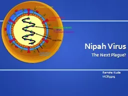
The Next Plague Ramsha Kudia MCB5505 Taxonomy Family Paramyxoviridae Subfamily Paramyxovirinae Genus Avulavirus Newcastle disease virus Genus Henipavirus Hendravirus Nipahvirus Genus Morbillivirus ID: 566223

pigs virus bats nipah virus pigs nipah bats human outbreak symptoms disease genus fruit viruses sap rna bangladesh treatment

Share:

Link:

Embed:

Download Presentation from below link

Download Presentation The PPT/PDF document "Nipah Virus" is the property of its rightful owner. Permission is granted to download and print the materials on this web site for personal, non-commercial use only, and to display it on your personal computer provided you do not modify the materials and that you retain all copyright notices contained in the materials. By downloading content from our website, you accept the terms of this agreement.


Presentation Transcript

Slide1

Nipah Virus

The Next Plague?

Ramsha Kudia

MCB5505Slide2

Taxonomy

Family: Paramyxoviridae

Subfamily Paramyxovirinae

Genus Avulavirus

Newcastle disease virus

Genus Henipavirus

Hendravirus

Nipahvirus

Genus Morbillivirus

Measles virus

Rinderpest virus

Genus Respirovirus

Sendai virus

Human parainfluenza viruses 1 and 3, as well some of the viruses of the common cold

Genus Rubulavirus

Mumps virus

Human parainfluenza viruses 2 and 4

Subfamily Pneumovirinae

Bovine respiratory syncytial virus

Human respiratory syncytial virus

Murine pneumonia virusSlide3

Henipavirus

Only zoonotic paramyxoviruses and are highly pathogenic

Genome size 18.2 kb, about 15% larger than other family members Biosafety level 4 because of pathogenicity, high mortality, and lack of treatment

The genus is able to infect a broad range of animal species, which is uncommon among the family

High mutation ratesSlide4

Virion Structure

Non-segmented, negative sense, single stranded RNA

EncapsulatedPolymorphic, but usually spherical or filamentousHelical nucleocapsid Slide5

Viral Genome

The genome contains 6 genes, which code for 9 proteins

Leader sequence at 3’ end and trailer at 5’ endN = nucleocapsid gene

protects from nuclease digestion

P = phosphoprotein

binds to the N and L proteins and forms part of the RNA polymerase complex

the P gene utilizes multiple reading frames to make different accessory proteins, which are not essential for replication, but may aid in survival

C protein regulates viral RNA synthesis and may be a virulence factor

M = matrix protein

organizes and maintains virion structure

F = fusion protein

Fusion requires a neutral pH

G = glycoprotein, attachment bind to sialic acid on the cell surface and facilitate cell entryL = Large polymerasecatalytic subunit of RNA dependent RNA polymeraseSlide6

Replication and Transcription

Replication occurs in cytoplasm

The virus attaches to host surface receptors by G glycoproteinEnvelope fuses with plasma membrane and the ribonucleocapsid is released into the cytoplasm

viral RNA dependent RNA polymerase binds to the encapsidated genome at the leader region

The polymerase sequentially transcribes each genes by recognizing start and stop signals flanking viral genes

mRNAs are capped and polyadenylated by the L protein during synthesis

Replication starts when there is enough nucleoprotein to encapsidate the new virion

Matrix proteins line the cytoplasmic side of the plasma membrane

The ribonucleocapsid interacts with the matrix protein and buds off, obtaining its envelope from the plasma membraneSlide7

History

The virus was first discovered in 1999 in Malaysia

There have been 12 outbreaks since thenNamed after cityWhen the disease first appeared, people thought it was Japanese Encephalitis (JE)

Authorities spent 5 months vaccinating people against JE and killing mosquitoes, which was a waste of time

There were features that made it different from JE; for example, it infected adults more often then children

There was also a disease in pigs going around

Some believed it was related to Hendravirus

When the disease spread out of Ipoh, it was discovered the pigs were transmitting disease

After determining cause, 1,000,000 pigs were shot and buried, destroying half the country's pig market

Found that one of the original locations of the outbreak in pigs was near the forest where there were fruit bats

The farm was also surrounded by fruit trees, which offered food for bats

Pigs had been infected by bats, which caused a problem because pigs could be stopped from crossing borders, but bats could notSlide8

geographic distribution of Nipah virus overlaps with that of Pteropus genus of batsSlide9

Animal Pathology

Most pigs suffered from respiratory illness, while neurological symptoms were less common

Nipah outbreaks in pigs and other domestic animals (horses, goats, sheep, cats and dogs) were first reported during the initial Malaysian outbreak in 1999

Many pigs had no symptoms, but others developed acute feverish illness, labored breathing, and neurological symptoms such as trembling, twitching and muscle spasms

Generally, mortality was low except in young piglet

The incubation time for pigs 4-14 days

Pigs are infectious during incubation

Pigs spread disease by coughingSlide10

Natural Host

Predominately family Pteropodidae – fruit bats

10 genera and 23 species of bats in a large part of Asia and Africaaggregate with a density of more than 3,000 bats/m2, in population of up to several million individual animals

long distance flyers, with some species travelling up to 640 km during seasonal migration

Migratory bats have been shown to exchange novel viruses with non-migratory onesSlide11

Geographic distribution of seropositive bats is growingSlide12

Pathogenesis in Humans

Targets microvascular endothelial cells

At autopsy, microscopic evaluation revealed widespread vasculitis, endothelial cell destruction and focal perivascular necrosis in small vessels in the lung, heart, an kidney with the most severe damage observed in the vessels of the CNS

Microvasucular blood vessels of several organs exhibit syncytia or multinucleated giant endothelial cells, accompanied by vascular inflammation

Not a feature of other viral encephalides

Greatest viral load seen in the CNSSlide13

Symptoms

Incubation 4-18 days

The onset of symptoms ranged from 24 hours to 1 monthSymptomatic to nonsymptomatic ratio 3:1

Some symptoms similar to influenza

Fever

Muscle pain

Headache

Pneumonia and systemic infection in rare cases

Seizures, abnormal pupillary reflexes, absent doll's eye reflex, profound tachycardia, hypertension, tremor, dysarthria

Reduced level of consciousness

Inflammation of brain

Disorientation

Coma Encephalitis can be acute or late onsetAcute can result in relapseSlide14

Transmission

Bat-to-pig

Half-eaten fruit from batsPig-to-many animalsDirect contact or by aerosols

Pig-to-human

From direct contact with pigs

Bat-to-human

Humans eating fruit with bat saliva

Bat urine, feces, saliva

Human-to-human

Possibly through aerosol

Contamination with human excretions

Spread in healthcare settingsSlide15

Diagnosis

serum neutralizationenzyme-linked immunosorbent assay (ELISA)

polymerase chain reaction (PCR) assayimmunofluorescence assayvirus isolation by cell cultureSlide16

Epidemiology

There have been outbreaks almost every year since it was discovered

Is a problem in Bangladesh Slide17

Outbreaks in of the Nipah Virus have seasonal pattern in South Asia

Limited geographical distribution

Transmission in Bangladesh also showed human-to-human transmissionSlide18

Current Nipah Virus Outbreak

Bangladesh Bans Sale of Palm Sap After an Unusually Lethal Outbreak

By DONALD G. McNEIL Jr.

Published: March 21, 2011

Bangladesh is suffering an outbreak of deadly Nipah virus, causing the government to adopt an unusual prevention tactic: a ban on the sale of fresh palm sap.

The virus, carried by bats, was identified only in 1999.  It causes dangerous brain inflammation in humans and is infectious. The Bangladeshi outbreak is unusually lethal

, killing 35 of the 40 people known to have been infected

.

The first known outbreak of Nipah virus was in Malaysia, where most victims raised or butchered pigs that were the source of infection. The pigs are believed to have rooted beneath bat colonies in trees, eating food contaminated by droppings. But

the Bangladesh outbreak happened without a swine vector

.

Bangladeshis

like drinking date palm sap, which is gathered “in a way similar to maple syrup collection,” said Dr. Jonathan H. Epstein, a veterinarian with the EcoHealth Alliance, which is helping Bangladesh track the virus. 

Gatherers called gachis climb high into the trees, shave the bark with machetes and hang clay pots on the trunks to collect the sap at night. Large fruit bats called Indian flying foxes are attracted and lap up the running sap, sometimes fouling the pots with

their saliva, urine or feces.

Many people in the tropics leave palm sap to ferment into wine — and fermentation might kill the virus.  But most Bangladeshis are Muslim, and do not drink alcohol, Dr. Epstein said.

The government health agency is also trying to persuade the gachis to put what he called “bamboo skirts” over the mouths of their collecting jars.

“The gachis like them,” he said. “They keep the stuff pure, so they can sell it for more.”Slide19

Prevention and Treatment

Pig farmers should quarantine suspected animals and avoid direct contact

The virus can easily be killed with bleach or detergents like sodium hypochoriteFruit should be washed and peeled before consumption

Boil date palm oil

Avoid contact with infected humans as it is possible to be infected by aerosols

There is no vaccine or treatment for Nipah Virus

Treatment is mostly focused on managing fever and the neurological symptoms

Ribavarin is used to treat symptoms such as nausea, vomiting, and convulsions

Vaccine is being developed

Recombinant sub-unit vaccine formulation protects against lethal Nipah virus in catsSlide20

Potential For Bioterrorism

Cause fear and panicNo effective treatment

Economic lossCan spread through aerosols or foodSlide21

References

Fong, I. W., and Ken Alibek.

New and Evolving Infections of the 21st Century. New York, NY: Springer, 2007. 279-93. Hsu, V. "Nipah and Hendra Viruses." Perspectives in Medical Virology 16 (2006): 179-99.

Knipe, David M. "Fields Virology by David M Knipe and Peter M Howley | Free EBooks Download - EBOOKEE!" Web. Apr. 2011.

Strauss, James H., and Ellen G. Strauss. Viruses and Human Disease. San Diego: Academic, 2002. 147-55.

Tabor, Edward. Emerging Viruses in Human Populations. Amsterdam: Elsevier, 2007. 179-94.

"ViralZone: Henipavirus." ExPASy Proteomics Server. Web. Apr. 2011. <http://expasy.org/viralzone/all_by_species/85.html>.

"WHO | Nipah Virus (NiV) Infection." Web. Apr. 2011. <http://www.who.int/csr/disease/nipah/en/index.html>.

"WHO | Nipah Virus." Web. Apr. 2011. <http://www.who.int/mediacentre/factsheets/fs262/en/>.

Wolf, Mike C., Oscar A. Negrete, and Benhur Lee. "Pathobiology of Henipavirus Entry: Insights into Therapeutic Strategies." Future Virology 2.3 (2007): 267-82. Slide22

Any Questions?