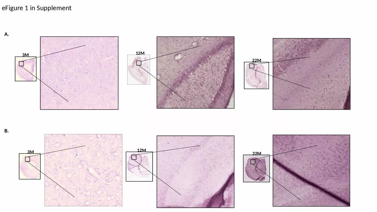
/
12M 3 M 22M 3 M 22M 12M eFigure 12M 3 M 22M 3 M 22M 12M eFigure

12M 3 M 22M 3 M 22M 12M eFigure - PowerPoint Presentation

martin
martin . @martin
Follow
0 views
Uploaded On 2024-02-16

12M 3 M 22M 3 M 22M 12M eFigure - PPT Presentation

1 in Supplement A B Acetylation HSF1 reprobe 3M 12M 24M 3M 12M 24M Acetylation HSF1 reprobe eFigure 2 in Supplement A B 21 37 51 3 1 32 919596 100 93 106119 124 ID: 1046372

dwarf hsf1 wild type hsf1 dwarf type wild ames mice efigure spots hippocampus western supplement acetylated spot shows blot

Share:

Link:

Embed:

Download Presentation from below link

Download Presentation The PPT/PDF document "12M 3 M 22M 3 M 22M 12M eFigure" is the property of its rightful owner. Permission is granted to download and print the materials on this web site for personal, non-commercial use only, and to display it on your personal computer provided you do not modify the materials and that you retain all copyright notices contained in the materials. By downloading content from our website, you accept the terms of this agreement.


Presentation Transcript

1. 12M3M22M3M22M12MeFigure 1 in SupplementA.B.

2. AcetylationHSF1 reprobe3M12M24M3M12M24MAcetylationHSF1 reprobeeFigure 2 in SupplementA.B.213751313291,95,96, 10093106,119, 124110,118, 126114,122,132, 137109171,173, 178,181,183,188175,184,192168159202197193195

3. eTable 1 in Supplement

4. eFigure 1 In Supplement. Immunohistochemical Staining of HSF1 in Ames wild type and Dwarf Mice Hippocampus. Immunohistochemistry in the 3M, 12M and 22M old Ames wild type and Dwarf brain tissue. The hippocampus region shows age-dependent increase in HSF1 level in Ames wild type mice (eFigure 1A) and increase in Dwarf mice hippocampus (eFigure 1B).eFigure 2 In Supplement. Western blot of HSF1 Acetylation in Ames wild type and Dwarf Mice Brain Tissue. eFigure 2A and 2B shows a representative western blot analysis of two-dimensional (2D) gel electrophoresis showing separated acetylated proteins and HSF1 from 3M, 12M and 22M old hippocampus for Ames wild type and Dwarf mice. eFigure 2A shows HSF1 acetylated spots and their corresponding number which is common in all three age groups in both Ames wild type and Dwarf mice hippocampus. eTable 1 In Supplement. Spot volume comparisons of Acetylated HSF1 proteins detected by 2D acetyl lysine and HSF1 western blots of WT and Dwarf Fresh WCE samples. Reference spot numbering, pI, MW, and spot volumes are given for the acetylated HSF1 protein spots. A total of 39 spots out of 208 spots detected by the acetyl-lysine antibody matched spots in the corresponding HSF1 western blot.

Related Contents


Next Show more