/
DNA TESTING STRATEGIES AIMED AT PREVENTING HEREDITARY NONPOLYPOSI DNA TESTING STRATEGIES AIMED AT PREVENTING HEREDITARY NONPOLYPOSI

DNA TESTING STRATEGIES AIMED AT PREVENTING HEREDITARY NONPOLYPOSI - PDF document

mary
mary . @mary
Follow
342 views
Uploaded On 2022-10-14

DNA TESTING STRATEGIES AIMED AT PREVENTING HEREDITARY NONPOLYPOSI - PPT Presentation

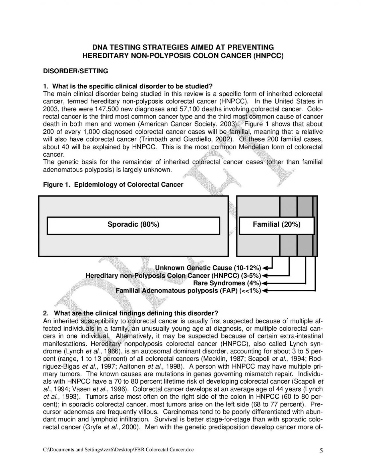
DISORDERSETTING 1 What is the specific clinical disorder to be studied The main clinical disorder being studied in this review is a specific form of inherited colorectal cancer termed hereditar ID: 960048

colorectal cancer hnpcc dna cancer colorectal dna hnpcc testing family msi criteria sequencing individuals mutations tumor mutation percent age

Share:

Link:

Embed:

Download Presentation from below link

Download Pdf The PPT/PDF document "DNA TESTING STRATEGIES AIMED AT PREVENTI..." is the property of its rightful owner. Permission is granted to download and print the materials on this web site for personal, non-commercial use only, and to display it on your personal computer provided you do not modify the materials and that you retain all copyright notices contained in the materials. By downloading content from our website, you accept the terms of this agreement.


Presentation Transcript

DNA TESTING STRATEGIES AIMED AT PREVENTING HEREDITARY NON-POLYPOSI DISORDER/SETTING 1. What is the specific clinical disorder to be studied? The main clinical disorder being studied in this review is a specific form of inherited colorectal cancer, termed hereditary non-polyposis colorectal cancer (HNPCC). In the United States in 2. What are the clinical findings defining this disorder? et al ) ) 2003, there were 147,500 new diagnoses and 57,100 deaths involving colorectal cancer. rectal cancer is the third most common cancer type and the third most common cause of cancer death in both men and women (American Cancer Society, 2003). 200 of every 1,000 diagnosed colorectal cancer cases wilbe familial, meaning that a relative will also have colorecta cancer (Trimbath and Giardiello, 2002). Of these 200 familial cases, about 40 will be explained by HNPCC. This is the most common Mendelian form of colorectal The genetic basis for the remainder of inherited colorectal cancer cases (other than familial adenomatous polyposis) is largely unknown. Epidemiology of Colorectal Cancer Unknown Genetic Cause (10-12%Hereditary non-Polyposis Colon Cancer (HNPCC) (3-5%Rare Syndromes (4%Familial Adenomatous polyposis (FAP) (%An inherited susceptibility to colorectal cancer is usually first suspected because of multiple af­fected individuals in a family, an unusually young age at diagnosis, or multiple colorectal can­cers in one individual. Alternatively, it may be suspected because of certain extra-intestinal manifestations. Hereditary nonpolyposis colorectal cancer (HNPCC), aso called Lynch syn­drome (Lynch ., 1966), is an autosomal dominant disorder, accounting for about 3 to 5 per­cent (range, 1 to 13 percent) of all colorectal cancers (Mecklin, 1987; Scapoli ., 1994; Rod-Sporadic (80%Familial (20% ., 1997; Aaltonen ., 1998). A person with HNPCC may have multiple pri­mary tumors. The known causes are mutations in genes governing mismatch repair. Individu­als with HNPCC have a 70 to 80 percent lifetime risk of developing colorectal cancer (Scapoli ., 1996). Colorectal cancer develops at an average age of 44 years (Lynch ., 1993). Tumors arise most often on the right side of the colon in HNPCC (60 to 80 per­cent); in sporadic colorectal cancer, most tumors arise on the left side (68 to 77 percent). Pre­cursor adenomas are frequently villous. Carcinomas tend to be poorly differentiated with abun­dant mucin and lymphoid infiltration. Survival is better stage-for-stage than with sporadic colo­rectal cancer (Gryfe et al., 2000). Men with the genetic predisposition develop cancer more of- C:\Documents and Settings\zzz6\Desktop\FBR Colorectal Cancer.doc 5 ___________________

_________________________________________________________ ___________________________________________________________________________ ten (80 percent) than women (40 percent) during their lifetime (Mitchell ., 2002). However, women with HNPCC are also at risk for malignancies involving the endometrium (39 to 60 per­cent) and ovary (9 percent) by age 70 (Cruz-Correa and Giardiello, 2002) (Table 1). For both men and women, there is an increased relative risk of transitional cell carcinoma of the ureter and renal pelvis and adenocarcinoma of the stomach, small bowel, and biliary system, ranging from 3 to 25 times the risk in the general population (Vasen ., 1990; Watson and Lynch, ., 1995). hereditary non-polyposis colorectalExtracolonic Disorders Associated with Hereditary Nonpolyposis Colorectal (HNPCCLesions Café au lait spots Sebaceous gland adenomas & carcinomasOvary ., 2001 Turcot’s syndrome is characterized by glioblastoma associated with HNPCC Muir-Torre syndrome is a variant of HNPCC characterized by sebaceous gland tumors and/or keratoacanthomas. What is the clinical setting in which testing is to be performed? This colorectal cancer ACCE Review has as its main aim to evaluate the efficacy of identifying HNPCC) mutations among individuals with newly diagnosed colorectal cancer and then testing for the mutaton in their frst degree relatives, for the purpose of offering preventive measures. At present, testing is readily available for identify­MLH1MSH2), that are responsible for up to 90 per­The high cost of such testing and the relatively low frequency of HNPCC mutations among individuals with newly diagnosed colorectal cancer precludes its use in all such cases. Instead, preliminary evaluations including family history of colorectal cancer and microsatellite instabilty (MSI) testing (see Question 6) have been developed to identify cases in which mutation testing might be warranted. In 1990, the Amsterdam criteria for diagnosis of HNPCC were proposed to identify index cases for research purposes (Vasen ., 1991). take account of cancers in extracolonic sites, the Amsterdam criteria II were later proposed. The Amsterdam criteria have been criticized as too restrictive for clinical use. ria, called the Bethesda criteria (Table 2), have been developed for selecting patients whose tumors are suitable for MSI testing (Umar et al., 2004). The Bethesda criteria are more sensi­tive, but less specific, than the Amsterdam criteria. C:\Documents and Settings\zzz6\Desktop\FBR Colorectal Cancer.doc 6 __________________________________________________________________________ ____________________________________________________________________________ Tumors for Mi

crosatellite Instability a (Testing is indicated using any one of the following criteria) Individuals with cancer in families that meet the Amsterdam criteria Individuals with 2 HNPCC-related cancers, including synchronous and metachronous colorectal cancer or associated extracolonic cancers b iagnosed at ibriform) on et alIndividuals with colorectal cancer and a first-degree relative with colorectal cancer and/or HNPCC-related extracolonic cancer and/or a colorectal adenoma; 1 of the cancers d and the adenoma diagnosed at age Individuals with colorectal cancer or endometrial cancer diagnosed at age Individuals with right-sided colorectal cancer with an undifferentiated pattern (solid/crhistology diagnosed at age Individuals with signet-ring cell-type colorectal cancer diagnosed at age Individuals with adenomas diagnosed at age 40 years From Giardiello ., 2001. Endometrial, ovarian, gastric, hepatobiliary, small bowel, or transitional cell carcinoma of the renal pelvis or ureter. Guidelines for age of cancer diagnosis have been adapted to ­can Gastroenterological Association (AGA) Medical Position Statement (Bethesda cri­Solid/cribriform defined as poorly differentiated or undifferentiated carcinoma com­posed of irregular, solid sheets of large eosinophilic cells and containing small gland­like spaces. The American Gastroenterological Association (AGA) recommends genetic testing for affected individuals in families meeting the Amsterdam or Bethesda criteria and first-degree adult rela­If a deleterious mutation is found in an affected family member, genetic testing ofno deleterious mutation is found in the affected person (or if an affected family member is not available for evaluation) testing an at-risk family member can provide a positive, but not a defini­tively negative, result. Web sites listing current recommendations for the AGA can be found at www.gastro.org and for the PDQ Cancer Gap in Knowledge The proportion of individuals that has a positive family history by the Bethesda Criteria, but are found not to have HNPCC, has not been reported. 4. What DNA test(s) are associated with this disorder? Preliminary family history questions. Asking questions about family history using the Amster­dam or Bethesda criteria represents the first level of preliminary evaluation of individuals with C:\Documents and Settings\zzz6\Desktop\FBR Colorectal Cancer.doc 7 newly diagnosed colorectal cancer to determine whether further testing is warranted. The Be­thesda criteria (Table 2) identify what clinical information is needed to assess risk, including who in the family has had what kind of cancer, at what age, how they are related to the index patient, whether the pati

ent’s colon cancer is right- or left-sided, its histology, and whether the patient has had adenomas. All individuals satisfying the Amsterdam criteria will automatically satisfy the Bethesda criteria. Preliminary laboratory testing. When an index case satisfies the Bethesda criteria, the next step 11. rect transcription errors. Errors are For the MSI cells. test discussed below. iMLH1 or l Disadvantages are the fact that experience is MLH1DNA Testing in the process involves testing the tumor for MSI. MSI is found in the DNA of colorectal cancers of individuals with mismatch repair gene mutations, but not in the DNA of adjacent normal colo­rectal mucosa or in DNA from other normal tissues of the same individual. Microsatellites are DNA sequences, usually consisting of 1 to 5 nucleotide repeats. For example, the most com­mon human microsatellite is a dinucleotide repeat of C and A, (CA)MSI refers to a gain or loss in the number of repeats in DNA in a tumor, compared to the number of repeats in the same region in non-tumor DNA from the same individual. MSI is recognized by observing mul­tiple bands on electrophoresis of PCR products using tumor DNA where fewer bands are seen using PCR products of non-tumor DNA. The multiplicity of bands results from the failure to cor­Such “infidelity” exposes the important role of the gene products of the mismatch repair genes in recognizing and correcting errors in DNA transcription. most likely in regions of short repeated sequences (microsatellites) which may induce slippage of DNA polymerase, resulting in insertion or deletion of the repeated units. This instability is also referred to as the “mutator phenotype” or “replication error phenotype (REP)”. test to have a negligible false negative rate, the sample must have a high proportion of tumor To ensure this high proportion, a pathologist is needed to separate tumor cells from non­tumor cells on tumor sections. MSI testing is the most commonly used preliminary laboratory test, and emphasis will be placed on this test rather than the alternative immunohistochemical Some laboratories use an alternative immunohistochemical (IHC) technque to detect mismatch repair gene products. IHC can be done on tumor samples to determine whether MSH2 gene products are present. It constitutes an alternative method to MSI testing for select­ing patients likey to have a detectable mutation. Advantages of IHC include low cost and an indication as to which gene to sequence first. needed in evaluation of the fluorescent pattern obtained and that a lack of protein is often due to somatic hypermethylation of the MLH1 promoter, rather than to an alteration in the DNA

sequence (i.e., false positive results). When DNA sequencing was done manually, it was common (among index cases with positive MSI test results) to employ a simpler procedure first and to completely sequence only those samples found to be abnormal. These simpler procedures included single strand conformation polymorphism analysis, denaturing gradient gel electrophoresis, Southern blotting, and more recently DHPLC. The most sensitive of these DNA-based procedures is DHPLC, the only method detecting 100 percent of mutations in a comparison of four methods (Eng However, most clinical laboratories now use automated DNA sequencing, and one laboratory (Myriad Genetic Laboratories) has also automated the large number of polymerase chain reac­tions required. Thus, the less sensitive methods have given way to the highly sensitive DHPLC as the method of choice and, with greater use of automated sequencing, there is a trend to C:\Documents and Settings\zzz6\Desktop\FBR Colorectal Cancer.doc 8 ____________________________________________________________________________ ____________________________________________________________________________ DNA sequencing cannot be performed on tumor tissue (see Question 15, for further explana­Once MSI has been identified in tumor tissue in an index case, the next recommended step is to obtain a sample (usually blood) for DNA sequencing. The known causes of HNPCC are mutations in one of several DNA mismatch repair genes (Table 3). These genes normally maintain fidelity of DNA replication by correction of nucleotide mispairs and small insertions or deletions generated by misincorporation or slippage of DNA polymerase during transcription. Sequencing is performed customarily only for count for about 90 percent of the mutations detectable via sequencing (Cruz-Correa and Giardi­ et alTable 3. Sequencing Gene MLH1 3p21 50.9 MSH2 2p16 37.6 MSH6 2p16 9.6 PMS1 2q31 0.3 PMS2 7q11 1.6 ello, 2002; Lynch and de la Chapelle, 2003). The commercially available DNA tests for identify­ing mutations associated with HNPCC are listed in Table 4 (Giardiello ., 2001). A review of uncommon mutations causing HNPCC is maintained at www.nfdht.nl\database\mdbchoice.htm. Mismatch Repair Genes: Site of Mutation in Hereditary Non-Polyposis Colon Chromosome Location Proportion of all HNPCC Mutations Associated with In­dividual Genes (%)* *Only a proportion of these mutations is detectable using tests available for clinical purposes HNPCC Mutations Not Detected by Conventional DNA sequencing detect all disease-causing mutations in the HNPCC genes listed in Table 3. Large deletions are missed and may constitute 20 to 25 percent of all disease-causing mutations (de la C

hapelle, 2003a; Wagner 2004). methods can be used to find large deletions, if DNA-based detection methods reveal no abnor­mality (Wahlberg ., 1999). RNA-based methods involve synthesizing and sequencing com­plementary DNA (cDNA), representing the whole coding region. A mutation involving a deletion spanning an intron-exon junction is detected as a shortened fragment. However, major disad­vantages are that defective alleles may be only weakly expressed or not expressed, and alter­native transcripts may confuse the analysis (Kohonen-Corish ., 1996). In sequencing, the normal allele often obscures the mutant allele. “Conversion technology” pro­vides haploid cells for DNA analysis by fusing the patient’s lymphocytes to an established mouse fibroblast line and, through the serial spontaneous loss of human chromosomes, isolat­ing cells that are haploid for the chromosome of interest. In one study of 22 HNPCC MSI­positive patients, conventional sequencing of diploid cell DNA identified a mutation in only 12 patients, whereas sequencing of hybrid haploid cells revealed a mutation in all 22 (Yan et al Multiplex probe ligation tag assay (MLTA) is an automated procedure that can identify some of the mutations that are not identified by more standard methodologies. These more elaborate tests are suitable for research purposes, but are not practical for use in clinical practice. C:\Documents and Settings\zzz6\Desktop\FBR Colorectal Cancer.doc 9 ____________________________________________________________________________ ___________________________________________________________________________ c Table 4. Commercially Available Genetic Tests for Hereditary Non-Polyposis Colon Can­ a Analysis Method Unknown Known MSH2, MLH1 $1540 $800-$3000 $200-$500 MSH2, MLH1 b + sequencing $260 MSH2, MLH1 c $800 $250 MSH2, MLH1 $750 $500 MSI testing of tumor Polyacrylamide gel Electrophoresis $300 to $500 without cancer can be offered testing with the expectation of a definitive result. Such testing, From Giardiello et al., 2001. SSCP, single-strand conformation polymorphism See www.genetests.org for details concerning specific laboratories Once a pathogenic mutation is detected in a family member with a relevant cancer, relatives which has been called single-site testing, is simpler and less expensive ($200 to $500) than the comprehensive testing of the index family member. Are preliminary screening questions employed? One study tested a cohort of colorectal cancers in Ohio and Finland using MSI testing, followed by DNA sequencing for those with positive MSI results (de la Chapelle, 2001, Altonen, 1998). that study, 86 percent of persons found to have an HNPCC

mutation were documented to have Thus, 14 percent of individuals (one in seven) who have colorectal cancer and an HNPCC mutation will have a negative family history. In practice, a policy that a b as to its reliincluded MSI testing for all colorectacers) are useful to identify a subset of patients for MSI testing. cancer cases would have a low yield and a relatively high cost per HNPCC patient identified. For that reason, preliminary questions regarding family his­tory (e.g., the Bethesda criteria – Questions 3 and 4) of colorectal cancer (and associated can- Since the family history is so important for ability. Many patients lack accurate Danish study compared the family history reporIn 39 percent of cases, the proband lacked the family history information necessary to determine whether the family met the Amsterdam criteria (Katballe ., 2001). Another study compared patient statements about their relatives with the death certificates, pathology reports, and the self-reports of relatives. The patients re­ported 89 percent of colon cancers in first-degree relatives and only 57 percent of colorectal cancers in second-degree relatives (Anton-Culver, 2003). 6.  Is it a stand-alone test or is it one of a series of tests? 7.  If it is part of a series of tests, are all tests performed in all instances (parallel) or are some tests performed only on the basis of other results (series)? Taking a family history and performing microsatellite instability analysis of tumor cells are both considered here to be preliminary tests. The term “susceptibility” is used to acknowledge the C:\Documents and Settings\zzz6\Desktop\FBR Colorectal Cancer.doc 10 fact that some family members with an HNPCC mutation identified as a consequence of finding a mutation in an affected relative will not develop colon cancer within their lifetime. An inherited susceptibility to colorectal cancer is generally determined as the result of a series of tests. In a colorectal cancer patient with no or few polyps, HNPCC is the first consideration, and MSI test­ing is the usual next step among index cases satisfying the Bethesda criteria. If the MSI test is abnormal, but no mutation is found on DNA sequencing, some laboratories perform special pro­cedures, such as Southern blotting or RNA analysis, to look for large deletions or large inser­tions. Laboratories differ as to the protocol followed. Some laboratories do DNA sequencing directly, while others perform another laboratory test and sequence only if that other test reveals Sequencing per se is generally not an option on a laboratory’s testing requisi­ C:\Documents and Settings\zzz6\Desktop\FBR Colorectal Cancer.do