/
Three or more adjacent ribs Three or more adjacent ribs

Three or more adjacent ribs - PDF document

megan
megan . @megan
Follow
345 views
Uploaded On 2022-09-22

Three or more adjacent ribs - PPT Presentation

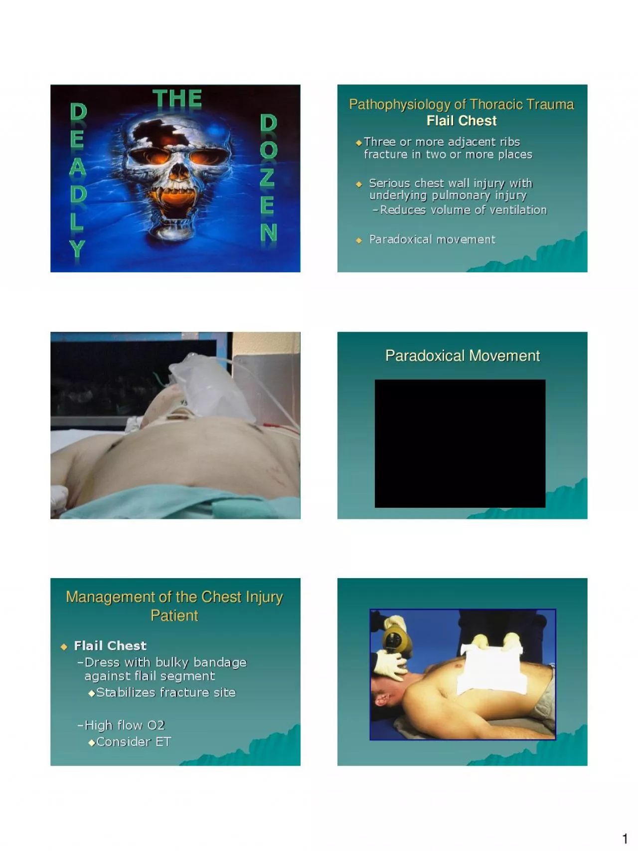
1 fracture in two or more places Serious chest wall injury with underlying pulmonary injury Reduces volume of ventilation Paradoxical movement Pathophysiology of Thoracic Trauma Flai ID: 955162

chest trauma injury thoracic trauma chest thoracic injury pathophysiology patient signs management symptoms pneumothorax rupture wound blood myocardial high

Share:

Link:

Embed:

Download Presentation from below link

Download Pdf The PPT/PDF document "Three or more adjacent ribs" is the property of its rightful owner. Permission is granted to download and print the materials on this web site for personal, non-commercial use only, and to display it on your personal computer provided you do not modify the materials and that you retain all copyright notices contained in the materials. By downloading content from our website, you accept the terms of this agreement.


Presentation Transcript

1  Three or more adjacent ribs fracture in two or more places  Serious chest wall injury with underlying pulmonary injury – Reduces volume of ventilation  Paradoxical movement Pathophysiology of Thoracic Trauma Flail Chest Inspiration Expiration Paradoxical Movement  Flail Chest – Dress with bulky bandage against flail segment  Stabilizes fracture site – High flow O2  Consider ET Management of the Chest Injury Patient 2  Free passage of air between atmosphere and pleural space  Air replaces space for lung tissue  Air will be drawn through wound if wound is 2/3 diameter of the trachea or larger Pathophysiology of Thoracic Trauma Open Pneumothorax Pathophysiology o

f Thoracic Trauma Open Pneumothorax  Signs & Symptoms – Penetrating chest trauma – Sucking chest wound – Frothy blood at wound site – Severe Dyspnea – Hypovolemia Sucking Chest Wound  Open Pneumothorax – High flow O 2 – Cover site with sterile occlusive dressing taped on three sides – Progressive airway management if indicated Management of the Chest Injury Patient Sealing a Wound 3 Sealing a Wound  Closed chest  Progression from Simple or Open pneumothorax  Excessive pressure reduces effectiveness of v/q Pathophysiology of Thoracic Trauma Tension Pneumothorax  Dyspnea  Hypoxia/cyanosis 

JVD  Hyperinflation of injured side of chest  Hyperresonance of injured side of chest Pathophysiology of Thoracic Trauma Pulmonary Injuries Tension Pneumothorax Signs & Symptoms 4  Tension Pneumothorax – Confirmation – Pleural Decompression  2 nd intercostal space in mid - clavicular line  Consider multiple decompression sites if patient remains symptomatic – Large over the needle catheter Management of the Chest Injury Patient  Accumulation of blood in the pleural space  Serious hemorrhage may accumulate 1,500 mL of blood – Mortality rate of 75% – Ventilation/Perfusion mismatch – Shock  Typically accompanies a pneumothorax Pathophysiology of Thoracic Trauma H

emothorax Hemothorax  Hemothorax – High flow O 2 – Shock Management  Positioning, warmth  2 large bore I.V.’s – EVALUATE BREATH SOUNDS for fluid overload Management of the Chest Injury Patient 5  30 - 75% of patients with significant blunt chest trauma  Typical MOI – Rib fx – Deceleration  Chest impact on steering wheel  Micro - hemorrhage may account for 1 - 1 ½ L of blood loss in alveolar tissue Pathophysiology of Thoracic Trauma Pulmonary Contusion  Right Atrium and Ventricle is commonly injured  Electrical disturbances due to irritability of damaged myocardial cells  Progressive problems – Hematoma – Myocardial necrosis – Dysrhythmias – CHF &/or

Cardiogenic shock Pathophysiology of Thoracic Trauma Myocardial Contusion  Bruising of chest wall  Tachycardia and/or irregular rhythm  Associated injuries – Rib/Sternal fractures  Chest pain unrelieved by oxygen Thoracic Trauma Cardiovascular Injuries Myocardial Contusion Signs & Symptoms  Myocardial Contusion – Monitor ECG  Alert for dysrhythmias  ALS / BLS Management of the Chest Injury Patient 6  Restriction to cardiac filling caused by blood or other fluid within the pericardium  Occurs in % of all serious chest trauma – V ery high mortality – 200 ml of blood Pathophysiology of Thoracic Trauma Pericardial Tamponade Pericardium  Dyspnea – Normal br

eath sounds  Pale/ cyanotic  Beck’s Triad – JVD – Muffled heart tones – Hypotension Thoracic Trauma Cardiovascular Injuries Pericardial Tamponade Signs & Symptoms  Pericardial Tamponade – High flow O 2 – I.V. access – “Diesel Therapy” Management of the Chest Injury Patient  Occurs almost exclusively with extreme blunt thoracic trauma  Secondary due to necrosis resulting from AMI  Signs & Symptoms – Severe rib or sternal fracture – Possible signs and symptoms of cardiac tamponade – Absence of vital signs Pathophysiology of Thoracic Trauma Myocardial Aneurysm/Rupture 7 Myocardial Rupture  Aorta most commonly injured in severe blunt or penetrating trauma

– 85 - 95% mortality  Injury may be confined to areas of aorta attachment  Signs & Symptoms – Rapid and deterioration of vitals – Pulse deficit between right and left upper or lower extremities Pathophysiology of Thoracic Trauma Traumatic Aortic Rupture Aortic Rupture  Aortic Aneurysm/Rupture – AVOID rough handling – Initiate I.V. therapy en - route  Mild hypotension may be protective – Keep patient calm Management of the Chest Injury Patient  Herniation of abdominal organs into thorax  MOI – High pressure blunt chest trauma – Penetrating trauma  Signs & Symptoms  Restriction of ipsilateral lung  Displacement of mediastinum  Bowel sounds may be not

ed in thorax Pathophysiology of Thoracic Trauma Traumatic Rupture of the Diaphragm 8 Management of the Chest Injury Patient  Diaphragmatic rupture – Oxygen administration  Positive pressure ventilation will worsen the injury – Rapid transport – NG tube placement (MC, protocol)  30% mortality  Contents in esophagus/stomach may move into mediastinum  Subcutaneous emphysema  Pain, hoarseness, dysphagia, respiratory distress, shock Pathophysiology of Thoracic Trauma Traumatic Esophageal Rupture Management of the Chest Injury Patient  Esophageal injury – Fluid replacement for shock – Rapid Transport – Watch airway  50% of patients with injury die within 1

hr of injury  Signs & Symptoms – Tachypnea, tachycardia – Cyanosis – Hemoptysis – Massive subcutaneous emphysema – Suspect/Evaluate for other closed chest trauma Pathophysiology of Thoracic Trauma Tracheobronchial Injury 9 SUB - Q Air  Results from severe compressive forces applied to the thorax  Signs & Symptoms – Head & Neck become engorged with blood  Face and tongue swollen  Bulging eyes with conjunctival hemorrhage – JVD – Hypotension, Hypoxemia Pathophysiology of Thoracic Trauma Traumatic Asphyxia QUESTIONS ? FOR MORE INFORMATION…. Christopher Ebright B.Ed., NREMT - P EMS Education Coordinator National EMS Academy Covington, LA c.ebrightnremtp@gmail