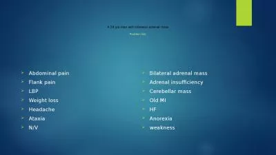
No conflict of interest to disclose Adrenal Glands AnatomyPhysiology HPA Axis Cortisol Adaptive response to stress Addison Disease Primary Adrenal Insufficiency Diagnosis Ongoing management ID: 908159
Download Presentation The PPT/PDF document "Betty Hulse Primary Adrenal Insufficienc..." is the property of its rightful owner. Permission is granted to download and print the materials on this web site for personal, non-commercial use only, and to display it on your personal computer provided you do not modify the materials and that you retain all copyright notices contained in the materials. By downloading content from our website, you accept the terms of this agreement.
Slide1
Betty Hulse
Primary Adrenal Insufficiency (Addison Disease): A Patient’s Perspective
Slide2No conflict of interest
to disclose
Slide3Adrenal Glands
Anatomy/Physiology
HPA Axis
Cortisol
Adaptive response to stressAddison Disease (Primary Adrenal Insufficiency)DiagnosisOn-going managementComplications
Topics
Slide4Upon completion of the session, the participant will be able to:
Describe the pathophysiology of primary adrenal insufficiency
Identify the diagnostic studies for investigation and diagnosis of primary adrenal insufficiency
Describe treatment and ongoing management of primary adrenal insufficiency
Objectives
Slide5Anatomy
Zona Glomerulosa: Mineralocorticoids (Aldosterone)
Zona
Fasciculata
: Glucocorticoids (Cortisol)
Zona Reticularis: Androgens (
Dihydroepiandrosterone
/DHEA, DHEA-S, test. precursor)
Medulla:
Catecholemines
(Epinephrine/Norepinephrine)
Slide6Hypothalamus
Corticotropin
-Releasing Hormone (CRH)
Anterior Pituitary
Adrenocorticotropic Hormone (ACTH)Adrenal CortexCortisolStress ResponsePerceived threat
SNS > Adrenal Medulla > epinephrine
Hypothalamus (CRH) > Ant. Pit (ACTH) >
Adrenal Cortex > Cortisol
Negative Feedback System
Protective levels of cortisol achieved
Signal Hypothalamus & Pituitary
HPA Axis
Slide7Epinephrine
(Adrenalin)
Stimulates ability to avoid harm from the threat (fight, flight)
BP, Heart rate, respirations, alertness, muscle tone
Cortisol Triggers actions that mediate the extra demands put on the bodyFuel & energy for recoveryIncreased glycogen productionGluconeogenesis from mobilized amino acids
Fatty acid mobilization for energy to muscles
Increased cardiac output and vascular tone
Increased Na+ retention
Anti-inflammatory effects
Threat resolves, cortisol mediated actions abate over time & body returns to resting state
Spikes in cortisol are superimposed on the circadian rhythm of cortisol secretion
Adaptive Response to Stress
Slide8Circadian Rhythm of Cortisol Secretion
Cortisol
production peaks in the early morning
, then gradually
declines over the course of the day. As we sleep, over the course of the night, cortisol production climbs again to its early morning diurnal zenith. The circadian rhythmicity of cortisol secretion is important ;
certain processes such as cell repair, immune restoration, and repletion of cellular redox potential are linked to the Circadian rhythm
maintained by the HPA axis.
Slide9Adrenal Insufficiency
Slide10Cortisol, Aldosterone, DHEA Production
Destruction of Adrenal Gland
Hemorrhage
TB Granulomas
Antibodies to adrenal cortex Genetic DisordersCongenital adrenal hyperplasiaDef in enzyme needed to produce cortisol & aldosteroneAdrenoleukodystrophyLong fatty acid chain build-up
Primary Adrenal Insufficiency
Slide11ACTH Production Failure
Destruction of pituitary gland
Pituitary tumors/cysts, surgery to remove tumors/cysts, radiation therapy, trauma, some inflammatory diseases
ACTH Production Suppression
Anticipate in pts taking = 7.5 mg prednisolone/day x 3 wksFeedback to pituitary gland to decrease ACTH production
Discontinuing steroid medication w/o tapering dose may cause adrenal crisis
Secondary Adrenal Insufficiency
Slide12Corticotropin
-Releasing Hormone Production
Hypothalamus
Tertiary Adrenal Insufficiency
Slide13Dysfunction or destruction of entire adrenal cortex
Insidious, progressive
Life threatening;
circulatory collapse
if not treated1st described by Thomas Addison in 1855TB Adrenalitis present in majority of patients
Most common cause today is
Autoimmune AI
(70-80%)
Prevalence in U.S. 40-60 cases/million people
F>M; no racial predilection
Most common age at presentation in adults: 30-50
yo
90% destruction of adrenal cortex by the time symptoms appear
Primary Adrenal Insufficiency
(Addison Disease)
Slide14Mineralocorticoid Deficiency (Aldosterone)
Aldosterone stimulates Na+ reabsorption & K+ excretion (mostly renal)
Renin-angiotensin-aldosterone feedback system
Deficiency results in urinary salt & water loss
Severe dehydrationDecreased circulatory volumeHypotension Androgen Deficiency
Deficiency can lead to fatigue, poor concentration & diminished feeling of well-being
Pathophysiology
Slide15Glucocorticoid Deficiency (Cortisol)
Disturbed carb, fat & protein metabolism
Insufficient gluconeogenesis
Hypoglycemia & decrease liver glycogen
Lipolysis for energy>>>weight loss Deficient neuromuscular function Results in weaknessDecreased resistance to infection, trauma, other stress
Myocardial weakness & dehydration
Reduced cardiac output
Pathophysiology
Slide16Weakness & fatigue
Hyperpigmentation
Diffuse tanning of exposed and, to lesser extent, unexposed areas, especially on pressure points (bony prominences), skin folds, scars, extensor surfaces
Black freckles & darkening of molesBluish black discolorations of mucous membranesAnorexia, abdominal pain, nausea, vomiting, diarrhea
Weight loss
Decreased tolerance to cold
Joint & muscle pain, muscle cramps
Dehydration, dizziness, syncope
Hypotension
Gradual onset, nonspecific nature of symptoms often lead to incorrect initial diagnosis (
ortho
/rheumatologic, cancer, GI, cardiac, infectious disease…)
Signs/Symptoms
Slide17Melanocytic stimulation factors & ACTH produced from same precursor
2ndary & tertiary adrenal insufficiency (no increase in ACTH, no hyperpigmentation)
Hyperpigmentation
Slide18Hyperpigmentation
Slide19http://www.slideshare.net/openmichigan/endocrine-photo-gallery
Slide2047 yowf
presents with following complaints of 4 months duration:
Joint pain of gradual onset that is steadily getting worse; pain is isolated mostly to hips and knees (experiences intense pain with rising from seated position; walking is painful at first but pain subsides within a minute or two)
Muscle cramping and spasm
Unintentional weight loss of 23 lbs (recently started a sedentary job, appetite is reported good but she has noticed that she gets full faster; no restrictions on what she eats. She eats what she wants & as much as she wants)Weakness that is becoming worse (increasingly difficult to rise from supine position especially if from floor level)
Patient Case - Betty Hulse
Subjective:
Slide21Intense fatigue (normal activity is very difficult)
pt
who normally exercises regularly has not been able to do so due to fatigue/weaknessGeneralized hyperpigmentation of the skin; reports becoming very tan with very little exposure to the sun
Hyperpigmentation of palmar creases & darkening of moles & scars
Episodes of presyncope (cannot stand for extended periods of time w/o feeling like she is going to pass out)Denies abdominal pain, nausea, vomiting, diarrhea; Denies night sweats, fevers, coughDenies tremors & heat intolerance
Subjective Continued:
Slide22Allergies: PCN
Rxn
: Rash as a child
Medical conditions: hypercholesterolemia
Medications: Lovastatin 10 mg PO qdSupplements/Herbals: Calcium 500 mg PO bidSurgeries: Left bunionectomy
, tonsillectomy
Immunizations: all up-to-date
PMH
Slide23Coronary artery disease
Stroke
HTN
Hyperlipidemia
Breast cancerPrimary biliary cholangitisUlcerative colitis
Hashimoto’s hypothyroidism
Family
Hx
Slide24Vitals
BP: 80/50
T: 98.6 pulse: 80 RR: 15 Ht: 5 ft 5 in
Wt
: 105 lbs BMI: 18HEENT, Neck, Heart, Lungs, Abdomen, Extremities, Pulses, Reflexes, Lymph NodesAll NormalSKIN: generalized tanning, palmar creases, extensor surfaces and scars are hyperpigmented
, moles are very darkly pigmented, mucus membranes do not show hyperpigmentation.
Physical Exam
Slide25Adrenal Insufficiency (hyperpigmentation, hypotension, weakness, weight loss)
Malignancy (rapid unexplained weight loss)
Rheumatologic disorder (joint pain, fatigue, weakness)
Hyperthyroidism (unintentional weight loss)
Differential Diagnosis
Slide26Neutrophils 45 %
Lymphocytes 50 %
H
Monocytes 2 %
Eosinophils 3 %CBCWBC 4,100 L
RBC 4.0
Hgb
13.5
Hct
41
MCV 92
MCHC 32
MCH 35
RDW 12
PLTS 121,000
L
Diagnostics
BMP
Glucose 80
Calcium 9.0
BUN 10
Creatinine 0.8
Na+ 140
K+ 3.4
L
Cl- 99
CO2 30
Slide27Thyroid Function Panel:
TSH 15.6
H
FT4 – wnl FT3 – wnlOther Labs drawn: Cortisol (A.M.), ACTH, Aldosterone, 21-hydroxylase Abs (results pending)CXR:
No abnormal findings
CT of Abdomen (with & w/o contrast):
Abdominal lymph nodes at upper limits of normal (0.8 mm); consistent with infectious or lymphoproliferative process
Diagnostics
Slide28Assessment:
Hypothyroidism
Possible Lymphoproliferative disorder
Plan:
Levothyroxine 50 ug PO qdReferral to Hematology/Oncology Specialist for further evaluationReturn visit or call with lab results
Pt is requesting consult with endocrinology
First available appointment is in 9 weeks
Assessment/Plan
Slide29Repeated CBC (no significant changes from previous results)
Hematologist/Oncologist not concerned about 0.8 mm LNs
No further
Heme
/Onc evaluation necessary$300
Heme
/
Onc
Visit
Slide30Patient did not feel well enough to wait 9 weeks for treatment
Self-referred to a newer endocrinologist; next available
appt
in 4 weeks
Transferred pertinent med records (including diagnostics) to endocrine’s officeEndocrine Evaluation:Reviewed Lab Results (Drawn at primary care):21-Hydoxylase Ab: positive
ACTH: elevated (
nl
for am draw is 10-60
pg
/ml)
Aldosterone: 3.7 (
nl
adult, upright is 4-31 ng/dl)
Cortisol: 3 mg (nl for am draw is 7-25 ug/dl)
Ordered ACTH (Cosyntropin) Stimulation Test
(definitive diagnostic study)
Follow-up
appt
in 2 weeks
Endocrine Consult
Slide31Protocol
Draw baseline serum cortisol level (8am)
Administer 250 ug
Cosyntropin
IMDraw serum Cortisol 60 minutes post ACTHDraw serum Cortisol 2 hours post ACTH2 Weeks Later:Feeling pretty sick, went home from work to lay down until
appt
Baseline cortisol: 0 ug/
dL
(8 am
nl
: 7-25
μg
/
dL)
Cortisol, 60-minute post ACTH result: 0.3 ug/dL
Cortisol, 2-hour post ACTH result: 0.1 ug/dL
(2h post-stim
nl
: ≥ 18
μg
/dL)
ACTH Stimulation Test
Slide32Cortisol Replacement
Hydrocortisone
BSA x 13 =1.52 x 13 = 19.8 mg/day
General dose range 15-25 mg PO
qd (2 – 3 divided doses)2/3rds of total daily requirement upon rising in a.m. (15 mg), rest in 2nd dose around 4pm (5 mg)Aldosterone ReplacementFludrocortisone
0.1-0.2 mg PO
qd
DHEA+-
Medic Alert Bracelet/Pendant
Emergency dose kit
Treatment
Hydrocortisone
Slide33Within 1
mth
after initiating tx
Vitals, focused H&P
Normal BP, weight gain, returning of energy, feeling strongerAnnual follow-up BMPTFTHgA1C%
Plasma renin activity & serum aldosterone if indicated
Some patients will report weight control problems & lack of a sense of well-being
Trial of DHEA 25-50 mg PO
qd
Follow up
Slide34Mineralocorticoid dose adjustments
Measure plasma renin activity & serum aldosterone
Better correlates with efficacy of replacement therapy
BUN level was elevated; e-
lytes were wnlsAdjusted fludrocortisone doseGlucocorticoid dose adjustments Continued excessive fatigue & increasing pigmentation after start of treatment suggests insufficient glucocorticoid replacement
Weight gain or facial plethora suggests glucocorticoid excess
Long-term over-replacement may be associated with decreased bone density
Regular measurements of bone mineral density
Long-Term Monitoring
Slide35Initial
fludrocortisone dose
0.1 mg/day (ankle edema, so decreased dose to 0.05)Drawn while upright, without Na++ restricted diet, normal ranges for age
>
15 4/12/18 Pl renin activity 11.1 ng/mL/h (0.5-4.0 ng/mL/hr) 4/12/18 Serum aldosterone 3.3 ng/dL (4.0-31.0 ng/dL)
Increased
fludrocortisone dose
to 0.1 mg/day & recheck levels
6/26/18
Pl renin activity
8.2
ng/mL/h (0.6-3.0 ng/mL/
hr
) 6/26/18 Serum aldosterone
<4.0 ng/dL (4.0-31.0 ng/dL) Increased fludrocortisone dose
to 0.2 mg/day & recheck levels
7/10/18
Pl renin activity
0.9 ng/mL/h (0.6-3.0 ng/mL/
hr
)
7/10/18
Serum aldosterone
<4.0 ng/dL (4.0-31.0 ng/dL)
Hold
fludrocortisone dose
at 0.2 mg/day
4/17/19
Pl renin activity
1.8 ng/mL/h (0.6-3.0 ng/mL/
hr
)
Mineralocorticoid Dose Adjustments
Slide36Increase oral dose of glucocorticoid (double dose) for two days
Illness (fever, vomiting, diarrhea, flu, infection)
, i
njury, bone fracture
Vomiting-if occurs within 30 minutes of taking medication, then take a double dose again immediately
If still vomiting, then use home emergency kit (IM injection) & seek medical advice
Fever of
>
104 F, take triple dose & call medical provider
Surgery (IV pre-op, then every 8
hrs
for 24-48
hrs
)
Consult endocrinology-dose depends on type of surgery & anticipated time under anesthesia
https://www.addisonsdisease.org.uk/newly-diagnosed-sick-day-rules
Stress Dosing – Adrenal Crisis Prevention
Slide37SoluCortef
(Act-O-Vial), syringes, needles
Train pt & family members
100 mg lyophilized hydrocortisone + diluent
Storage: 66-77 FProtect from lightReconstitute
Mix gentle until fully dissolved
Inject IM
Discard after 3 days
Dexamethasone solution
Ready to draw up & inject
4 mg/ml (steroid conversion below)
Glucocorticoid potency: Dexamethasone 25 = 1 Hydrocortisone
Dose equivalence: 0.75 mg Dexamethasone = 20 mg Hydrocortisone (4 mg Dexamethasone = 107 mg Hydrocortisone)
Prescribe Emergency Kit
Slide38Steroid Conversion Chart
http://emupdates.com/2009/11/24/steroid-potencyconversion-chart/
Slide39Cortisol stress production in functioning glands – 250-300 mg/24
hrs
Adrenal Crisis see pie chart for common triggers; vomiting & diarrhea most common
Life-threatening,
Requires Immediate TreatmentSigns/Symptoms:HypotensiveSyncopeHypoglycemicHypovolemiaShock & vascular collapse
IV fluids (with or w/o dextrose) – Rapid infusion 1
st
Liter;
addn’l
Liters next 24
hrs
IV hydrocortisone (do not delay) – 50-100 mg IV q 6-8
hrs
for 1-3 days
BP will not respond adequately to fluids or vasopressors alone
ID &
Tx
underlying condition that precipitated crisis
Once stable, resume normal dosing regimen
Adrenal Crisis
Slide40Polyglandular
autoimmune syndrome type I & type II
Autoimmune adrenal insufficiency
Increased risk of developing multiple autoimmune-mediated endocrine gland failure
Type II (relatively common)Hypothyroidism Diabetes mellitus type I HypoparathyroidismHypopituitarismType I (very rare)Hypothyroidism
Diabetes mellitus type I
Hypoparathyroidism
Hypogonadism
Concomitant disease: mucocutaneous candidiasis, celiac disease, pernicious anemia, vitiligo, alopecia, etc.
Complications
Slide41Slide42Medscape
http://emedicine.medscape.com/article/116467-overview
Merck Manual
http://www.merckmanuals.com/professional/endocrine_and_metabolic_disorders/adrenal_disorders/addison_disease.html?qt=adrenal%20insufficiency&alt=sh Mayo medical laboratorieshttp://www.mayomedicallaboratories.com/test-catalog/ Cleveland Clinic
http://www.clevelandclinicmeded.com/medicalpubs/diseasemanagement/endocrinology/diseases-of-the-adrenal-gland/
Current Medical Diagnosis
& Treatment
2021
https://www.addisonsdisease.org.uk/newly-diagnosed-sick-day-rules
References