/
Abnormal Length and Posi�on of the Sigmoid Colon and Its 
. Abnormal Length and Posi�on of the Sigmoid Colon and Its 
.

Abnormal Length and Posi�on of the Sigmoid Colon and Its . - PDF document

min-jolicoeur
min-jolicoeur . @min-jolicoeur
Follow
459 views
Uploaded On 2015-10-22

Abnormal Length and Posi�on of the Sigmoid Colon and Its . - PPT Presentation

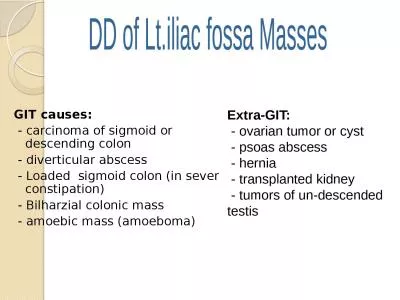
VOL10 NO 4 ISSUE 40 OCT DEC 2012 Page 95 Case Note Clinical Signix00660069cance Nayak SB George BM Mishra S Department of Anatomy Melaka Manipal Medical College Manipal Campus ID: 169356

VOL.10 NO.

Share:

Link:

Embed:

Download Presentation from below link

Download Pdf The PPT/PDF document "Abnormal Length and Posi�on o..." is the property of its rightful owner. Permission is granted to download and print the materials on this web site for personal, non-commercial use only, and to display it on your personal computer provided you do not modify the materials and that you retain all copyright notices contained in the materials. By downloading content from our website, you accept the terms of this agreement.


Presentation Transcript

VOL.10 | NO. 4 | ISSUE 40 | OCT  DEC 2012 Page 95 Case Note Abnormal Length and Posi�on of the Sigmoid Colon and Its Clinical Signi�cance Nayak SB, George BM, Mishra S Department of Anatomy, Melaka Manipal Medical College (Manipal Campus) Manipal University Karnataka, India. Corresponding Author Satheesha Nayak B. Department of Anatomy Melaka Manipal Medical College (Manipal campus) Interna�onal Centre for Health Sciences Karnataka, India Email: nayaksathish@yahoo.com Cita�on Nayak SB, George BM, Mishra S. Abnormal Length and Posi�on of the Sigmoid Colon and Its Clinical Sig ni�cance. ABSTRACT Sigmoid colon is one of the most variable parts of the large intes�ne. Here we report a very rare type of varia�on of the sigmoid colon. This varia�on was observed during the rou�ne dissec�ons for undergraduate medical students at Melaka Manipal Medical College (Manipal Campus) India in September 2012. In shaped loop in front of the descending colon and the le� kidney. It had a sigmoid mesocolon which covered the le� kidney. The sigmoid colon had an ascending and a descending limb. This posi�on of sigmoid colon and its mesocolon is dangerous as it can get twisted to form a volvulus. It also might cause confusions in radiologic and sigmoidoscopy techniques. KEY WORDS INTRODUCTION The sigmoid colon or pelvic colon is a part of the large intes�ne. It is about 15 inches in length and extends from the brim of the pelvis to the third piece of the sacrum where it becomes the rectum. It forms a loop which hangs down in the pelvis over the pelvic organs. As a part of the hindgut, the sigmoid colon is supplied by the branches of the inferior mesenteric artery. It is suspended by a peritoneum. Its le� limb is a�ached along the upper half of the le� external iliac artery and the right limb is a�ached to the posterior pelvic wall. The le� ureter passes behind the apex of the sigmoid mesocolon. We observed quite an unusual posi�on and rela�ons of the sigmoid colon which are of importance to various clinical prac��oners. CASE REPORT undergraduates, the varia�on of the sigmoid colon was observed. This varia�on was observed at Melaka Manipal Medical College (Manipal Campus) India in September 2012.The sigmoid colon was about 60cm long. It started as a con�nua�on of the descending colon at the le� iliac fossa and ascended upwards in front of the descending colon, �ll the le� colic �exure. It then made a U turn and pelvis and con�nued as the rectum (Figs 1, 2 and 3). The sigmoid mesocolon was re�ected onto the le� kidney and con�nued to the right as the le� layer of the mesentery of small intes�ne and con�nued on the le� over the descending colon (Fig 2). There was no pelvic limb for the sigmoid mesocolon. KATHMANDU UNIVERSITY MEDICAL JOURNAL Page 96 DISCUSSION Varia�ons in the length and posi�on of any part of the colon are of developmental origin and may lead to variety of acute and chronic pathological condi�ons. One of the most common varia�ons of the sigmoid colon is presence of its redundant loop. Komiyama et al in 1991 reported a 50-year-old Japanese male with excessively long distal part of the colon including the right-sided sigmoid colon. The redundant loop can cause various problems such as cons�pa�on, discomfort over the colon, indiges�on, loss of weight, insomnia, pain and tenderness in the right iliac fossa. 2,4 The symptoms of redundant colon may mimic symptoms of gastric ulcer, heart disease, chronic obstruc�on of bowel in addi�on to appendici�s. Redundant loop of sigmoid colon is prone to form a volvulus which can be diagnosed through a CT scan of the abdomen. Chandrika GT et al have reported a case of right sided sigmoid colon recently. In the case reported by them, he sigmoid mesocolon extended from right iliac fossa to the third sacral vertebra. The current case is unique because of the sigmoid colon extending to the le� lumbar quadrant of abdomen and covering the le� kidney. The a�achment of the sigmoid mesocolon was also peculiar. The N shaped loop formed by the descending colon and sigmoid colon might lead to confusions in interpreta�on of the barium enema radiographs. This course might cause di�cul�es in sigmoidoscopy as well. It might lead to cons�pa�on or compressive e�ect on the small intes�ne or even form a volvulus. Chances of developing a varicocele are greater with a sigmoid colon anomaly being reported here since it can directly compress the le� tes�cular vessels. The possible cause for this anomaly is the failure of the rearrangement of the parts of intes�ne a�er the reduc�on of the physiological umbilical hernia during the fetal life. Figure 1. Intes�nes seen a�er re�ec�on of the anterior abdominal wall. Note the high posi�on of sigmoid colon. Figure 2. Closer view of the sigmoid colon and sigmoid mesocolon (SMC). Note the ascending (ASC) and descending (DSC) limbs of sigmoid colon. M = mesentery of the small intes�ne. Figure 3. Le� lateral view of sigmoid colon (cadaver in supine posi�on). Note the descending colon (DC), ascending limb of sigmoid colon (ASC) and descending limb of sigmoid colon (DSC). VOL.10 | NO. 4 | ISSUE 40 | OCT  DEC 2012 Page 97 Case Note CONCLUSION Length and posi�on of parts of the colon may vary leading to variety of acute and chronic pathological condi�ons. REFERENCES 1. Pyrtek LJ, Jenney WL. Fixed retrocolic right sided dolichosigmoid colon. Ann Surg. 1960; 151: 268–273. 2. Gupta Indrajit, Majumdar Sudeshna, Mandal Subhra. A redundant loop of descending colon and right sided sigmoid colon. Int J Anat Var (IJAV). 2012; 5: 11–13. 3. Komiyama M, Shimada Y. A case of a right-sided sigmoid colon. Kaibogaku Zasshi. 1991; 66: 537–540. 4. Kantor JL. Anomalies of the colon: their roentgen diagnosis and clinical signi�cance résumé of ten years’ study. Radiology.1934; 23: 5. Kanagasuntheram R, Kin LS. Observa�ons on some anomalies of the colon. Singapore Med J. 1970; 11: 110–117. 6. Larochelle FD, Smith EE. The Dolichocolon. Radiology. June 1932; 18: 7. Chandrika Gurulingappa Teli, Gnanagurudasan. Right Sided Sigmoid Colon – Rare Case. Int J Biol Med Res. 2012; 3(2): 1842-1844.