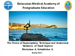
Dr Sayed Mostafa Hashemi Figure 1 a Facial profile of a child 15 years of age and b his father 37 years Proportional differences in facial and brain skull of the father and son ID: 259042
Download Presentation The PPT/PDF document "nasal septum deformity in children" is the property of its rightful owner. Permission is granted to download and print the materials on this web site for personal, non-commercial use only, and to display it on your personal computer provided you do not modify the materials and that you retain all copyright notices contained in the materials. By downloading content from our website, you accept the terms of this agreement.
Slide1Slide2
nasal septum deformity in childrenDr. Sayed Mostafa HashemiSlide3
Figure 1: (a) Facial profile of a child (1.5 years of age) and (b) his father (37 years).
Proportional differences in facial and
brain skull
of the father and son
.
The infant face shows smaller vertical dimensions, less frontal projection of the nose and a
largernasolabial
angle.Slide4
Evidence for timing of pediatric septoplastyBe´ jar et al. concluded from their study in the 1990s that they would encourage children with mild nasal obstruction to wait for surgery until after the nasal growth process is complete [16
].
They considered
this to be age 16 years for boys and age 14 years
for girls.Slide5
Consequences of conservative managementA recent study by D’Ascanio et al. performed a cephalometric study to compare 98 children (mean age 8.8 years, age range 7–12 years)
children (due to nasal septum deviations)
demonstrate facial
and dental anomalies in comparison to
nose-breathing controls
[4
].
most
septal deformities are bound to
worsen
after
the growth
of the nose and thus can cause or
increase sinusitis
and infections
of the upper airways and middle
ear and obstructive
sleep
apnea.Slide6
Figure 8: (a) 5-year-old girl with minimal deviation of the nose due to nasal trauma 1 year previously; (b) progressive deformity at the age of 8 years and (c) 15 years.Slide7
Growth of the nasal septumGrowth of the nasal septum occurs in two phases, with the cartilaginous septum reaching adult size by the time the child is 2 years old, and further enlargement due to growth of the bony septum.16Slide8Slide9
Figure 3: Radiographs of three anatomic specimens: 10 (a), 17 (b) and 30 (c) years of age. (A) septum cartilageSlide10
prevalence of nasal septum deformity in childrenThe overall prevalence of nasal septum deformity in children ranges from 0.93 to 55% and varies according to age and different types of nasal septum deformity classifications.1Slide11
EtiologyThe increase in age is associated with an increased rate of nasal septum deformities, probably because of the greater likelihood of suffering traumatic events.2passing through the birth canal can produce a traumatic event to the nasal septum. In fact,
Kawalski
and
Spiewak
found a 22.2% rate of septum deformity in children born by spontaneous birth, whereas this rate was reduced to only 3.9% in children born by caesarean birth.
For this reason, the importance of an
early diagnosis
of septum deformity in newborns has been underlined to enable immediate treatment and avoid possible worsening of respiratory function in adult age.4Slide12
Appropriate surgery base on animal studyin vitro studies appreciated the importance of the muco perichondrium on the survival of underlying septal cartilage and its contribution to skeletal growth
Bernstein showed that
submucous
resection of cartilage with preservation of a
muco
perichondrial
flap in young pups did not result in any growth disturbances
.
Functional
septoplasty
with
mucoperichondrium
preservation in ferrets also showed no differences in facial growth on
cephalometric
analysisSlide13
Clinical studies of pediatric septoplastyResults from animal studies provided clinicians with the confidence to perform septal surgery in children An
appreciation for
preservation of the mucoperichondrium appeared to
be paramount
for all surgeons performing this type of surgery
.
no wide cartilaginous resections must be made,
the areas
of contact between the septum, the
vomer
, and
the perpendicular
lamina of the
ethmoid
must be reconstituted,
and finally
, the remodeled cartilage must be repositioned.Slide14
warning Avoid incisions through the growing and supporting zones, in particular of the (spheno)ethmoido-dorsal zone
.
Posterior
chondrotomy
or separation of the
septum cartilage
from the perpendicular plate (in
particular the
dorsal part) should be avoided as this area is
of paramount
importance for support and
further growth
(length and height) of the nasal septum
and nasal
dorsum;Slide15Slide16
Effects of different technique on anthropometryAfter separating the patients into two groups, those treated by removing and repositioning of the quadrangular cartilage (external approach/extracorporeal septoplasty)
those treated by minimal septal resections (conservative
endonasal
approach),
it was noted that in both sexes the
nasolabial
angle of patients undergoing the extracorporeal
septoplasty
was significantly lower than that of patients undergoing conservative
septoplasty
therefore concluded that
septoplasty
performed by the
endonasal
approach does not interfere with the normal nasal growing process Slide17Slide18
Absolute and relative indications for pediatric septoplasty [3].Slide19
advocating the timing of septalDespite the majority advocating the timing of septal surgery to be 6 years and older, more clinical studies are required that may provide further evidence for correction of
septal deviations
in younger children, perhaps even at birth.
However, before
considering pediatric nasal septal surgery, a
thorough clinical
examination must be performed to ensure the
correct diagnosis
has been
madeSlide20
septal deviations at birthA long term follow up study by Sooknundun et al. supported closed reduction of nasal septal deviations at birth [23]. Results of this study revealed no untoward effects such as
nasofacial
disproportion
or retardation of facial growth
.
The authors
reported that
uncorrected septal deviation is accompanied by
statistically valid
symptoms such as upper respiratory tract infections, ear
pain and
discharge and that surgical correction of septal deviations
at birth
can prevent the need for
septoplasty
surgery at a later date
in addition
to preventing a number of airway related conditionsSlide21
4. ConclusionNumerous long term follow up studies have provided evidence that pediatric septoplasty can be performed without affecting nasal and facial growth.
Studies have also shown that
conservative management
of deviations of the nasal septum can lead to
facial asymmetry
.
Despite
the majority advocating the timing of
septal
surgery to be 6 years and older, more clinical studies are
required that
may provide further evidence for correction of
septal deviations
in younger children, perhaps even at birth
.
Slide22