/
J Ayub Med Coll Abbottabad 2014;26(2) J Ayub Med Coll Abbottabad 2014;26(2)

J Ayub Med Coll Abbottabad 2014;26(2) - PDF document

myesha-ticknor
myesha-ticknor . @myesha-ticknor
Follow
451 views
Uploaded On 2015-11-20

J Ayub Med Coll Abbottabad 2014;26(2) - PPT Presentation

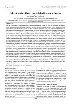
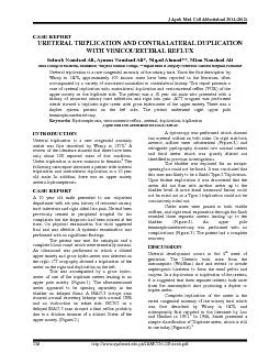
httpwwwayubmededupkJAMC26 2Sohrabpdf 258 CASE REPORT URETERAL TRIPLICATION AND CON TRALATERAL D UPLICATION WITH VESICOURETERAL REFLUX Sohrab Naushad Ali Aymon Naushad Ali Niqad Ahmad ID: 199388

http://www.ayubmed.edu.pk/JAMC/26 - 2/Sohrab.pdf 258 CASE REPORT URETERAL TRIPLICATION AND CON TRALATERAL

Share:

Link:

Embed:

Download Presentation from below link

Download Pdf The PPT/PDF document "J Ayub Med Coll Abbottabad 2014;26(2)" is the property of its rightful owner. Permission is granted to download and print the materials on this web site for personal, non-commercial use only, and to display it on your personal computer provided you do not modify the materials and that you retain all copyright notices contained in the materials. By downloading content from our website, you accept the terms of this agreement.


Presentation Transcript

J Ayub Med Coll Abbottabad 2014;26(2) http://www.ayubmed.edu.pk/JAMC/26 - 2/Sohrab.pdf 258 CASE REPORT URETERAL TRIPLICATION AND CON TRALATERAL D UPLICATION WITH VESICOURETERAL REFLUX Sohrab Naushad Ali, Aymon Naushad Ali*, Niqad Ahmad**, Mian Naushad Ali Shifa College of Medicine, Islamabad, *Khyber Medical College, ** Department of Surgery Northw est General Hospital, Peshawar Ureteral triplication is a rare congenital anomaly of the urinary tract. Since the first description by Warny in 1870, approximately 100 known cases have been reported in the literature, often accompanied by a variety of asso ciated anomalies in contralateral kidney. This report presents a case of ureteral triplication with contralateral duplication and vesicoureteral reflux (VUR) of the upper moiety on the triplicate side. The patient was a 10 year old male who presented with a history of recurrent urinary tract infections and right loin pain. ACT urogram was performed which showed a triplicate right ureter with gross hydroureter of the upper moiety. There was a duplex system present on the left side. The patient underwent righ t upper pole heminephroureterectomy. Keywords: Hydronephrosis, vesicoureteric reflux, ureteral, duplication, triplication J Ayub Med Coll Abbottabad 2014;26(2): 258 – 60 INTRODUCTION Ureteral triplication is a rare congenital anomaly which was first describe d by Wrany in 1870. 1 A review of the literature showed that there have been only about 100 reported cases of this condition. Ureter triplication is more common in females . 2 The following case report discusses a patient with ureteral triplication and contra lateral duplication in a 10 year old male. In addition, there was an upper moiety uretero - hydronephrosis. CASE REPORT A 10 year old male presented to our outpatient department with six year history of recurrent urinary tract infections and right sided loin pain. He had been previously treated in peripheral hospital for his complaints but the diagnosis had been missed at the time. On physical examination, the child appeared frail and was afebrile. A systemic examination was performed with no significant find ings. The patient was sent for urinalysis and a c omplete blood count which were essentially normal. An ultrasound was performed in which a dilated upper moiety and gross hydro - ureter was detected on the right. CT urography showed a triplication of the uret er on the right and duplication on the left. This was accompanied by a gross hydro - ureter of one of the triplicate ureters leading to an upper pole moiety (Fig ure - 1). The aforementioned ureter appeared to be opening separately in the bladder on delayed fi lms. A MAG - 3 isotope scan showe d normal excreting kidneys with normal GFR and no obstruction on either side. MCUG on a delayed MAG - 3 scan showed a faint reflux probably due to a dilution because of a dilated Ureter of the upper moiety. (Fig ure - 2.) A cystos copy was performed which showed two ureteral orifices on both sides. On right side both ureteric orifices were catheterized ( Fig ure - 3. ) and retrograde pyelography showed two normal ureters and third ureter which was grossly dilated not identified in previo us investigations. The bladder was explored for an ectopic opening but could not be found. It was concluded that this case was likely to be a Smith Type - 2 Triplication. Upon further exploration it was discovered that the ureter did not fuse with another u reter up to the bladder level. A more distal intramural fusion could not be ruled out so a Type - 1 triplication could not be conclusively ruled out. Guide wires were passed in both visible orifices, and right renal exploration through the flank revealed th ree separate ureters leading up to the bladder. (Fig ure - 4 ). An upper pole heminephroureterectomy was performed with no complications (Fig ure - 5 ). The patient had a complete recovery . DISCUSSION Ureteral development occurs in the 4 th week of gestation. The U reteric buds arise from the mesonephric (Wolffian) duct and extend to invade nephrogenic blastema to form the renal pelvis and calyces. In a duplication or triplication of the ureters, it is suggested that three separate ureteric buds arise from the mesone phric duct producing a duplex or triplex ureter. Complete triplication of the ureter is the rarest congenital anomaly of the urinary tract which was first described by Wrany in 1870, and subsequently first reported in the literature by Lau and Henline in 1 931. 3 In 1946, Smith presented a simple classification of Triplicate ureter, which is still in use today (Figure - 6). 4 J Ayub Med Coll Abbottabad 2014;26(2) http://www.ayubmed.edu.pk/JAMC/26 - 2/Sohrab.pdf 259 Figure - 1(a): – CT Urogram. Early film showed bilateral duplex kidneys and right side triplex ureter not opacified. (b): CT - urogram (Delay ed Film) outlined grossly dilated ureter of triplex right upper moiety. Figure - 2: Mag - 3 Scan Figure - 3: Cystoscopy Cystoscopy only showed two ureteric openings. 3 rd opening could not be localized, probably in ectopic location. Figure - 4: Retrograde pye lography through the identifiable openings opacified the lower and middle moieties, confirming that upper pole ureter is not connected with other two and probably in some remote ectopic location Figure - 5 (a): On exploration three separate ureters identif iable. Guide wires palpable in normal looking ureters. (b): Grossly dilated ureter followed to upper pole and partial Nephroureterctomy accomplished. (c): Full length specimen Type Smith Classification a. Triple ureters, (Complete triplication) Three separate ureters and three separate ureteral o rifices in bladder, urethra or ectopic location. (35%) b. Incomplete triplication Three ureters arise from the kidney. Two join on the way to the bladder and two orifices present in the bladder (21%) c. Trifi d ureter. All three ureters unite and drain through a single orifice (31%) d. Two ureters from the kidney. One divides into two to have three draining orifices (9%) Figure - 6: Smith Classification J Ayub Med Coll Abbottabad 2014;26(2) http://www.ayubmed.edu.pk/JAMC/26 - 2/Sohrab.pdf 260 Triplication of ureter has been reported with a variety of associated anomalies. Haluk Soylemez 5 reported a case with vesicoureteral reflux in a contralateral duplex system. H. Ander reported a case of triplex ureter in solitary kidney . 6 Mills reported a rather unique case with ureter triplication with a blind en ding bifid ureter . 7 This form of triplication is beyond the scope of the Smith classification. In some exceptional forms of presentation in cross ectopia 8 , Horseshoe kidneys 9 and bilateral triplicicity of ureters . 1 0 Very few cases of ureteral quadruplicati on have been reported . 11,12 Clinical signs of triplication are non - specific, and duplication and triplication are often incidental findings on investigation. They consist of abdominal or back pain, hematuria, fever and urine storage and voiding symptoms. 1 3 Triplication can occasionally be associated with urinary tract calculi, infection, obstruction and recurrent acute epidydimitis. 5 Diagnostic tools inlude ultrasonography, intravenous pyelography, CT urography, retrograde pyelography, MRU and cystoscopy. A large battery of tests may be needed to diagnose and classify the disease accurately. 13 REFERENCES : 1 . Mubarak MY , Zainun AR , Rohaya M . Ureteral triplication with contral ateral partial duplex system . Med J Malaysia 2009; 64 (3): 236 – 7 . 2 . Sánchez - de - Badajoz E , Ramos J , Burgos R . Ureteral Triplication with contralateral ureter duplicat ion . Urol Int 1992 ;4 8:2 1 7 – 8 . 3 . Splanger EB. Complete triplication of the ureter. Radiology 1963; 80 : 795 – 7 . 4 . Smith I. Triplicate ureter, Br J Surg 1946 ; 34:182 – 5 5 . Soyklemez H, Koplay M, Altunoduk B, Oguz F. Ureteral triplication and contralateral duplication with vesicuoureteral reflex . Balkan Med J 2011; 28:469 – 70 . 6 . Ander H , Ziylan O , Cayan S , Kadioğlu TC , Beşişik A - Ureteral triplication (Type I) associated with vesicoureteral refluex in solitary ki dney . Int Urol Nephrol 1997 ; 29 : 5 37 – 40 7 . I.W. Mills, G. M. Oades, M. D. Dinn . Ureteric triplication with a blind ending bifid ureter – BJU International 2000 ; 86(3) : 402 . 8 . Golomb J, Ehrlich RM. Bilateral ureteral triplication with crossed fused ectopic kidneys a ssociated with the VACTREL Syndrome. J Urol 1989 ; 141:1398 – 99 . 9 . Pode D, Shapiro A, Lebensart P. Unilateral triplication of the collecting system in a horshoe kidney. J Urol 1983 ; 130:533 – 4 . 10 . Shoda R, Ejiri S, Fujita T, Wada N. A case of bilateral ureteral trip licaton. Hinyokika Ki y o 1985 ; 31:475 – 81 . 11 . Kl inge L, K o bi a k R, Slongo T . Quadruplication of ureter in combination with a large ureter al cyst. Pediatr Surg Int 2001; 17(7):566 – 8 . 12 . Koszutski T, Kudela G, Mikosinski M, Utrata W. Quadruplication of dystopic kidney in combination with ureteral cyst. J Pediat Surg 2008 ; 43(12) : e 13 – 5 . 13 . Villanueva Peña A, De Diego Rodríguez E. Ureteral triplication, an unusual isolated presentation. Actas Urol Esp 2004;28(10):766 – 70 . Address for Correspondence: Dr. Aymon Naushad Ali, House 69, Street 6, Sector G - 2, Phase 2, Hayatabad, Peshawar, KPK, Pakistan. Cell: +92 - 300 - 8596269 Email: aymon.n@gmail.com