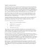
August 7 2013 Long Case HPI 50 yo woman from the Dominican republic presenting to pulmonary clinic for cough 45 years Symptoms may have started after a cold Worse in the summer no temporal relation to nightday ID: 697580
Download Presentation The PPT/PDF document "Marc Freiman Wednesday Pulmonary Confere..." is the property of its rightful owner. Permission is granted to download and print the materials on this web site for personal, non-commercial use only, and to display it on your personal computer provided you do not modify the materials and that you retain all copyright notices contained in the materials. By downloading content from our website, you accept the terms of this agreement.
Slide1
Marc FreimanWednesday Pulmonary ConferenceAugust 7, 2013
Long CaseSlide2
HPI
50
yo
woman from the Dominican republic presenting to pulmonary clinic for cough 4-5 years
Symptoms may have started after a cold
? Worse in the summer, no temporal relation to night/day
Dry, non-productive
DOE 2-3 city blocks, 2 flights of stairs
ROS - Denies HA, sinus congestion, heartburn, reflux. Denies chest pain, palpitations,
orthopnea
, PND or edemaSlide3
PMHx/Soc Hx
Vitiligo
Denies childhood asthma
From DR 3 years ago
Worked in paper shredding factory for 1 yr
Currently works in retail
Never smoker
No
EtOH
,
illicitsSlide4
Physical exam
Afeb
P 95 130/84 96% RA; BMI 30 (150lbs, 5’)
General: comfortable
Clear, no wheeze. ? Crackles at bases bilaterally
Neck: supple, no masses, neck nodes not
palpable
CV: RRR No
m/r/g
No cervical LAD, neck supple
No
desaturation
on exertionSlide5
CXRSlide6Slide7
Symptomatic treatment
B
enadryl
ChlorpheniramineSlide8
Return visit
Benadryl lets her sleep through the night
Still with continued cough
She climbed
3 flights of stairs
and became SOB but did not
desaturate
- minimal
sats
96% HR 120Slide9
PFTSlide10
PFTSlide11
CT ScanSlide12Slide13Slide14Slide15Slide16Slide17Slide18
CT read
LUNGS
: There are
multiple nodules
in both lungs measuring
up to 5 mm
.
Mosaic
attenuation is seen in both lungs most prominent in the lower lobes suggestive of small airways or small vessel disease. Slide19
Labs
CBC,
Chem
7
wnl
ANA, RF negative
TTE unremarkableSlide20
Chronic cough Slide21Slide22
Just kidding…Slide23
VATS biopsy
Had
bronchoscopy
w
BAL
VATS biopsy for right lung with RML and RLL biopsy
Nodule palpated in RMLSlide24
Bronchoscopy and VATS results
Middle lobe
lavage
cytology negative
Aerobic, anaerobic, fungal and AFB cultures negative
RIGHT
LOWER LOBE BIOPSY:
LUNG PARENCHYMA WITH CONGESTION, HEMORRHAGE AND HEMOSIDERIN LADEN MACROPHAGES.
NO TUMOR IDENTIFIED. Slide25Slide26Slide27
synaptophysinSlide28
chromograninSlide29
RML biopsy
IMMUNOHISTOCHEMICAL STUDIES PERFORMED ON PARAFFIN EMBEDDED TISSUE (BLOCK A2) SHOWS POSITIVE STAINING FOR
CHROMOGRANIN, SYNAPTOPHYSINMULTIPLE FOCI OF NEUROENDOCRINE TUMOR, TUMORLETS/ SMALL CARCINOID TUMOR. Slide30
DIPNECH
Diffuse idiopathic pulmonary
neuroendocrine
cell hyperplasiaSlide31
Overview of bronchopulmonary
neuroendocrine
tumors (BP-NET)
4 types
Typical
carcinoid
Atypical
Carcinoid
Large cell
neuroendocrine
carcinoma
Small cell
neuroendocrine
carcinomaSlide32
Diffuse Idiopathic Pulmonary Endocrine Cell Hyperplasia (DIPNECH)
Preneoplastic
Pulmonary
tumorlets
(<5mm)
Had been known to occur in:
ILD
B
ronchiolitis
obliterans
Patients living at
high altitudesSlide33
Purpose of the neuroendocrine cell in the lung?
Unknown
Arises from
Kulchitsky
cell
Thought to be involved as ‘airway sensors’
Mediate airway tone, pulmonary circulation, and control of breathing.
Act as both chemo and mechanoreceptors
Also likely involved in development of the lungSlide34Slide35
AJRCCM - demographics
Women – 92% (23/25)
Mean diagnosis 58 years old
Range 36-76
67% non-smokers (16/24)Slide36
PFTsSlide37
CT findings
Pulmonary nodules (63%, 15 pts)
Ground glass (29%, 7 pts)
Bronchiectasis
(21%, 5 pts)
Mosaic attenuation (17%, 4 pts)Slide38
Clinical course – AJRCCM 2011
92% of patients had symptoms
Cough, dyspnea
, wheezing
Symptoms lasted between days to years – average 8.6
years
Widely variable course has been described
Not clear exactly why some people deteriorate – known to produce
bombesin
and
fibrinogenic
cytokines
41% (7) stable without clinical deterioration
Oral
predniosne
given to 2 of these patients
24% (4) clinically declined and didn’t improve
35% (6) declined but showed improvement clinically
Oral prednisone used in addition to bronchodilators in 4 of 6
No deaths
1 patient with asthma history who died of sepsis found to have DIPNECH on autopsySlide39Slide40
Treatment
No formal evaluations of a treatment algorithm are available
Resection of dominant lesion
Oral/
inh
steroids
w
bronchodilators
Chemotherapy
Surgical lung resection
Presence of lymph nodes has not been associated
w
worse outcome
Lung transplantation (1pt, single lung, followed for 2 yrs)
Observation
?
Somatoastatin
analoguesSlide41
Somatostatin-receptor scintigraphy
(
OctreoScan
)
Tumors often express
somatostatin
Labeled
somatostatin
analog (
octreotide
)
Previously thought to be gold standard for diagnosis
Sn
approx 80-90%
Somatostatin
uptake may correspond to treatment response.
Somatostatin
targeted PET scan
Sn
as high as 100%, identified more lesions than SRS or CTSlide42
111In–DTPA–
pentetreotide
whole body
scintigraphy
(
Octreoscan
) showing an elective uptake of the
radiolabeled
octreotide
in the liver, expression of distant localization of the
neuroendocrine
tumor
of the lung (arrow).
Filosso
P L et al.
Eur
J
Cardiothorac
Surg
2002;21:913-917
© 2002 Elsevier Science B.V.Slide43
Detail of the
Octreoscan
showing the liver
metastase
.
Filosso
P L et al.
Eur
J
Cardiothorac
Surg
2002;21:913-917
© 2002 Elsevier Science B.V.Slide44
Patient’s octreotide scan
No uptake in the lungs
Increased uptake in the
cecum
Negative colonoscopySlide45
Further investigation
Predisposing factors?
? Hormonal component
Tend to be middle-aged females
Unclear if race/ethnicity plays a role
Incidence?
Treatment algorithm?Slide46
References
Ann Oncol (2001) 12 (9): 1295-1300
.
Davies SJ,
Gosney
JR,
Hansell
DM, et al. Diffuse idiopathic
pul
-
monary
neuroendocrine
cell hyperplasia: an under-
recognised
spec-
trum
of disease. Thorax. 2007;62:249-252.
Cameron CM, Roberts F, Connell J,
Sproule
MW. Diffuse idiopathic pulmonary
neuroendocrine
cell hyperplasia: an unusual cause of cyclical ectopic
adrenocorticotrophic
syndrome. Br J
Radiol
. 2011;84:e14-e17.
25.
Fessler
MB, Cool CD, Miller YE, Schwarz MI, Brown KK.
Idio
-
pathic
diffuse hyperplasia of pulmonary
neuroendocrine
cells in a patient with
acromegaly
.
Respirology
. 2004;9:274-277.
26. Pinchot SN,
Holen
K,
Sippel
RS, Chen H.
Carcinoid
tumors. Oncologist. 2008;13:1255-1269.
Bronchopulmonary
neuroendocrine
tumors.Gustafsson
BI, Kidd M, Chan A,
Malfertheiner
MV,
Modlin
IM
Brambilla
E, Travis WD, Colby TV,
Corrin
B,
Shimosato
Y. The new World Health Organization classification of lung
tumours
.
Eur
Respir
J. 2001;18:1059-1068.
Aubry
MC, Thomas CF
Jr
, Jett JR,
Swensen
SJ, Myers JL.
Signifi
-
cance
of multiple
carcinoid
tumors and
tumorlets
in surgical lung specimens: analysis of 28 patients. Chest. 2007;131:1635-1643.
Miller RR, Muller NL.
Neuroendocrine
cell hyperplasia and
obliter
-
ative
bronchiolitis
in patients with peripheral
carcinoid
tumors. Am J
Surg
Pathol
. 1995;18:653-658.
Sheerin
N, Harrison NK, Sheppard MN,
Hansell
DM,
Yacoub
M, Clark TJ.
Obliterative
bronchiolitis
caused by multiple
tumourlets
and
microcarcinoids
successfully treated by single lung
transplanta
-
tion
. Thorax. 1995;50:207-209.
Aguayo
SM, Miller YE, Waldron JA
Jr
, et al. Brief report: idiopathic diffuse hyperplasia of pulmonary
neuroendocrine
cells and airways disease. N
Engl
J Med 1992;327:1285–8.