/
Neural Control of Eye Movements Neural Control of Eye Movements

Neural Control of Eye Movements - PowerPoint Presentation

natalia-silvester
natalia-silvester . @natalia-silvester
Follow
343 views
Uploaded On 2019-06-23

Neural Control of Eye Movements - PPT Presentation

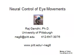
Raj Gandhi PhD University of Pittsburgh neg8pittedu 4126473076 wwwpitteduneg8 Biology of Vision November 9 2015 References httpwwwtutiscaSensesL11EyeMovementsL11EyeMovementsswf ID: 760090

Share:

Link:

Embed:

Download Presentation from below link

Download Presentation The PPT/PDF document "Neural Control of Eye Movements" is the property of its rightful owner. Permission is granted to download and print the materials on this web site for personal, non-commercial use only, and to display it on your personal computer provided you do not modify the materials and that you retain all copyright notices contained in the materials. By downloading content from our website, you accept the terms of this agreement.


Presentation Transcript

Slide1

Neural Control of Eye Movements

Raj Gandhi, Ph.D.University of Pittsburghneg8@pitt.edu 412-647-3076www.pitt.edu/~neg8

Biology of Vision

November 9, 2015

Slide2

References

http://www.tutis.ca/Senses/L11EyeMovements/L11EyeMovements.swf

Principles

of Neural Science

,

Kandel

, Schwartz &

Jessell

(2000)

The Neurology of Eye Movements

, Leigh & Zee (1999)

Neuroanatomy through Clinical Cases

,

Blumenfeld

(2002)

Slide3

SaccadesVergenceSmooth pursuitVestibulo-ocular reflex (VOR)Optokinetic response/nystagmus (OKR/OKN)

Types of Eye Movements

http://www.tutis.ca/Senses/L11EyeMovements/L11EyeMovements.swf

Slide4

Six extraocular muscles operate as three agonist/antagonist pairs to move each eye.Lateral / medial recti – horizontal movementsSuperior / inferior recti – vertical movements; small contribution to torsionSuperior oblique / inferior oblique – torsion (cyclorotation of the orbit) and, to a smaller extent, vertical movements

Superior oblique

Trochlea

Lateral rectus

Superior rectus

Optic nerve

Inferior rectus

Inferior oblique

levator

Inferior rectus

Insertion of

inferior oblique

Lateral rectus

Tendon of

superior oblique

Sup. rectus (cut)

levator palpebrae (cut)

common tendinous ring

optic

chiasm

optic

nerve

Medial rectus

Superior oblique

trochlea

Extraocular muscles

(Modified from Kandel & Schwartz, Principles of Neural Science, 2nd ed., Elsevier Science Publishing, 1985)

Insertion ofsuperior rectus

http://www.tutis.ca/Senses/L11EyeMovements/L11EyeMovements.swf

Slide5

Slide6

PPRF

(paramedian pontine

reticular formation)

Abducens nuc.

Medial longitudinal

fasciculus (MLF)

Oculomotor nuc.

Trochlear nuc.

lateral rectus

medial

recti

abducens nerve

oculomotor

nerve

Modified from Fig. 13.12 of

Blumenfeld, Neuroanatomy

Through Clinical Cases,

Sinauer, 2002.

Control of horizontal eye rotation

Slide7

PPRF

(paramedian pontine

reticular formation)

Abducens nuc.

Oculomotor nuc.

Trochlear nuc.

lateral rectus

medial

recti

abducens nerve

oculomotor

nerve

Modified from Fig. 13.12 of

Blumenfeld, Neuroanatomy

Through Clinical Cases,

Sinauer, 2002.

Control of horizontal eye rotation

Effects of lesion of abducens nerve ?

Medial longitudinal

fasciculus (MLF)

Slide8

Sixth nerve (abducens) palsy

Slide9

Modified from Fig. 13.3 of

Blumenfeld, Neuroanatomy

Through Clinical Cases,

Sinauer, 2002.

Oculomotor Nuclear Complex

bilateral

ipsilateral

ipsilateral

ipsilateral

bilateral

contralateral

contralateral

Slide10

Gandhi’s three monkeys

Slide11

Main Sequence Properties

Eye Movements: Saccades

Slide12

Neural Control of Saccades

Both cortical and subcortical regions contribute to the control of saccades. In the brainstem, neurons in the

pontine

reticular formation (Pon RF) and mesencephalic reticular formation (MRF) respectively control the horizontal and vertical/torsional components of saccades.

Modified from Kandel et al.

Slide13

Abducens neurons discharge at a tonic rate during fixation, burst during ipsiversive eye movements, and decrease or cease activity during contraversive eye movements.

The eye position (during fixation) is directly proportional to the discharge rate of abducens neurons.

Slide14

Slide15

Slide16

Slide17

Key points of saccadic system

Direct (velocity) and indirect (neural integrator) pathways

http://www.tutis.ca/Senses/L11EyeMovements/L11EyeMovements.swf

Slide18

Omnipause neurons:

-- monosynpatically inhibit EBNs

-- tonic discharge rate during fixation and cease activity during saccades, functioning in anti-phase with EBNs

Slide19

Slide20

Visual cortex topography

Slide21

Superior Colliculus (SC)Topographical organization

a major subcortical player:

SUPERIOR COLLICULUS

Slide22

Superior Colliculus (SC)

Topographical organization

Slide23

Slide24

Superior Colliculus (SC)

The SC is a laminar structure separated functionally into superficial, intermediate and deep layers.

A target presented at 20-deg to the right of fovea (black dot) will excite middle-to-caudal cells in the superficial and intermediate/deep layers of the SC.While the superficial layers respond to presentation of a visual target, the intermediate and deep layers elicit a motor burst with or without a visual response. The sensory response is not limited to visual stimuli, and the motor output is not limited to saccades.

Slide25

Temporal features of saccade-related activity

“Visual” burst

“Motor” burst

Slide26

Superior Colliculus

Each neuron in the intermediate layers of the SC discharges during saccades of a restricted amplitude and direction. The cell discharge is weaker for movements of other metrics. The region for which a SC neuron discharges is called the movement field.The topographic map of movement fields in the intermediate and deep layers coincides with the (visual) response fields of the superficial layers.

Optimal Direction

Different Amplitudes

Optimal Amplitude

Different Directions

Slide27

The # of spikes discharged by this representative neuron is plotted against direction (middle) for saccades of optimal amplitude and against amplitude (left) for saccades in the optimal direction. This cell discharged most vigorously for 10-deg horizontal (rightward) saccades…note recording is in the left SC (right panel).Appreciate that the # of spikes cannot indicate saccade amplitude and direction. It is the location of the neuron on the SC map (left panel) that determines the movement vector. Thus, neurons in the SC use a spatial or place coding scheme.

Superior Colliculus

Optimal Direction

Different Amplitudes

Optimal AmplitudeDifferent Directions

Slide28

Key points of saccadic system

Direct (velocity) and indirect (neural integrator) pathways

Spatial to temporal transformation

Slide29

If each SC neuron discharges for a restricted range of saccades, then a population of SC cells is active for any given saccade.

The executed saccade is a weighted

contribution

of the movement vectors encoded by each neuron in the ensemble of active neurons.

Slide30

Scatter plot of the number of boutons per 100 fibers (ordinate) deployed in the PPRF.

Dashed vertical lines

separate sections that belong to different animals.

Small open circles

indicate the number of boutons observed in adjacent individual 75 µm sections, whereas

large solid circles

indicate the average for the animal indicated. The

inset

is a plot of the average number of boutons deployed in the PPRF per 100 fibers per section (

B

; ordinate) from each one of the injection sites versus the size of the horizontal component of the characteristic vector of the saccades evoked from the same site ( H; abscissa). Error bars indicate the SEM. The

solid line

is the linear regression line through the data and obeys the equation displayed.

(Moschovakis et al., J Neurosci, 1998).

Slide31

Key points of saccadic system

Direct (velocity) and indirect (neural integrator) pathways

Spatial to temporal transformation

Vector

decoding

mechanisms in “spatial” structures

Slide32

SC neurons deliver desired eye movement command.

Omnipause neurons (OPNs) that preserve fixation cease their tonic activity.Excitatory burst neurons (EBNs) discharge a high-frequency burst (pulse) to drive the eyes at high velocity.Nucleus prepositus hypoglossi (nph), or the neural integrator, neurons integrate the pulse of EBNs into a tonic response.Extraocular motoneurons (abducens, in this example) sum the outputs of EBNs and neural integrator. The high frequency burst quickly moves the eyes to an eccentric location and the tonic activity maintains the new location.OPNs resume activity to end saccade.

Brainstem control of saccades

Slide33

Stimulation of the OPNs during a saccade stops the ongoing movement in midflight. Shortly after stimulation offset, a resumed saccade is executed to bring the eyes near the desired location. The resumed saccade can be generated even when a visual target is not continuously illuminated. This

interrupted saccade

also demonstrates that saccades are under feedback control.

The feedback is not based on visual or proprioceptive cues. Instead, a

corollary discharge

of the instantaneous eye movement is used to control the saccadic eye movement.

Slide34

Copyright ©2003 The American Physiological Society

Barton, E. J. et al. J Neurophysiol 90: 372-386 2003

Inactivation of EBN region

Slide35

Feedback control

Slide36

Key points of saccadic system

Direct (velocity) and indirect (neural integrator) pathways

Spatial to temporal transformation

Vector

decoding

mechanisms in “spatial” structures

Feedback control maintained by corollary discharge, not sensory feedback