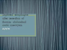
resection of thoraco abdominal aorta aneurysm 23910 Case presentation A 50 years old male was transferred from other hospital One day before referal he was admitted to that hospital because of severe ID: 393565
Download Presentation The PPT/PDF document "Ruptured esophagus after" is the property of its rightful owner. Permission is granted to download and print the materials on this web site for personal, non-commercial use only, and to display it on your personal computer provided you do not modify the materials and that you retain all copyright notices contained in the materials. By downloading content from our website, you accept the terms of this agreement.
Slide1
Ruptured esophagus after resection of thoraco abdominal aorta aneurysm
23/9/10Slide2
Case presentationA 50 years old male was transferred from other hospital.One day before
referal
, he was admitted to that hospital because of severe
epigastric
pain, the doctor suspected peptic perforation. Exploratory laparotomy was done and found retroperitoneal hematoma in upper part of
abdoment
. The wound was closed and the patient was transferred.Slide3
CT angiogram was done after surgery, it showed large thoraco abdominal aortic aneurysm – Crawford type 5
CAG was done, it was normal.Slide4
Chest X ray admission daySlide5
Left thoracotomy incision was made through 6th intercostal space. The aneurysm extended from mid thoracic to just above celiac artery. The maximal diameter was 10 cm.
Hematoma surrounded the aneurysm.
Left femoral vein was exposed but cannula could not be passed into right atrium. So, the aorta was cross clamped just above the aneurysm. It was opened, clots evacuated, and transected above celiac artery.
A 22 mm
dacron
graft was anastomosed to distal aorta first , and proximal anastomosis was completed. Aortic clamps were released. The operation and his post operative course was uneventful. Until post operative day 14, he complained of dysphagia and
vomitting
. He was febrile and chilled.Slide6Slide7
Chest CT scan showed presence of air in aneurysmal sac surrounding the graftEmergency left thoracotomy was done.A large amount of pus and food particles surrounding the graft.
Longituidinal
necrosis of lower esophagus about 2 cm long.Slide8Slide9Slide10
Esophagectomy was done with closure of cardia.
Debridement and excision of aneurysmal wall and cleansing of graft.
Open diaphragm and mobilize
omental
flap.
Cover the graft with
omental
flap.
Gastrostomy, feeding
jejunostomy
and cervical
esphagectomy
were done.Slide11
cultureAortic wall Streptococcal
fecalis
Pus
Steptococcus
fecalis
Yeast cell
lactobacilli.Slide12
CT scan after chest re thoracotomySlide13Slide14Slide15Slide16
Postoperative course after rethoracotomy
Clinically well, he became afebrile and
he tolerated
jejunostomy
feeding fairly.
Two weeks later reopened
midline laparotomy was done, while mobilizing the
cardia
, there was pus from the aneurysmal sac, the
omental
flap covered the graft well and no necrotic tissue, the space was cleaned and
irrgated
.
A
redivac
drain was left in the aneurysmal sac and the abdomen was closed
Post operatively he was well, tolerated
jejunostomy
feeding, two weeks later the
redivac
drain was removed and IV antibiotic continued for total two months.
He was discharged to the referring hospital.Slide17
CT after re laparotomySlide18Slide19
Near complete disappearance of perigraft fluid before esophageal bypassSlide20Slide21
2nd admission3 months after previous admission, he was well and wanted to drink and eat by mouth.
Repeated CT chest showed small about of fluid in the aneurysmal sac, much smaller than before.
Right thoracotomy was done and total thoracic
esophagectomy
was done
One week later, the
abdomen was
reopened, left side colon was mobilized preserving left colic vessel and the colon was pulled up via retrosternal space and anastomosed to cervical esophagus.
Cologastrostomy
,
colo
-colostomy were done.Slide22
There was superficial abdominal wound infection. His post operative course was uneventful otherwise.Oral diet was resumed on 8
th
post operative
day after contrast study showed no leakage no obstruction.Slide23
discussionEsophageal necrosis after surgery of descending thoracic aortic aneurysm is not rare.Mechanisms
ruptured aneurysm caused pressure on the esophagus
surgery excluded aortic branches to esophagus,
ischemic
necrosisSlide24
Prognosis is almost always fatal due to sepsis,
mediastinitis
prosthetic
graft infection
extra anatomical bypass – difficult or impossible
infection involving suture lines – aortic anastomotic dehiscence -> fatal hemorrhageSlide25
Omental graft bactericidal
blood supply and white blood cell to combat bacteria
cover the prosthetic graft is an important strategy to combat graft infection
filling the space surrounding the graft
prevent reinfection