/
Vaccine immunology Vaccine immunology

Vaccine immunology - PDF document

natalia-silvester
natalia-silvester . @natalia-silvester
Follow
444 views
Uploaded On 2015-08-11

Vaccine immunology - PPT Presentation

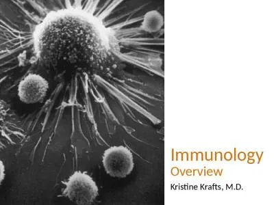
Section 1 leading to conditions such as allergy autoimmunity or even premature death are being raised The objective of this chapter 19 Vaccine immunology 2 Chapter 2 The conjugation of bacterial ID: 105395

Section 1: leading conditions such

Share:

Link:

Embed:

Download Presentation from below link

Download Pdf The PPT/PDF document "Vaccine immunology" is the property of its rightful owner. Permission is granted to download and print the materials on this web site for personal, non-commercial use only, and to display it on your personal computer provided you do not modify the materials and that you retain all copyright notices contained in the materials. By downloading content from our website, you accept the terms of this agreement.


Presentation Transcript

vulnerable populations, including very young, elderly, and immunosuppressed populations, also largely relies on a better understanding of what supports or limits vaccine ef!cacy under special circumstancesÑat the population and individ-ual levels. Lastly, the exponential development of new vaccines raises many questions that are not limited to the targeted diseases and the potential impacts of their prevention, but that address the speci!c and nonspeci!c impacts of such vaccines on the immune system and, thus, on health in general. These immune-related concerns have largely spread into the population, and questions related to the immunological safety of vaccinesÑthat is, their capacity for triggering 2 nonÐantigen-speci!c responses possibly leading to allergy, autoimmunity, or even premature deathÑare being raised. Certain Òoff-targets effectsÓ of vaccines have also been recog-nized and call for studies to quantify their impact and identify the mechanisms at play. The objective of this chapter is to extract from the complex and rapidly evolving !eld of immu-nology the main concepts that are useful to better address these important questions.HOW DO VACCINES MEDIATE PROTECTION?Vaccines protect by inducing effector mechanisms (cells or molecules) capable of rapidly controlling replicating patho-gens or inactivating their toxic components. Vaccine-induced immune effectors (Table 2.1) are essentially antibodiesÑproduced by B lymphocytesÑcapable of binding speci!cally to a toxin or a pathogen. A recently identi!ed critical subset of vaccine-induced CD4+ Th cells are follicular T-helper (Tfh) cells: they are spe-cially equipped and positioned in the lymph nodes to support potent B-cell activation and differentiation into antibody-secreting-cells4 and were identi!ed as directly controlling anti-body responses and mediating adjuvanticity.5Ð7 Another important subset are T-helper 17 (Th17) cells which essen-tially defend against extracellular bacteria that colonize the skin and mucosa, recruiting neutrophils and promoting local in"ammation.8,9 These effectors are controlled by regulatory T cells (Tregs) involved in maintaining immune tolerance.10 Most antigens and vaccines trigger B- and T-cell responses, such that there is no rationale in opposing vaccines favoring antibody production (Òhumoral immunityÓ) and T-cell responses (Òcellular immunityÓ). In addition, CD4+ T cells are required for most antibody responses, whereas antibodies exert signi!cant in"uences on T-cell responses to intracellular pathogens.11What Are the Main Effectors of Vaccine Responses?The nature of the vaccine exerts a direct in"uence on the type of immune effectors that are elicited and that mediate protec-tive ef!cacy (Table 2.2 Neutralizing viral replication (e.g., preventing viral binding and entry into cells)Ð Promoting Activating Indirectly Follicular Mycobacterium tuberculosis)Ð Th2 effector cells producing IL-4, IL-5, IL-13, and responding to extracellular pathogens (bacteria and helminths)Ð Th9 effector cells producing IL-9 and also responding to extracellular pathogensÐ Th17 effector cells producing IL-17, IL-22, and IL-26 and contributing to mucosal defense (Streptococcus pneumoniae, Bordetella pertussis, Mycobacterium tuberculosis)BOX 2.1 Main Immunological DeÞnitions (Continued)ISOTYPE SWITCHING PATTERN RECOGNITION RECEPTORSGermline-encoded receptors sensing the presence of infection via the recognition of conserved microbial pathogen-associated molecular patterns and triggering innate immune responses.REGULATORY T CELLST cells that on activation differentiate into cells that express speciÞc cytokines (IL-10, transforming growth factor [TGF]-"/surface markers) and act to suppress activation of the immune system through various mechanisms, maintaining immune homeostasis and tolerance to self-antigens.RESIDENT MEMORY T CELLSEffector memory T cells residing in speciÞc tissues (lungs, gut, skin) and conferring an immediate-early line of defense against viral and bacterial pathogens.SOMATIC HYPERMUTATIONA process that introduces random mutation in the variable region of the B-cell receptor (i.e., immunoglobulin) locus at an extremely high rate during B-cell proliferation. This mechanism occurs through the inßuence of the activation-induced cytidine deaminase enzyme and generates antibody diversiÞcation.T LYMPHOCYTESCells that originate in the thymus, mature in the periphery, become activated in the spleen/nodes if their T-cell receptors bind to an antigen presented by an MHC molecule and they receive additional costimulation signals driving them to acquire killing (mainly CD8+ T cells) or supporting (mainly CD4+ T cells) functions.T-INDEPENDENT B-CELL RESPONSESDifferentiation pathway of B cells, mainly elicited by polysaccharides, that takes place in the marginal zone and extrafollicular areas of the spleen/nodes. Its hallmarks are to be rapid (days), while eliciting the transient (months) production of antibodies of low afÞnity without inducing immune memory.T-DEPENDENT B-CELL RESPONSESDifferentiation pathway of B cells elicited by protein antigens that recruit T and B cells into GCs of the spleen/nodes. Its hallmarks are to be slow (weeks), while eliciting long-lasting (years) production of antibodies of high afÞnity and immune memory.TOLL-LIKE RECEPTORS vaccines but achieved by glycoconjugate vaccines, which may prevent nasopharyngeal colonization or nonbacteremic pneu-monia21 in addition to invasive diseases.Under most circumstances, inactivated vaccines do not elicit suf!ciently high and sustained antibody titers on mucosal surfaces to prevent local infection. It is only after having infected mucosal surfaces that pathogens encounter vaccine-induced IgG serum antibodies that neutralize viruses, opso-nize bacteria, activate the complement cascade (see Table 2.1 Vaccine TypeSerum IgGMucosal IgGMucosal IgA Table 2.2). Live oral or nasal vaccines, such as rotavirus, oral polio, nasal in"uenza, or cholera vaccines, induce serum IgA and secretory IgA, which also help limit viral shedding on mucosal surfaces.Under certain circumstances, however, passive antibody-mediated immunity is inef!cient (tuberculosis). There is con-clusive evidence that T cells are the main effectors of BCG, even though speci!c T-cell frequency and cytokine expression pro!les do not correlate with protection in BCG-immunized infants,15,22 or in zoster immunized adults.23,24 However, there is indirect evidence that vaccine-induced T cells contribute to the protection conferred by other vaccines. CD4+ T cells seem to support the persistence of protection against clinical pertus-sis in children primed in infancy, after vaccine-induced anti-bodies have waned, ! Through these pattern-recognition recep-tors, among which Toll-like receptors ful!ll an essential role (Table 2.3 these host cells sense the potential danger when they encounter a pathogen and become activated (Fig. 2.2). They modulate the expression of their surface molecules and produce proin"ammatory cytokines and chemokines,34Ð37 which result in the extravasation and attraction of monocytes, granulocytes, and natural killer cells and the generation of an in"ammatory microenvironment (see Fig. 2.1) in which monocytes differentiate into macrophages and immature DCs become activated.38 This activation modi!es the expression of Patrolling DCs are also numerous in well-vascularized muscles, which is the preferred route of injection for nonlive vaccines. They are fewer in adipose tissues, such that subcutaneous Demonstrated Ligands in Vaccines 5Ð7The development of this GC reaction requires a couple of weeks, such that hypermutated IgG antibodies to protein vaccine antigens ! The situ-ation is less clear-cut in humans, where IgG1 antibodies fre-quently predominate regardless of the polarization of T-cell help. The extrafollicular reaction is rapid, and IgM and low- gated to a protein carrier driving effective Tfh differentiation that PS-speci!c B cells are driven toward GC responses, receive optimal cognate help from carrier-speci!c Tfh cells, and differentiate into higher-af!nity antibody-producing cells, longer-lived plasma cells, and/or memory B cells. Protein anti-gens exhibit markedly distinct carrier propertiesÑregardless of their capacity to induce B- and Th-cell responses.77,78 That these differences may reect differences in Tfh induction is a likely hypothesis.79,80 The limited number of potent carrier proteins implies that an increasing number of conjugate vac-cines rely on the same carriers (e.g., CRM B cells capable of long-term survival and thus requiring later boosting. Optimal recall and anamnestic responses require longer intervals of at least 3 to 4 months, with longer intervals associated with generally greater responses (see below).Age at immunization also modulates vaccine antibody persistence, which is shorter at the two extremes of life (see sub-sequent text). Certain conditions may also limit the persistence of vaccine antibody responses because of enhanced catabo-lism (as in HIV)118 or the loss of antibodies in the urinary or digestive tract. The identi!cation of the mechanisms that support or limit the persistence of vaccine antibody responses represents a major challenge.What Are the Hallmarks of B-Cell Memory Responses? Memory B cells are generated during primary responses to T-dependent vaccines.50,119 They persist in the absence of anti-gens but do not produce antibodies (i.e., do not protect), unless reexposure to antigen drives their differentiation into antibody-producing plasma cells. This reactivation is rapid, such that booster responses are characterized by the rapid increase to higher titers of antibodies that have a higher af!n-ity for antigens than do antibodies generated during primary responses (Table 2.6).Memory B cells are generated in response to T-dependent antigens, during the GC reaction, in parallel to plasma cells (Fig. 2.5).50,119,120 At their exit of GCs, memory B cells acquire migration properties toward extrafollicular areas of the spleen and nodes. This migration occurs through the bloodstream, in which postimmunization memory B cells are transiently present on their way toward lymphoid organs.It is essential to understand that memory B cells do not produce antibodiesÑthat is, they do not protect. Their partici-pation in vaccine ef!cacy requires an antigen-driven reactiva-tion that may occur in response to endemic pathogens, to colonizing or cross-reacting microorganisms (Ònatural boostersÓ), or to booster immunization. The activation of memory B cells results in their rapid proliferation and differentiation into plasma cells that produce very large amounts of higher- immune system indeed resides in its highly polymorphic nature, enabling suf!cient immunological diversity to over-come a high number of diverse pathogens. This diversity impacts vaccine responses.93 Probing how host genetic markers may result in variations of vaccine-induced responses is expected to identify gene polymorphisms that predict the like-lihood of successful or adverse vaccine outcome, whereas epi-genetic studies may help reveal how environmental in"uences affect innate and adaptive immune responses.93 This work is still in its infancy, but holds great promise, especially when combined with novel systems vaccinology approaches.Immune competence obviously affects vaccine antibody responses, which are limited at the two extremes of life (see subsequent text), and by the presence of acute or chronic diseases, acute or chronic stress, and a variety of factors affect-ing innate and/or B- and T-cell immunity.Few nonlive vaccines (e.g., hepatitis A and human papil-lomavirus [HPV] vaccines) induce high and sustained anti-body responses after a single vaccine dose, even in healthy young adults. Primary immunization schedules therefore usually include at least two doses, optimally repeated at a minimal interval of 3 to 4 weeks (longer intervals enhancing rather than reducing the responses) to generate successive waves of primary B-cell and GC responses. These priming doses may occasionally be combined into a single ÒdoubleÓ dose, such as for hepatitis A or B and for HPV immunization.97Ð101 Fig. 2.3). This is illustrated by the accuracy of mathematical models predicting the kinetics of antiÐhepatitis B surface antigen (HBsAg),110 antiÐhepatitis A,111 or anti-HPV112,113 antibodies.A few determinants of the persistence of vaccine antibody responses (see Table 2.5) have been identi!ed. The nature of the vaccine has a crucial role: only live attenuated viral vac-cines or virus-like particles induce antibody responses that persist for several decades, if not lifelong, in absence of sub-sequent antigen exposure and reactivation of immune memory. In contrast, the shortest antibody responses are elicited by PS antigens, which fail to trigger Tfh/GC responses and thus do not elicit high-af!nity plasma cells capable of reaching the BM survival niches. Antibody persistence may also be modulated ¥ Differentiate higher postbooster anti-HBsAg responses are observed in people with high (e.g., $100 IU/L) rather than intermediate (10Ð99 IU/L) anti-HBsAg after their primary vaccination.127,128 This is likely to re"ect the induction of a larger pool of memory B cells.The dose of antigen is also an important determinant of memory B-cell responses (see Table 2.7). At priming, higher antigen doses generally favor the induction of plasma cells, whereas lower doses may preferentially drive the induction of immune memory.129 Closely spaced primary vaccine doses may be bene!cial for early postprimary antibody responses but not for postbooster antibody responses, as illustrated with meningococcal group C glycoconjugates.130 As a rule, acceler-ated schedules in which a 4- to 6-month window is not included between priming and boosting result in signi!cantly lower booster responses (see Table 2.7). At the time of boosting, a higher antigen content raises stronger booster responses, presumably by recruiting more memory B cells into the response. This is illustrated by higher antibody responses of children immunized with a higher-antigen-dose pertussis vaccine131 or primed with a glycoconjugate vaccine and boosted with a higher concentration PS (20Ð50 %g of PS) when com-pared with the glycoconjugate (1Ð3 %g of PS) vaccines. to generate signi!cantly higher antibody levels than primary immunization. Should this not be the case, the effective generation or persistence of memory B cells should be questioned.The reactivation, proliferation, and differentiation of ! The requirement for boosters to confer long-term vaccine pro-tection is also well illustrated for pertussis, for which boosters are required to extend protection beyond childhood.153 An interesting observation is that vaccine-induced memory per-sists following pertussis immunizationÑas illustrated by anamnestic responses to a booster doseÑbut is not suf!cient for protection. Yet the incubation period of pertussis exceeds receptors. Consequently, people with residual antibodies to a given antigen may show only a limited increase of their anti-body responses.The persistence of memory B cells is of utmost importance for long-term vaccine ef!cacy. Antigen persistence may extend for prolonged periods on the surface of FDCs (see Table 2.7) and contribute to the duration of immune memory. activated by local in"ammation, which provide the signals required for their migration to draining lymph nodes (see Fig. 2.1). During this migration, DCs mature and their surface expression of molecules changes. Simultaneously, antigens are processed into small fragments and displayed at the cell surface in the grooves of MHC (human leukocyte antigen [HLA] in humans) molecules. As a rule, MHC class I molecules present peptides from antigens that are produced in the cytosol of infected cells, whereas phagocytosed antigens are essentially displayed on MHC class II molecules.160Ð163 Thus, mature DCs reaching the T-cell zone of lymph nodes display MHCÐpeptide complexes and high levels of costimulation molecules at their surface.164 CD4+ T cells recognize antigenic peptides displayed by class II MHC molecules, whereas CD8+ T cells bind to class I MHC-peptide complexes (Fig. 2.6).165 Their recognition is restricted to short peptides (8Ð11 [CD8+] or 10Ð18 [CD4+] amino acids) displayed on speci!c MHC class I or II molecules, respectively. Antigen-speci!c T-cell receptors may bind only to speci!c MHC molecules 4 to 7 days. An interesting hypothesis is that as Bordetella per-tussis bacteria essentially remain on the mucosal surfaces, anti-gens may fail to efciently reach the vaccine-induced B and T cells residing in the lymph nodes. For example, the prompt reactivation of immune memory is not suf!cient to control polio viral replication in the digestive tract.154Live attenuated viral vaccines (measles, rubella) are consid-ered the prototype inducers of lifelong immunity, although prolonged immunity is also induced by certain nonlive vac-cines (hepatitis A, HPV, inactivated poliovirus vaccine, rabies). This derives in part from the induction of sustained antibody responses, which, however, tend to slowly decline in the absence of recurrent exposure,155 and might eventually result in a growing proportion of seronegative vaccinated young adults, including women of childbearing age. Whether the reactivation of immune memory will be suf!cient to curtail the replication process and confer protection against measles, rubella, or varicella, and whether adult booster doses may become needed after microbial control, are essential ques-tions. The resurgence of mumps outbreaks in fully vaccinated young adults may re"ect the induction of low numbers of memory B cells156 and demonstrates that secondary vaccine failure may occur even with live attenuated vaccines.157 The questions, which are central to sustained vaccine ef!cacy, are for DCs, to which they provide signals (CD40L, etc.) resulting in further activation, for B cells (see Fig. 2.2) and for CD8+ cytotoxic T cells (see Fig. 2.6 and Table 2.8). They are elicited by each vaccine type, except plain PS, which are not properly displayed by MHC molecules. Thus, the demonstration of postimmunization CD4+ T-cell responses does not imply a direct role in vaccine efcacy. CD4+ T-cell activation by DCs triggers their differentiation along distinct differentiation pathways.164,166 By default, DCs essentially trigger the induc-tion of Th2-type CD4+ T cells producing IL-4, IL-5, and IL-13, which are implicated in the defense against extracellular pathogens such as helminths.167 More potently activated DCs release IL-12p70, which induces the differentiation into Th1 cells that essentially produce IFN-! and tumor necrosis factor (TNF)-# and, thus, contribute to the elimination of intracel-lular pathogens directly (cytokine responses) and indirectly through macrophage activation and support to CD8+ T-cell differentiation (see Fig. 2.6).168 Th1 and Th2 cells support B-cell activation and differentiation during extrafollicular responses, whereas Tfh CD4+ cells provide critical help to GC B cells (see Fig. 2.3).169 Under certain conditions, activated TABLE 2.8 T-Cell Responses to VaccinesTypeMechanisms (Presumed)FunctionCD4+ T-helper cellsTh1IFN-! productionExtrafollicular B-cell helpTh1Cell contact, IFN-!Activation of CD8+ T cellsTh1/Th2Cell contact, CD40LDendritic cell activationTh2IL-4, IL-5, IL-13Extrafollicular B-cell helpTh2Cell contact, IL-4Suppression of CD8+ T cellsTh17IL-17, IL-21, IL-22Mucosal inßammationCD4+ follicular T-helper cellsTfh1IFN-!Germinal center B-cell helpTfh2IL-4, IL-5, IL-13Germinal center B-cell helpCD4+ regulatory T cellsMultiple mechanismsSuppression of CD4+/CD8+ responsesCD8+ T cellsIFN-!, TNF-#Killing of infected cellsMemory T cellsEffector memory T cellsTh1/Th2 cytokines, perforin, granzymeRapid secondary effectors responses in peripheryCentral memory T cellsIL-2, IL-10, CD40LDelayed activation/proliferation in lymph nodesTissue-resident memory T cellsTh1/Th2 cytokines, perforin, granzymeTissue localization enabling immediate-early reactivationIFN, interferon; IL, interleukin; Th, T-helper; TNF, tumor necrosis factor.DCs may also release IL-23, supporting the induction of in"ammatory Th17 cells by TGF-" and IL-6.Numerous factors in"uence the preferential differentiation of CD4+ T cells toward the Th1, Th2, Tfh, or Th17 pathway.170 The main determinant of CD4+ T-cell differentiation is the extent and type of DC activation by the innate system,164 although a recent observation suggests that polarized CD4+ T-cell responses may result from preferential expansion rather than priming. 190Antigen persistence essentially controls the proportion of Tcm and Tem memory cells (see Table 2.9): Tcm cells pre-dominate when antigen is rapidly cleared, whereas Tem/Trm cells become preponderant when antigen persists, such as in chronic infections.174,180,181 This is a challenge for novel non-replicating vaccines that should induce and maintain suf!cient Tem/Trm cells for immediate clearance in infected tissues. The long-term persistence of memory T cells is well established. this response is not associated with antibody responses. Vaccination with tetanus toxoid was found to expand speci!c and bystander memory T cells but !cult to study. Studies in which vaccines rou-tinely administered to human infants were administered at various stages of the postnatal maturation to infant mice indi-cated that the same limitations of antibody responses are seen in both humans and mice, re"ecting similar postnatal con-straints.236 These animal models showed that limitations of antibody responses in early life result from the limited and delayed induction of GCs in which antigen-speci!c B cells proliferate and differentiate. This was ! cines can have off-target effects. The epidemiological studies on this subject have been done mainly by a group working in Guinea-Bissau and their thesis is that live vaccines (including BCG, measles, and oral polio vaccine [OPV]) can reduce mor-tality caused by respiratory viral infections, whereas killed ! ; hepatitis B vaccine induces lower primary IFN-! responses and higher secondary Th2 responses in early life than in adults260; and tetanus-speci!c IFN-! CD4+ T-cell Aging affects the magnitude and the persistence of antibody responses to protein vaccines, as reby lower serum antibodies to in"uenza,290,291 For example, studies in aged mice have convincingly demonstrated the existence of age-related changes in FDCs.303,304 The limited ability of aged subjects to generate high-afnity antibody responses also re"ects changes in their antibody repertoire.304,305Age-associated changes in T-cell responses are re"ected by a progressive decline in na•ve T cells, re"ecting declining thymic output. This is associated with a marked accumulation of large CD8+ clones presumably resulting from prior infec-tions. These large T-cell clones (e.g., elicited in response to cytomegalovirus) have reached a state of replicative senes-cence, and homeostatic mechanisms negatively in blunted if anticarrier immunity is required for immunogenicity (e.g., for CRM197 conjugates) and maternal antibodies inter-fere with its induction.275 Maternal antibodies were reported as inhibiting cotton-rat B-cell responses by interaction with the inhibitory/regulatory Fc!RIIB receptor on antigen-speci!c B cells. The extent to which this mechanism accounts for the inhibition of human infant responses remains unde!ned.The inhibitory in"uence of maternal antibodies is depen-dent on the antibody titer and re"ects the ratio of maternal antibodies to vaccine antigen.90 This was elegantly demon-strated in a study in which Israeli infants were immunized with hepatitis A vaccine at 2, 4, and 6 months. Maternal antibodies usually allow a certain degree of priming (i.e., of induction of memory B cells) through yet unde!ned mechanisms. As a rule, the blunting of infant antibody responses by maternal antibodies disappears after boosting. Importantly, maternal antibodies do not exert their inhibitory in"uence on infant T-cell responses, which remain largely unaffected or even enhanced.283Ð285 This is best explained by the fate of maternal antibodyÐvaccine antigen complexes: immune complexes are taken up by macrophages and DCs, dissociate into their acidic phagolysosome compartment, and are processed into small peptides. These peptides S, Lopez S, Obermoser G, et al. Induction of Gavillet B, Eberhardt CS, Auderset F, et al. MF59 medi-ates its B cell adjuvanticity by promoting T follicular helper cells and thus germinal center responses in adult and early life. Pediatrics. 2001;108:E81.27. Ausiello CM, Lande R, Urbani F, et al. Cell-mediated immunity and antibody responses to Bordetella pertussis antigens in chil-dren with a history of pertussis infection and in recipients of an acellular pertussis vaccine. J Infect Dis. 2000;181:1989-1995.28. Ausiello CM, Lande R, Urbani F, et al. Cell-mediated immune responses in four-year-old children after primary immunization with acellular pertussis vaccines. Infect Immun Immune response to measles vaccine in 6 month old infants in Papua New Guinea. . 2015;33(suppl 2):B14-B20.37. Maisonneuve C, Bertholet S, Philpott DJ, De Gregorio E. Unleashing the potential of NOD- and Toll-like agonists as vaccine adjuvants. Proc Natl Acad Sci USA. 2014;111(34):12294-12299.38. Iwasaki A, Medzhitov R. Regulation of adaptive immunity by the innate immune system. Science. 2010;327:291-295. IC, Toellner KM, Cunningham AF, et al. Extrafollicu-lar antibody responses. Immunol Rev. 2003;194:8-18.54. Deenick EK, Hasbold J, Hodgkin PD. Decision criteria for resolv-ing isotype switching con"icts by B cells. Eur J Immunol. 2005;35:2949-2955.55. De Silva NS, Klein U. Dynamics of B cells in germinal centers. Nat Rev Immunol. 2015;15(3):137-148.56. Linterman MA, Vinuesa CG. T follicular helper cells during immunity and tolerance. Prog Mol Biol Transl Sci. 2010;92:207-248.57. Crotty S. Follicular helper CD4 T cells (T(FH)). Annu Rev Immunol. 2011;29:621-663.58. Rasheed MA1, Latner DR, Aubert RD, et al. Interleukin-21 is a critical cytokine for the generation of virus-speci!c long-lived plasma cells. J Virol Immaturity of PR, Galli G, Castellino F, et al. In"uenza vaccine immunology. Immunol Rev. 2011;239:167-177.87. Kracker S, Durandy A. Insights into the B cell speci!c process of immunoglobulin class switch recombination [published online ahead of print February 13, 2011]. Immunol Lett ometani K, Kurosaki T. Differentiation and maintenance of long-lived plasma cells. Curr Opin Immunol. 2015;33:64-69.105. Halliley JL, Tipton CM, Liesveld J, et al. Long-lived plasma cells are contained within the CD19(-)CD38(hi)CD138(+) subset in human bone marrow. Immunity. 2015;43(1):132-145.106. Winter O, Moser K, Mohr E, et al. Megakaryocytes constitute a functional component of a plasma cell niche in the bone marrow. Blood. 2010;116:1867-1875.107. Chu VT, Frohlich A, Steinhauser G, et al. Eosinophils are required for the maintenance of plasma cells in the bone marrow. Nat Immunol. 2011;12:151-159. D, Vaz AR, Miller E. Antibody avidity as a surrogate marker of successful priming by Haemophilus in!uenzae type b conjugate vaccines following infant immunization. J Infect Dis. 1998;177:1112-1115.127. Zanetti AR, Mariano A, Romano L, et al. Long-term immunoge-nicity of hepatitis B vaccination and policy for booster: an Italian multicentre study. Lancet. 2005;366:1379-1384.128. Duval B, Gilca V, Boulianne N, et al. Comparative long term immunogenicity of two recombinant hepatitis B vaccines and the effect of a booster dose given after !ve years in a low ende-micity country. Pediatr Infect Dis J. 2005;24:213-218. . 2004;364:365-367.142. Pichichero ME. Booster vaccinations: can immunologic memory outpace disease pathogenesis? Pediatrics. 2009;124:1633-1641.143. Young BW, Lee SS, Lim WL, et al. The long-term ef!cacy of plasma-derived hepatitis B vaccine in babies born to carrier mothers. J Viral Hepat. 2003;10:23-30.144. Lin YC, Chang MH, Ni YH, et al. Long-term immunogenicity and ef!cacy of universal hepatitis B virus vaccination in Taiwan. J Infect Dis. 2003;187:134-138.145. Whittle HC, Maine N, Pilkington J, et al. Long-term ef!cacy of continuing hepatitis B vaccination in infancy in two Gambian villages. Lancet. 1995;345:1089-1092.146. Makela PH, Kayhty H, Leino T, et al. Long-term persistence of immunity after immunisation with Haemophilus in!uenzae type b conjugate vaccine. Vaccine. 2003;22:287-292.147. Weinberg GA, Einhorn MS, Lenoir AA, et al. Immunologic priming to capsular polysaccharide in infants immunized with Haemophilus in!uenzae type b polysaccharideÐNeisseria meningiti-dis outer membrane protein conjugate vaccine. J Pediatr. 1987;111:22-27.148. McVernon J, Johnson PD, Pollard AJ, et al. Immunologic memory in Haemophilus in!uenzae type b conjugate vaccine failure. Arch Dis Child. 2003;88:379-383.149. Lee YC, Kelly DF, Yu LM, et al. Haemophilus in!uenzae type b vaccine failure in children is associated with inadequate production of high-quality antibody. Clin Infect Dis. 2008;46:186-192.150. Ramsay ME, McVernon J, Andrews NJ, et al. Estimating Hae-mophilus in type b vaccine effectiveness in England and Wales by use of the screening method. J Infect Dis. 2003;188:481-485.151. McVernon J, Andrews N, Slack MP, et al. Risk of vaccine failure after Haemophilus in type b (Hib) combination vaccines with acellular pertussis. Lancet. 2003;361:1521-1523.P, Borrow R, Miller E, et al. Meningococcal serogroup C conjugate vaccine is immunogenic in infancy and primes for memory. J Infect Dis. 1999;179:1569-1572.153. Lee GM, Lebaron C, Murphy TV, et al. Pertussis in adolescents and adults: should we vaccinate? Pediatrics. 2005;115:1675-1684.154. Abbink F, Buisman AM, Doornbos G, et al. Poliovirus-speci!c memory immunity in seronegative elderly people does not protect against virus excretion. J Infect Dis. 2005;191:990-999.155. Davidkin I, Peltola H, Leinikki P, etDuration of rubella immunity induced by two-dose measles, mumps and rubella (MMR) vaccination: a 15-year follow-up in Finland. Vaccine. 2000;18:3106-3112.156. Latner DR, McGrew M, Williams N, et T cell clones primed by pathogens or vaccines. Science. 2015;347(6220):400-406.172. Duthie MS, Windish HP, Fox CB, et al. Use of de!ned TLR ligands as adjuvants within human vaccines. Immunol Rev. 2011;239:178-196.173. Yewdell JW, Haeryfar SM. Understanding presentation of viral antigens to CD8 T cells in vivo: the key to rational vaccine design. Annu Rev Immunol. 2005;23:651-682.174. Robinson HL, Amara RR. T cell vaccines for microbial infections. Nat Med. 2005;11(4 suppl):S25-S32.175. Campbell DJ, Koch MA. Phenotypical and functional specializa-tion of FOXP3+ regulatory T cells. Nat Rev Immunol. 2011;11:119-130.176. Boer MC, Joosten SA, Ottenhoff TH. Regulatory T-Cells at the Interface between Human Host and Pathogens in Infectious Diseases and Vaccination. Front Immunol. 2015;6:217.177. Wing JB, Sakaguchi S. Foxp3+ T(reg) cells in humoral immunity. Int Immunol. 2014;26(2):61-69.178. Dannull J, Su Z, Rizzieri D, et al. Enhancement of vaccine-mediated antitumor immunity in cancer patients after depletion of regulatory T cells. J Clin Invest. 2005;115:3623-3633.179. Perret R, Sierro SR, Botelho NK, et al. Adjuvants that improve the ratio of antigen-speci!c effector to regulatory T cells enhance tumor immunity. P, Bansal A. Human immunode!ciency virus vaccines. Infect Dis Clin North Am. 2014;28(4):615-631.186. Zhou Y, Sullivan NJ. Immunology and evolvement of the adeno Curr Top Microbiol Immunol. 2005;293:89-114.189. Schenkel JM, Masopust D. Tissue-resident memory T cells. Immu-nity. 2014;41(6):886-897.190. Zens KD, Farber DL. Memory CD4 T cells in in"uenza. Curr Top cells but antibody production remains vaccine-speci!c. Blood. 2006;107:2806-2813.199. Huang SS, Platt R, Rifas-Shiman SL, et al. Post-PCV7 changes in colonizing pneumococcal serotypes in 16 Massachusetts com-munities, 2001 and 2004. [published correction appears in Pedi-atrics 117:593Ð594, 2006]. Pediatrics. 2005;116:e408-e413.200. Mayer S, Laumer M, Mackensen A, et al. Analysis of the immune response against tetanus toxoid: enumeration of speci!c T helper cells by the Elispot assay. Immunobiology. 2002;205:282-289.201. Wraith DC, Goldman M, Lambert PH. Vaccination and autoim-mune disease: what is the evidence? Lancet. 2003;362:1659-1666.202. Bacchetta R, Gregori S, Roncarolo MG. CD4 immune system? Pediatrics. 2002;109:124-129.209. Stowe J, Andrews N, Taylor B, et al. No evidence of an increase of bacterial and viral infections following measles, mumps and rubella vaccine. Vaccine. 2009;27:1422-1425.210. Aaby P, Jensen H, Samb B, et al. Differences in female-male mortality after high-titre measles vaccine and association with subsequent vaccination with diphtheria-tetanus-pertussis and inactivated poliovirus: reanalysis of West African studies. Lancet. 2003;361(9376):2183-2188.211. Aaby P, Martins CL, Garly ML, et al. Non-speci . 2013;31(30):3098-3103.218. Jensen KJ, Larsen N, Biering-Sorensen S, et al. Heterologous immunological effects of early BCG vaccination in low-birth-weight infants in Guinea-Bissau: a randomized-controlled trial. J Infect Dis. 2015;211(6):956-967.219. Kleinnijenhuis J, J Innate Immun. 2014;6(2):152-158.223. Kleinnijenhuis J, dose of Haemophilus in type B conjugate vaccine in early life. Arch Dis Child Fetal Neonatal Ed. 2004;89:F269-F271.239. Gans HA, Arvin AM, Galinus J, et al. De!ciency of the humoral immune response to measles vaccine in infants immunized at age 6 months. JAMA. 1998;280:527-532.240. Vazquez M, LaRussa PS, Gershon AA, et al. Effectiveness over time of varicella vaccine. JAMA. 2004;291:851-855.241. Siegrist CA. Mechanisms by which maternal antibodies in"uence infant vaccine responses: review of hypotheses and de!nition of main determinants. Vaccine. 2003;21:3406-3412.242. Jones C, Pollock L, Barnett SM, et al. The relationship between concentration of speci!c antibody at birth and subsequent response to primary immunization. Vaccine. 2014;32(8):996-1002.243. Amenyogbe N, Levy O, Kollmann TR. Systems vaccinology: a promise for the young and the poor. Philos Trans R Soc Lond B Biol Sci. 2015;370(1671), pii: 20140340. ME, Voloshen T, Zajac D, et al. Avidity maturation of antibody to Haemophilus in ) T-cell immunity. Front Immunol. 2014;5:393.259. Vekemans J, Ota MO, Wang EC, et al. T cell responses to vaccines in infants: defective IFNgamma production after oral polio vac-cination. Clin Exp Immunol. 2002;127:495-498.260. Ota MO, Vekemans J, Schlegel-Haueter SE, et al. Hepatitis B Infect Dis J nal antibody. J, Delassus S, Dyall R, et al. Clonal expansions of B lymphocytes in old mice. Regulation of aged humoral immune defense against pneumococcal bacteria by IgM memory B cell. J Immunol. 2005;175:3262-3267.299. Luscieti P, Hubschmid T, Cottier H, et al. Human lymph node morphology as a function of age and site. J Clin Pathol. 1980;33:454-461.300. Lottenbach KR, Mink CM, Barenkamp SJ, et al. Age-associated differences in immunoglobulin G1 (IgG1) and IgG2 subclass antibodies to pneumococcal polysaccharides following vaccina-tion. Infect Immun. 1999;67:4935-4938.301. Burns EA, Lum LG, Seigneuret MC, et al. Decreased specic anti-body synthesis in old adults: decreased potency of antigen-speci!c B cells with aging. Mech Ageing Dev. 1990;53:229-241.302. Linterman MA. How T follicular helper cells and the germinal centre response change with age. Immunol Cell Biol. 2014;92(1):72-79.303. Aydar Y, Balogh P, Tew JG, et al. Follicular dendritic cells in aging, a Òbottle-neckÓ in the humoral immune response. Ageing Res Rev. 2004;3:15-29.304. Zheng B, Han S, Takahashi Y, et al. Immunosenescence and germinal center reaction. Immunol Rev. 1997;160:63-77.305. Song H, Price PW, Cerny J. Age-related changes in antibody repertoire: contribution from T cells. Immunol Rev. 1997;160:55-62.306. Kang I, Hong MS, Nolasco H, et al. Age-associated change in the frequency of memory CD4+ T cells impairs long term CD4+ T cell responses to in"uenza vaccine. J Immunol. 2004;173:673-681.307. Kovaiou RD, Weiskirchner I, Keller M, et al. Age-related differ-ences in phenotype and function of CD4+ T cells are due to a phenotypic shift from naive to memory effector CD4+ T cells. Int Immunol