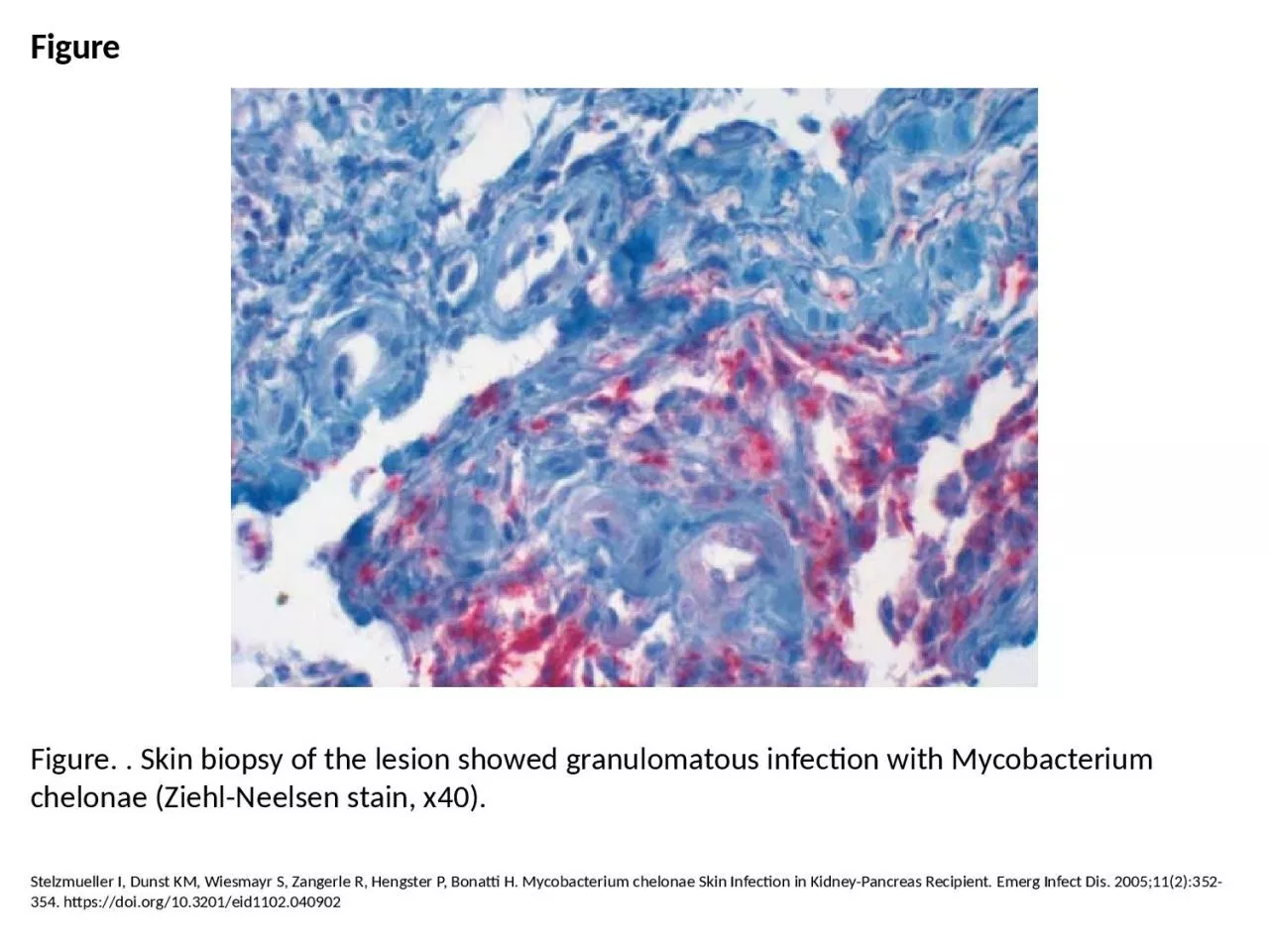
PPT-Figure Figure. . Skin biopsy of the lesion showed granulomatous infection with Mycobacterium

Author : natalie | Published Date : 2024-01-13

Stelzmueller I Dunst KM Wiesmayr S Zangerle R Hengster P Bonatti H Mycobacterium chelonae Skin Infection in KidneyPancreas Recipient Emerg Infect Dis 2005112352354

Presentation Embed Code

Download Presentation

Download Presentation The PPT/PDF document "Figure Figure. . Skin biopsy of the lesi..." is the property of its rightful owner. Permission is granted to download and print the materials on this website for personal, non-commercial use only, and to display it on your personal computer provided you do not modify the materials and that you retain all copyright notices contained in the materials. By downloading content from our website, you accept the terms of this agreement.

Figure Figure. . Skin biopsy of the lesion showed granulomatous infection with Mycobacterium: Transcript


Stelzmueller I Dunst KM Wiesmayr S Zangerle R Hengster P Bonatti H Mycobacterium chelonae Skin Infection in KidneyPancreas Recipient Emerg Infect Dis 2005112352354 httpsdoiorg103201eid1102040902. Kathleen O’Hanlon, M.D.. Professor, Family and Community Health. JCESOM/Marshall University. November 2014. Goals of this Presentation include discussion of the following:. Indications & contraindications of . Presenting Author: Dr. . Sariya. . Mohammadi. . J. 2. nd. year resident (. G. en Surgery). JSS Medical College, Mysore. Co- Author: Dr. . Raxith. S.R. Senior Resident. Dept. of Gen Surgery. JSS Medical College, Mysore. Follow-Up Melanoma. - lignancy usually mandate further surgical intervention. If the lesion is a thin melanoma (less than 0.75-mm thick) and the specimen was from an area of average thickness for t infec-A 42-year-old man presented with a history of pain-ment with short-course antibiotics (amoxicillin,fish for the past 2 years and cleaned it regularly himself.pea-size lesions were also expresse Coscolla M, Lewin A, Metzger S, Maetz-Rennsing K, Calvignac-Spencer S, Nitsche A, et al. Novel Mycobacterium tuberculosis Complex Isolate from a Wild Chimpanzee. Emerg Infect Dis. 2013;19(6):969-976. https://doi.org/10.3201/eid1906.121012. Niang M, Brouqui P, Raoult D. Epidemic Typhus Imported from Algeria. Emerg Infect Dis. 1999;5(5):716-718. https://doi.org/10.3201/eid0505.990515. Meissner EG, Bennett JE, Qvarnstrom Y, da Silva A, Chu EY, Tsokos M, et al. Disseminated Microsporidiosis in an Immunosuppressed Patient. Emerg Infect Dis. 2012;18(7):1155-1158. https://doi.org/10.3201/eid1807.120047. Riyesh T, Karuppusamy S, Bera BC, Barua S, Virmani N, Yadav S, et al. Laboratory-acquired Buffalopox Virus Infection, India. Emerg Infect Dis. 2014;20(2):324-326. https://doi.org/10.3201/eid2002.130358. Kay MK, Perti TR, Duchin JS. Tattoo-associated Mycobacterium haemophilum Skin Infection in Immunocompetent Adult, 2009. Emerg Infect Dis. 2011;17(9):1734-1736. https://doi.org/10.3201/eid1709.102011. Brown T, Nikolayevskyy V, Velji P, Drobniewski F. Associations between Mycobacterium tuberculosis Strains and Phenotypes. Emerg Infect Dis. 2010;16(2):272-280. https://doi.org/10.3201/eid1602.091032. Angelakis E, Edouard S, La Scola B, Raoult D. Bartonella henselae in Skin Biopsy Specimens of Patients with Cat-Scratch Disease. Emerg Infect Dis. 2010;16(12):1963-1965. https://doi.org/10.3201/eid1612.100647. Scrimgeour EM, Johnston WJ, Al Dhahry SH, El-Khatim HS, John V, Musa M. First Report of Q Fever in Oman. Emerg Infect Dis. 2000;6(1):74-79. https://doi.org/10.3201/eid0601.000114. Cassar O, Blondot M, Mohanna S, Jouvion G, Bravo F, Maco V, et al. Human Herpesvirus 8 Genotype E in Patients with Kaposi Sarcoma, Peru. Emerg Infect Dis. 2010;16(9):1459-1462. https://doi.org/10.3201/eid1609.100381. Ternavasio-de la Vega H, Ángel-Moreno A, Hernández-Cabrera M, Pisos-Álamo E, Bolaños-Rivero M, Carranza-Rodriguez C, et al. Skin and Soft Tissue Infections (Patera Foot) in Immigrants, Spain. Emerg Infect Dis. 2009;15(4):598-600. https://doi.org/10.3201/eid1504.081457.

Download Document

Here is the link to download the presentation.
"Figure Figure. . Skin biopsy of the lesion showed granulomatous infection with Mycobacterium"The content belongs to its owner. You may download and print it for personal use, without modification, and keep all copyright notices. By downloading, you agree to these terms.

Related Documents