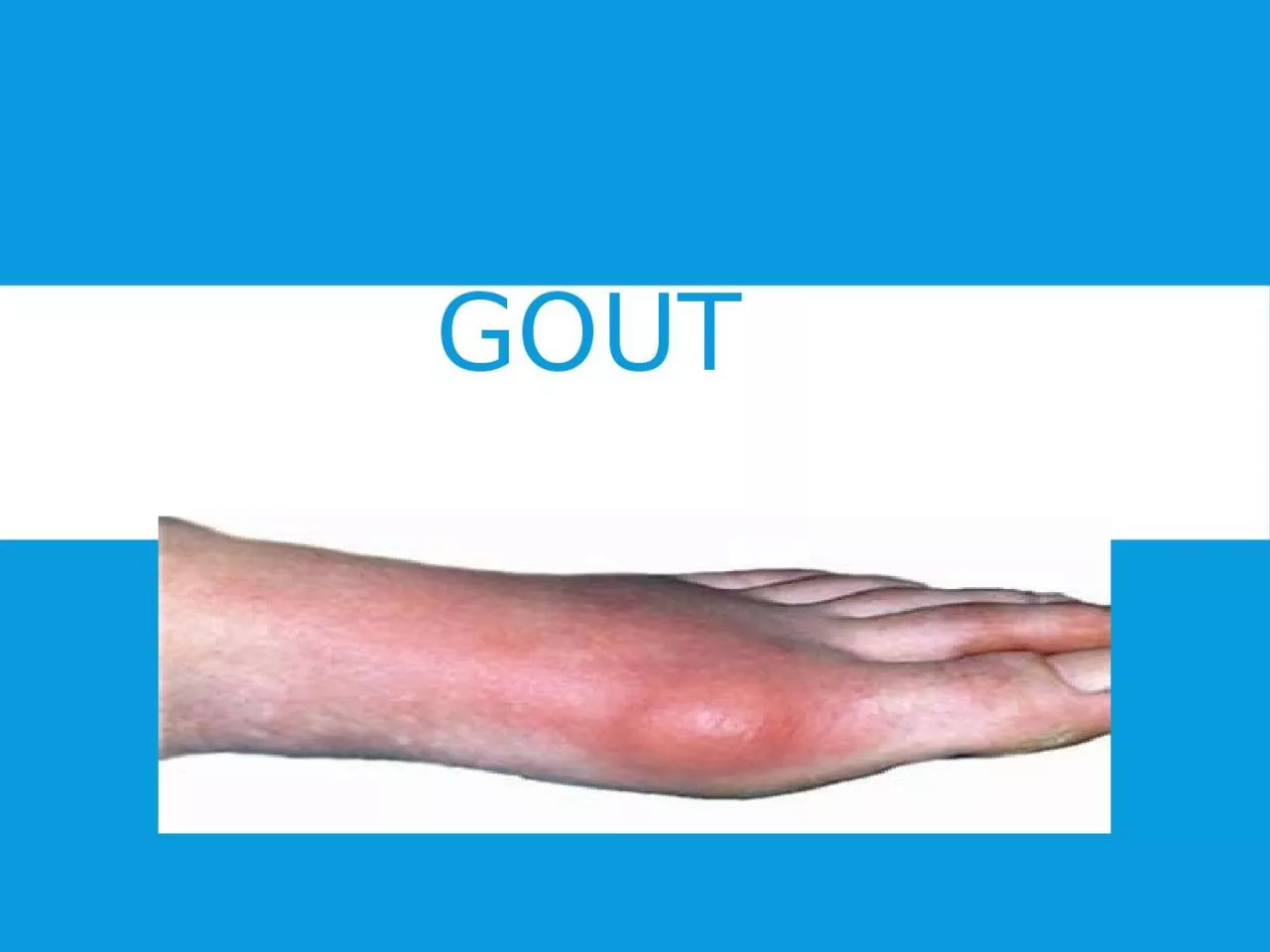
purine bodies in blood pre gout Whereas the kidney is unable to separate this combination of uric acid and purine bodies Then the uric acid salts accumulate in the blood PTO ID: 926945
Download Presentation The PPT/PDF document "Gout PATHOLOGY excessive concentration..." is the property of its rightful owner. Permission is granted to download and print the materials on this web site for personal, non-commercial use only, and to display it on your personal computer provided you do not modify the materials and that you retain all copyright notices contained in the materials. By downloading content from our website, you accept the terms of this agreement.
Slide1
Gout
Slide2PATHOLOGY
excessive concentrations of uric acid and some purine
bodies in blood pre gout.
Whereas, the kidney is unable to separate this combination of uric acid and
purine
bodies.
Then the uric acid salts accumulate in the blood.
PTO..
Slide3PATHOLOGY
Then, the crystal deposits in joints, tendons and surrounding tissue , resulting in an attack of gout.
Gout may be confirmed by the presence of crystals in the joint fluid .
Blood uric acid level may be normal during an attack.
PTO..
Slide4CLINICAL FEATURES
The disease is commonly seen in the first metatarsophalangeal
joint and
metacarpophalangeal
joints .
It can occur over the age between 30 -40 years.
The deposition of
biurate
of soda occur around the affected joints ,
The joint changes include
subchondral
cysts ,
osteophytes
formation and in the later stages , reduction in the joint space.
Slide5Sign and symptoms :-
Symptoms can include joint pain, gout pain is often intense , inflammation and redness .
The effected joint will often becomes
red,swallen,and
tender to touch.
During the acute phase, the involved joint has signs and symptoms of acute inflammation with a hard and tender nodule known as ‘ chalk stones’ or ‘
tophi
’.
Fever often accompanies
excrucitating
night
paiin
, but the involved joint is
comparitively
by
painfree
during day.
Slide6FOOT
Slide7CLINICAL FEATURES
Fever often accompanies excruciating night pain , but the involved joint is comparatively painfree
during the day.
There will be decreased mobility as gout progresses , it may limit the Range of motion .
If in case , gout occurs before the age of 30 there can be a genetic metabolism disorder ,
( this condition is rare ).
Slide8Pathophysiology :-
Gout is caused by disorders of
purine
metabolism resulting in
elivated
levels of
puric
acid
->7mg /dl in men
->6mg /dl in women
Prolonged
hyperuricemia
leads to formation of monosodium
urate
monohydrate crystals, the joint at the base of the big toe is affected in about half of the cases , it may also result in ‘
tophi
’.
It can also effect the kidney.
Slide9Predisposing factor of gout :-
1) gender –male > female ,
2) family history ,
3) Previous attack ,
4) obesity,
5)
alchohal
6) diet
Purine
rich
7)
dieuritics
8)renal insufficiency ,
9) rich diet ,
10) hypertension ,
11) diabetic conditions.
Slide10Differential diagnosis :-
X-ray , which is generally normal ,( erosion is seen in chronic stage ),
Joint aspiration –polarising light microscopy of crystals ,
A synovial fluid , gram stain and culture may be performed.
Slide11X-RAY
Slide12Complications of gout:-
Gout ‘ tophi
’
prsenting
as nodules on the finger and helix of the ear ,
tophi
on the toe and ankle , tophus on the knee.
Gout complicated by rupture of
tophi
, the
exudate
of which tested
positiive
for uric acid crystals.
Slide13COMPLICATIONS
Gout can present in multiple ways , although the most common in a recurrent attack of acute inflammatory arthritis ( a red tender hot , swollen joints ).
The metatarsal
phalangeal
joints at the base of big toe is affected most often , accounting for half of the cases , other joints such as the heels, knees, wrists , and fingers, may also be effected .
Joint pain usually begins during night and peaks within 24 hours of onset.
Slide14JOINTS AFFECTED
Slide15Management of gout :-
There are three main goals of the medical management of gout –
1) terminate acute attack
2) prevent re-
occurance
3) correct , prevent
furthur
damage from
hyperuricemia
.
Slide16Chronic Tophaceous gout :-
If gout is untreated,
Other joint involvement,
Formation of
tophi
-collection of crystals in soft tissues,
Bone erosions at joint –” punched-out”.
Erosion
on x-ray.
Slide17CHRONIC TOPHACEOUS GOUT
Slide18CHRONIC TOPHACEOUS GOUT
Slide19Pseudogout
Pseudogout
is usually present in the older age group and affects knee and wrist joints.
It can be
polyarticular
with an evidence of calcification of the cartilage (
chondro-calcinosis
).
There is a deposition of calcium pyrophosphate crystals .
Slide20HAND
Slide21FOOT
Slide22Treatment :-
1) conservative treatment ,
2) physiotherapy treatment-
Conservative treatment :-1) NSAIDS,
2)
colchicine
,( along with analgesics) ,
probencid
,
3)
glucocorticoids
,
4)
indomethacin
75-200 mg.
Lithium ionization is sometimes done in between the acute attacks.
This
iontophoresis
forms soluble lithium
urate
in place of insoluble sodium
urate
.
Slide23Physiotherapy treatment :-
1) ultrasound treatment ,( to reduce inflammation and pain ).
2) icing ( to calm the joint ) ..
Cryotherapy
in the form of crushed ice packs .
3) strengthening to the muscles,
4)
proprioception
exercises – which assists in maintaining the joint sense of position .
5) stretching exercises.
Slide24EXERCISES
Slide25T/T
Treatment plan will be the same in gout and psudogout
.
AIM of treatment :- to give relaxation , to improve the range of motion , to reduce the level of pain and inflammation .
Patient should be
adviced
to bring changes in
sedantry
lifestyle ( like in weight reduction ).
Thank you.