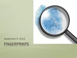
/
  Development of analytical fingerprints   Development of analytical fingerprints

  Development of analytical fingerprints - PowerPoint Presentation

newson
newson . @newson
Follow
345 views
Uploaded On 2020-08-27

  Development of analytical fingerprints - PPT Presentation

for the quality control of snake venoms Max Mousseron Institute University of Montpellier France Université FHB in Abidjan Ivory Coast 1 March 1415 2016 London UK André ID: 804683

naja batch analytical lachesis batch naja lachesis analytical muta pdadmac snake venoms fingerprint venom sodium capillary analysis conditions bge

Share:

Link:

Embed:

Download Presentation from below link

Download The PPT/PDF document "  Development of analytical fingerprint..." is the property of its rightful owner. Permission is granted to download and print the materials on this web site for personal, non-commercial use only, and to display it on your personal computer provided you do not modify the materials and that you retain all copyright notices contained in the materials. By downloading content from our website, you accept the terms of this agreement.
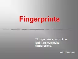

Presentation Transcript

Slide1


Development of analytical fingerprints

for the quality control of snake venoms

Max Mousseron Institute, University of Montpellier, France Université FHB in Abidjan, Ivory Coast

1

March 14-15, 2016 London, UK

André

Sawa

Kpaïbe

,

Gaëlle

Coussot

, Yoann Ladner, Michèle C.

Aké

, Catherine Perrin

Slide2

2

WHY SNAKE VENOMS?

Slide3

3

Natural active substances : 30-40 % new drugs

(LEEM) Raw materials : mineral / microbiological /plant / animal

AnimalplantInsulin

Bee venom

Conus

Snake

venom

MorphineQuinine

Artemisine

Ginkgo biloba

Snake

venoms

:

new drugs

to

treat diseases

microbiological

mineral

Bacteria YeastSodium bicarbonate zinc chloride

Insulin

Morphine

Artemisine

Sodium bicarbonate

Slide4

4

SNAKE VENOMS

Slide5

Cardiovascular

effects

captopril isolated from

Bothrops

jacaraca

Full

strain

mimetic

Gynecological

disorders

Behavioural

disorders

Homoeopathy

pills

of Lachesis mutaVENOMS5

raw

substance, extracted

and/or modified compound

Complex media

Single molecule

Slide6

Lachesis

muta6 Complex

molecules: peptides and proteins (90% weight) + mineral / carbohydrates / lipids…

-

Toxins =>

Poisoning

- Enzymes (≤

25 identified)

Intragender

variability

protein

composition

of 3

species

of

Lachesis

genderLachesis melanocephala

Lachesis

acrochorda

ENZYMES

(20-150

KDa)VAP:

vasoactive peptide

SVMP:

serine venom metalloproteinase

CTL: C-type

lectin-like protein

svVEGF: snake venom

vascular endothelial growth

factorSP: serine proteinase

LAO: L

amino acid oxydase

PLA2: phospholypase A2

CRISP: cysteine-rich secretory

protein

COMPOSITION

Slide7

7

Genetic composition +++Environment Very important variability

DRUG EFFICACITY

Quality Control

of raw material ???

Snake

diet

Geographic localisation

VARIABILITY

Slide8

8

?

Fingerprint

Biological

Variability

Analytical

methods

QUALITY CONTROL ???

Slide9

9

QUALITY CONTROL

SDS-PAGE :

poor resolution with lack of reproducibility (sample preparation artifacts, migration time and staining variability

), do not take into account biological variability

Slide10

10

ANALYTICAL FINGERPRINTING

Slide11

11

ANALYTICAL FINGERPRINT VENOMS STRAINS

Within batches-Inter batches comparison

BATCH 1

BATCH 2

BATCH 3

Slide12

12

BATCH 1

BATCH 2BATCH 3Inter batches comparison of the same

species chemometric treatment

To obtain

the specific fingerprint

ANALYTICAL FINGERPRINT VENOMS STRAINS

Slide13

13

Specific

analytical fingerprint of the same speciesRoutine => reference for the CQ of that

snake venom

ANALYTICAL FINGERPRINT VENOMS STRAINS

Slide14

14

Specific

fingerprints using

Capillary Electrophoresis for the control quality of

snake

venoms

Slide15

15

Naja

naja (ELAPIDEAE)

Lachesis muta (VIPERIDEAE)

Reptiles

S

quamates

Scolecophidia

Class

Order

S

erpentes

Suborder

Henophidia

Caenophidia

Colubridae

Atractaspididae

Elapidae

Viperidae

Infra-

order

Familly

Boidae

Aniliidiae

Uropeitidae

Xenopeltidae

Acrochordidaedae

Leptotyphlopidae

Typhlopidae

Anomalepidididae

STUDIED STRAINS

Slide16

4 mg

10 g/L

4°C, 11 500 g, 10 min

400µL

10g/L, 50µL

- 20°C

5g/L , 100µL

CE

analysis

Freeze

-

dried

product

from

raw

material

16

European

Pharmacopeia

/

U.S

Pharmacopeia

SAMPLE PREPARATION

Slide17

Neutral

Cationic

PEOPDADMAC

17Neutral: Poly(ethylene

oxyde) [PEO], Hydroxypropyl

Cellulose [HPC]

EOF

is supposed

to be zero

,

proteins migration in the capillary is only related to their effective mobility

Cationic

:

Poly(

diallyldimethylammonium

chloride

) [PDADMAC]

EOF

is directed to the anode, its mobility is opposite to protein mobility;its large amplitude forces the proteins to migrate to the anodeCoatings: chimical modification of capillary surface to prevent

protein adsorption

Nehmé

R., Perrin C., Cottet H. et al

, Electrophoresis 2008, 29, 3013- 3023.

CE ANALYSIS :

coating

the

capillary surface

Slide18

Comparison of

Lachesis

muta venom (same batch) electropherograms obtained with coated capillary (PEO, HPC

and PDADMAC )PDADMAC coating is well

adapted to

venom analysis

µ

eff

(cm

2

V

-1

S

-1

)

Abs(UA)

PDADMAC

HPC

PEO

CE conditions

: BGE sodium phosphate (pH2; I: 50 mM)  E : 212 V/cm  ; hydrodynamic injection: 6,1 nL (5 g/L);  = 214

nm

CE ANALYSIS :

coating

the

capillary

surface

Slide19

pH

selection

of electrolyte separation (efficacity, mobility)

2.0 / 3.0 / 4.0 / 7.0 / 9.2co-ion selection of electrolyte

separation

(efficacity

)

- sodium -

triethanolamine (trolamine

)

-

-

amino

caproic

acid

-

bis-triscounter-ion selection of electrolyte separation (efficacity, mobility) -Phosphate

-Citrate

19

CE ANALYSIS : Background Electrolyte (BGE)

Slide20

Capillary

effective Length : 40cm Temperature : 35°C

ionic strength : 30 mM

Electric field

: 212V/cm

Detection: 214 nm

Inject sample

: 7.81nl

BGE: sodium phosphate, pH 2.0

Coating

: PDADMAC, pH 8.0

20

Electrophoretic

parameters

selection (capillary length, temperature, ionic strength… .)3 Batches of Lachesis muta 30

analyzes per batch

CE ANALYSIS : optimal conditions

Slide21

CHEMOMETRIC TREATMENT

21

Collaboration

with:Dr Christelle Reynes and Pr Robert Sabatier

Slide22

Analytical

conditions

: BGE phosphate de sodium pH 2.0

I: 30 mM /

Coating

PDADMAC /

Leff

50 cm /

T

35°C /

E

212V/cm /

214nm/

injection

7, 81 nl Lachesis muta Batch 1Lachesis muta Batch 3Lachesis muta Batch 2

FEO

CHARACTERISTIC

PEAKS AND

RSD (%)

PEAKS

P

1

P

2

P

3

P

4

P

5

P

6

P

7

P

8

P

9

RSD(%)

0.30

0.24

0.27

0.31

0.31

0.36

0.49

0.43

0.04

ELECTROPHORETIC PROFILES IN CZE –

Lachesis

muta

Slide23

DATA PRE-PROCESSING

23

Batch 1

Batch 2Batch 3Raw data (per batch) – correcting

baselines

Slide24

Al

i

gnment of batch signals

:24Batch 1

Batch 2

Batch 3

DATA PRE-PROCESSING

Slide25

Signals

alignment – Recognition of common peaks 25

Specific part ?

variable part ?

Batch 1

Batch 2

Batch 3

EXTRACTION AND PEAKS ALIGNMENT

Slide26

Individual

extraction of signals peaks

26EXTRACTION AND PEAKS ALIGNMENT

Slide27

Emp

reint

e « Lachesis »

27SPECIFIC PART OF FINGERPRINT

Slide28

Fingerprint

« Lachesis muta »

28To apply to the quality control of Lachesis muta venom

Analyses of other venoms

USING THE SPECIFIC PART OF THE FINGERPRINT

Slide29

Analytical

conditions

: BGE phosphate de sodium pH 2.0

I: 30 mM /

Coating

PDADMAC /

Leff

50 cm /

T

35°C /

E

212V/cm /

214nm/

injection

7, 81nl Batch 1Batch 2

ELECTROPHORETIC PROFILES IN CZE –

Naja

naja

Slide30

30

No

enough peak resolution

Improved analytical conditions Sample

Preparation

FEO

Analytical

conditions

:

BGE

phosphate de sodium pH 2.0

I

:

30

mM

/

Coating

PDADMAC / Leff

5

0 cm / T

35°C /

E 212V/cm /

214nm/ injection

7, 81nl

ELECTROPHORETIC PROFILES IN CZE –

Naja

naja

Slide31

31

SAMPLE PREPARATION –

Naja naja

5g/L CE analysis

Fragments

(Digest)

Proteolysis

step

Incubation time

Proteolysis

buffer

Temperature

Denaturation

step

Slide32

32

Incubation

30min Incubation 3h

Incubation 12hBefore incubation

FEO

FEO

Conditions d’analyse:

BGE

phosphate de sodium pH 2.0 I

:

30

mM

/

Recouvrement

PDADMAC /

Leff

50 cm /

T

35°C / E 212V/cm /

214nm/

injection 7, 81nl

ELECTROPHORETIC PROFILES IN CZE –

Digest of Naja naja

Slide33

33

Before

incubationAfter incubation

FEO

ELECTROPHORETIC PROFILES IN CZE –

Digest of Naja naja

Chemometric

treatment => many

batches

=>

Integrated

in-line

reactor

for

proteolysis

step

=> D-PES methodology

Slide34

34

ACKNOWLEDGMENTS

André Sawa Kpaibe

Dr Yoann LadnerDr Silvia MasDr Marie-Dominique BlanchinPr Catherine PerrinPr Michèle C. Aké