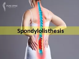
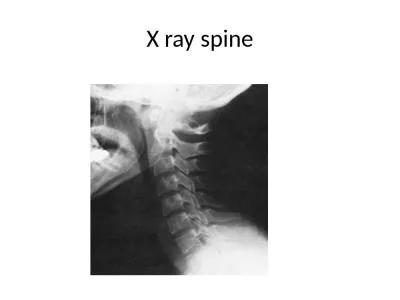
Waleed Awwad MD FRCSC Assistant professor consultant spine surgeon History Spondylolisthesis Displacement of one vertebra in relation to another vertebra below Incidence Affects 57 of US population ID: 914516
Download Presentation The PPT/PDF document "Spine m ade easy Spondylolisthesis" is the property of its rightful owner. Permission is granted to download and print the materials on this web site for personal, non-commercial use only, and to display it on your personal computer provided you do not modify the materials and that you retain all copyright notices contained in the materials. By downloading content from our website, you accept the terms of this agreement.
Slide1
Spine made easy
Spondylolisthesis
Waleed
Awwad
.
MD, FRCSC
Assistant professor consultant spine surgeon
History
Slide3Spondylolisthesis
Displacement of one vertebra in relation to another vertebra belowIncidence:Affects 5-7% of US population85% at L510% at L4
Natural history:Progression observed in childrenAdults, 8-30% present after 4th decade
Slide4Spondylolysis
Spondylolisthesis
Slide5ClassificationEtiology:Wiltse classification
Marchetti-Bartolozzi classificationSeverity:Meyerding classification
Slide6Marchetti-Barhetti-Bartolozzi classificationDevelopmental
High dysplastic with lysis or with elongationLow dysplastic with lysis or with elongationAcquiredTraumatic (due to acute or stress fracture)
After surgery (caused by direct or indirect surgery)Pathological (due to local or systemic pathology)Degenerative (found in primary or secondary degenerative conditions)
Slide7Wiltse classification
Slide8Wiltse classificationType I dysplasia (congenital).
Slide9Dysplastic changesProximal sacral rounding
Trapezoidal L5Vertical sacrumJunctional kyphosisCompensatory hyper-lordosis
Slide10Wiltse classificationType II isthmic.
Slide11Wiltse classificationType III degenerative.
Slide12Wiltse classificationType IV post traumatic.
Slide13Wiltse classificationType V pathological.
Slide14Wiltse classificationType VI iatrogenic
Slide15Meyerding grading system
Slide16Assessment X-rays:Anteroposterior
LateralOblique views
Slide17Assessment X-rays:Anteroposterior
Lateral
Slide18Assessment
X-rays:AnteroposteriorLateral
Oblique views
Pedicle “
eye
”
Ascending process
(ears)
Transverse process
(nose)
Spondylolysis
(collar)
Descending process
(legs)
Slide19Assessment X-rays:Anteroposterior
LateralOblique viewsCT scan
Slide20Assessment X-rays:Anteroposterior
LateralOblique viewsCT scanBone scan
Slide21Assessment X-rays:Anteroposterior
LateralOblique viewsCT scanBone scanMRI
Slide22“Coronal”
“Sagittal”
Spinal canal stenosis: facet joint orientation
Slide23Spinal canal stenosis: facet joint orientation
Facet orientation > 45 degrees is 25 times more likely to develop degenerative spondylolisthesis most commonly at L4/5
Women: Men
= 5:1
African-American women > Caucasian women
Slide24Buttress
Sagittal orientation of facet joints
Spinal canal stenosis: facet joint orientation
Slide25Buttress
Decompression and removal of this buttress can create instability
when load is applied
Spinal canal stenosis: facet joint orientation
Slide26Spinal canal stenosis: facet joint orientation
Coronal orientation of the facet joints enables decompression of neural
elements without creating instability
Slide27Spinal canal stenosis: synovial cysts
Indicates presence of significant joint and synovial pathology
Need to excise
synovium
or immobilize the segment in order to prevent recurrence
Slide28Spinal canal stenosis: facet joint fluid or gas
Fluid or gas in the facet joint of a patient indicates the presence of instability
Fluid
Gas
Slide29Standing
Spinal canal stenosis: facet joint fluid or gas
Supine
Slide30Spinal canal stenosis: intervertebral disc height
Slide31Spinal canal stenosis: intervertebral disc height
Slide32Undercutting
facetectomy
Spinal canal stenosis: intervertebral disc height
Slide33Expect loss of disc height over time
Spinal canal stenosis: intervertebral disc height
Slide34Adequate decompression
initially
Recurrence of foraminal
compromise over time
Spinal canal stenosis: intervertebral disc height
Slide35Spinal canal stenosis: intervertebral disc height
Slide36Assessment—
Meyerding Classification
Slide37Standard method of measurement
Assessment—slip angle
Method used when inferior end plate of L5 is irregular
Slide38MeasurementSlip angle
Slide39MeasurementSlip angleNormally Negative or 0
Slide40MeasurementSacral inclinationNormally > 30 degrees
Slide41MeasurementPelvic incidencePI =
PT + SSMean children 47 degreesMean adults 57 degreesLow PI loss of lordosisHigh exaggerated lordosis
Slide42High PT Low SS
Low PT High SS
Slide43Sagittal alignmentStance
GaitHead over pelvisHips and knees
Slide44Risk factors for progressionYoung age (progression is rare after 20 years)
FemaleLigamentous laxicity> 50% slippage> 10% slip angleL5 - S1 instability
Trapeaoidal L5Dome- shaped upper sacrumLess likely to progress with decreased disk space and an Anterior sacral lip
Slide45TreatmentMajority can be managed nonoperatively:
NSAIDSPhysical therapyPars interarticularis
injectionFacet injections above defect (communicates with defect)
Nerve root blocks for root symptoms
Slide46Indications for Surgery:Persistant back pain which interfere with activities of daily living
Symptomatic with failed conservative treatmentSignificant progressionGrade III or higher with >55 degrees slip angleNeurologic deficitsAcute traumaticTreatment
Slide47TreatmentSurgery may be indicated to treat persistent radiculopathy and/or back pain when origin of pain is localized to the spondylolisthesis level
PLF results in good outcome if fusion in situ performedWhere reduction is undertaken, 360o fusion preferred
Slide48Surgical optionsFusion in situ
Fusion in situ and decompressionDecompression and reductionPosterior lateral fusion and anterior column support
Slide49Gill laminectomyNerve root decompressionRemoval of lamina of the affected level
May lead to increased instabilityRadicular symptoms may persist unless decompression is accompanied by fusion to stabilize the segment in order to prevent ongoing neural irritation
Slide50High gradeGaines resection
Sacral osteotomyFibular strut - BohlmanIncreased risk of neurological compromise (L5 nerve root) with attempted reduction
Slide51Take-home messagesThe majority of patients with a spondylolysis or listhesis
are asymptomaticInitial treatment should focus on activity modification and core stabilizationSurgery indicated for patients with:
Failure of nonoperative treatmentSignificant or progressive deformity
Neurological compromiseThe aim of surgery is to:
Decompress neural elements and preserve functionReduce lumbosacral kyphus
Achieve fusion
Slide52Slide53Slide54Slide55Slide56Slide57Slide58