PPT-MAGNETIC RESONANCE (MR) SAFETY
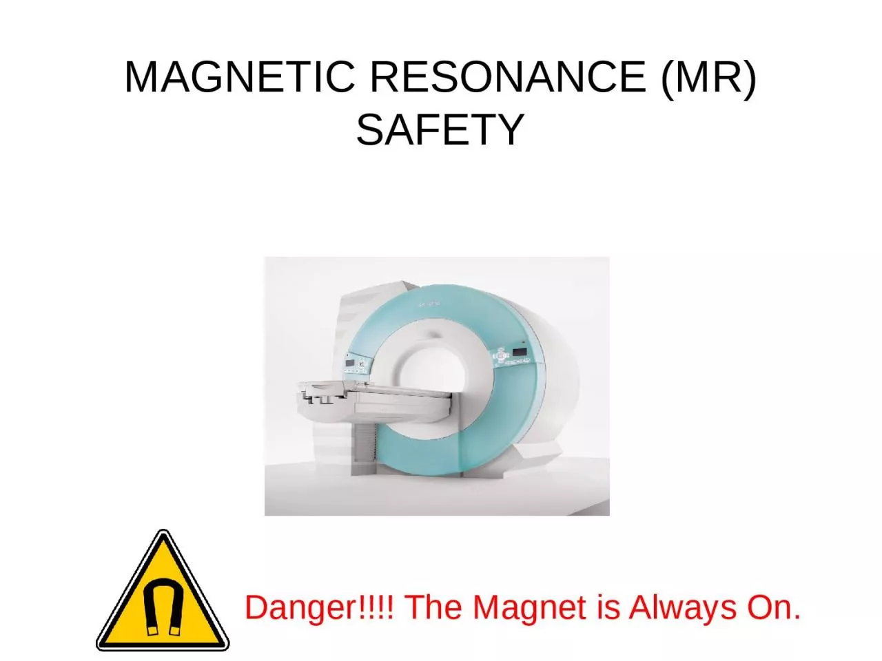
Danger The Magnet is Always On Who Should Take This Course Individuals who frequent the MR area such as those who transport patients those who clean the area support
Download Presentation
"MAGNETIC RESONANCE (MR) SAFETY" is the property of its rightful owner. Permission is granted to download and print materials on this website for personal, non-commercial use only, provided you retain all copyright notices. By downloading content from our website, you accept the terms of this agreement.
Presentation Transcript
Transcript not available.