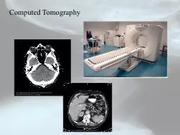
PDF-Magnetic Resonance Imaging MRI and Computed Tomography CT Scan
Author : layla | Published Date : 2022-08-26
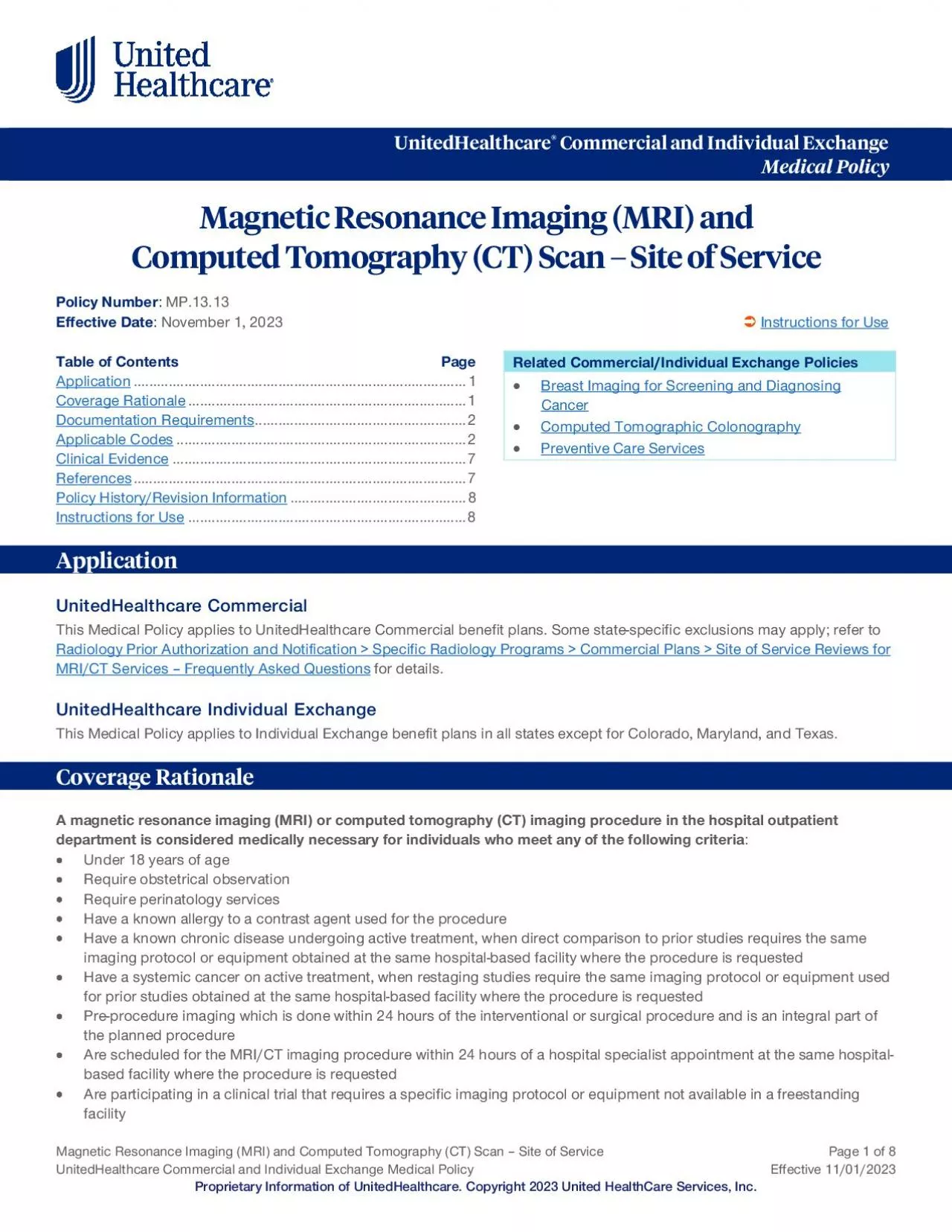
150 Site of Service Page 1 of 8 UnitedHealthcare Commercial Utilization Review Guideline Effective 04012022 Proprietary Information of UnitedHealthcare Copyright
Presentation Embed Code
Download Presentation
Download Presentation The PPT/PDF document "Magnetic Resonance Imaging MRI and Compu..." is the property of its rightful owner. Permission is granted to download and print the materials on this website for personal, non-commercial use only, and to display it on your personal computer provided you do not modify the materials and that you retain all copyright notices contained in the materials. By downloading content from our website, you accept the terms of this agreement.
Magnetic Resonance Imaging MRI and Computed Tomography CT Scan: Transcript
Download Rules Of Document
"Magnetic Resonance Imaging MRI and Computed Tomography CT Scan"The content belongs to its owner. You may download and print it for personal use, without modification, and keep all copyright notices. By downloading, you agree to these terms.
Related Documents