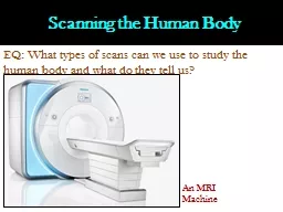
/
 Scanning the Human Body EQ: What types of scans can we use to study the human body and  Scanning the Human Body EQ: What types of scans can we use to study the human body and

Scanning the Human Body EQ: What types of scans can we use to study the human body and - PowerPoint Presentation

pasty-toler
pasty-toler . @pasty-toler
Follow
344 views
Uploaded On 2020-04-02

Scanning the Human Body EQ: What types of scans can we use to study the human body and - PPT Presentation

An MRI Machine I Computerized Axial Tomography CATCT CT scanning adds many Xray images with the aid of a computer to generate crosssectional views of a patients anatomy CT scans are performed to analyze the internal structures of various parts of the ID: 774620

brain body mri magnetic brain body mri magnetic tomography dye images scanning cat pet scans resonance eeg waves radioactive

Share:

Link:

Embed:

Download Presentation from below link

Download Presentation The PPT/PDF document " Scanning the Human Body EQ: What types ..." is the property of its rightful owner. Permission is granted to download and print the materials on this web site for personal, non-commercial use only, and to display it on your personal computer provided you do not modify the materials and that you retain all copyright notices contained in the materials. By downloading content from our website, you accept the terms of this agreement.


Presentation Transcript

Slide1

Scanning the Human Body

EQ: What types of scans can we use to study the human body and what do they tell us?

An MRI Machine

Slide2

I. Computerized Axial Tomography (CAT/CT)

CT scanning adds

many X-ray

images with the aid of a computer to generate cross-sectional views of a patient's anatomy.

CT scans are performed to analyze the internal structures of various parts of the

body to view traumatic injuries

CAT Scans can be used to view injuries or abnormalities in the brain to determine how to treat people appropriately

Slide3

I. Computerized Axial Tomography (CAT/CT)

Slide4

II. Electroencephalogram (EEG)

EEG’s use small flat metal disks (electrodes) attached to the head to monitor the electrical impulses within the brain

Information is taken down in a series of wavy lines

This test is often used to test for epilepsy or other brain disorders that are caused by misfiring of neurons within the brain

Slide5

II. Electroencephalogram (EEG)

WTH!!! I thought Mom said I was getting an Egg!!!

Slide6

III. Magnetic Resonance Imaging (MRI)

Uses

a magnetic field and radio waves to create detailed images of the organs and tissues within your body

.

T

he

magnetic field temporarily realigns hydrogen atoms in your body. Radio waves cause these aligned atoms to produce very faint signals, which are used to create

cross-sectional

MRI images — like slices in a loaf of

bread

An fMRI

(functional MRI) is used to detect activity (or lack thereof) in various parts of the brain

If a part of the brain is not functioning or not functioning well, then a person could have some disorders

Slide7

III. Magnetic Resonance Imaging (MRI)

Slide8

IV. Positron Emission Tomography (PET)

In a PET scan, radioactive dye is injected into the body

This dye will collect in your organs including the brain

The subject is placed into a tunnel-shaped scanning machine that takes pictures of the radioactive dye in the brain

This creates a 3D image of the brain

This scan is primarily used in psychology to find areas of the brain that are placated due to Alzheimer's or other ailments

Slide9

IV. Positron Emission Tomography (PET)