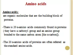
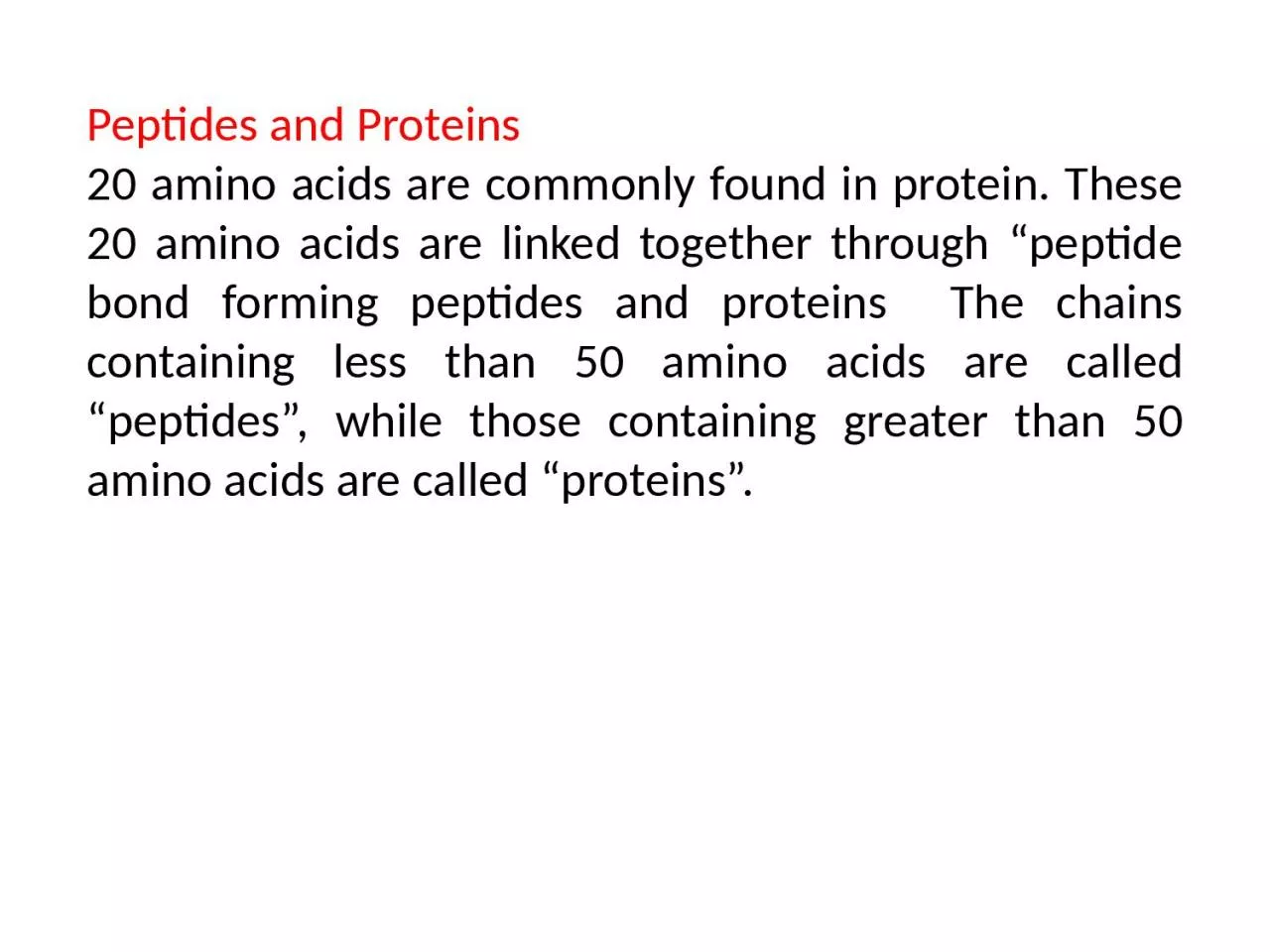
Peptide bond formation αcarboxyl group of one amino acid with side chain R1 forms a covalent peptide bond with αamino group of another amino acid with the side chain R2 by removal of a molecule of water The result is Dipeptide ie Two amino acids linked by one peptide bond ID: 916284
Download Presentation The PPT/PDF document "Peptides and Proteins 20 amino acids ar..." is the property of its rightful owner. Permission is granted to download and print the materials on this web site for personal, non-commercial use only, and to display it on your personal computer provided you do not modify the materials and that you retain all copyright notices contained in the materials. By downloading content from our website, you accept the terms of this agreement.
Slide1
Peptides and Proteins
20 amino acids are commonly found in protein. These 20 amino acids are linked together through “peptide bond forming peptides and proteins The chains containing less than 50 amino acids are called “peptides”, while those containing greater than 50 amino acids are called “proteins”.
Slide2Peptide bond formation
:
α-carboxyl group of one amino acid (with side chain R1) forms a covalent peptide bond with α-amino group of another amino acid
(
with the side chain R2) by removal of a molecule of water. The result is : Dipeptide ( i.e. Two amino acids linked by one peptide bond). By the same way, the dipeptide can then forms a second peptide bond with a third amino acid (with side chain R3) to give
Tripeptide
. Repetition of this process generates a polypeptide or protein of specific amino acid sequence
.
Slide3Peptide Bond
Slide4Peptide bond formation: - Each polypeptide chain starts on the left side by free amino group of the first amino acid enter in chain formation . It is termed (N- terminus). - Each polypeptide chain ends on the right side by free COOH group of the last amino acid and termed (C-terminus).
Fullname:
Alanyl
tyrosyl
aspartyl
glycine
Slide5Examples on Peptides:
1- Dipeptide
(two amino acids joined by one peptide bond): Example: Aspartame which acts as sweetening agent being used in replacement of cane sugar. It is composed of aspartic acid and phenyl alanine.
2-Tripeptides
(3 amino acids linked by two peptide bonds). Example: GSH which is formed from 3 amino acids: glutamic acid, cysteine and glycine. It helps in
protects
against
free radical which
causes cell damage.
Slide63-
octapeptides
: (8 amino acids) Examples: Two hormones;
oxytocine
and vasopressin (ADH).
4- Oligopeptide
: short polymer of residues linked by peptide bonds; up
to10-20
residues.
5- polypeptides
: longer polymer of residues linked by peptide bonds
;
larger sizes.
6- Protein
: one or more polypeptide chains
Notes
:
•
Residue – an amino acid (or peptide unit) in an
oligopeptide
, polypeptide or protein
•
Biological polymers are associated with biological function Proteins
Slide8Identification of N-terminal Residue
(a)
N-terminal residue can be identified by using a reagent that bond covalently with its α-NH2 group. Because the bond is stable to hot acid hydrolysis, the derivative of the N-terminal residue can be identified by chromatographic procedures after the protein has been
hydrolysed
.
Two reagents are commonly used
Slide91- Sanger’s
reagent:
The reagent contains
1-fluoro-2,4-dinitrobenzene (FDNB)
. It reacts with free –NH2 group in an alkaline medium.
Slide10The
compound so formed can be isolated after protein hydrolysis and identified
.
Sanger
was first to sequence a polypeptide. He determined the complete primary structure of the hormone insulin.
Slide112. Reaction with
Dansyl
Chloride:
The N-terminal–NH2 group can also combine with
Dansyl
chloride(1-dimethyl aminonaphthalene-5-sulphonyl chloride) to form a fluorescent
dansyl
derivative which can be isolated and identified.
Slide12(b)
Edman
reaction:
A similar reaction with –NH2
groupc
an occur with the reagent phenyl isothiocyanate and thus enables the identification of the N-terminal amino acid.
Slide13Slide14Sequenator
Edman
and
G.
Begg
have perfected an automated
amino acid
sequenator
for carrying out sequential degradation of peptides by the
phenylisothiocyanate
procedure (
Edman’s
reaction).Automated amino acid sequencers now widely used
, which
permit very rapid determination of the amino
acid sequences
of polypeptides
upto
100 amino acid approximately. Amino acids are determined sequentially
from N-terminal
end. The
phenyl
thiohydantoin
amino
acid
liberated
is
identified by high performance
liquidchromatography
(HPLC).
Slide15Proteins
Proteins are the most abundant molecules in living cells, constituting 40% - 70% of their dry weight. Proteins are built from amino acid monomers.
Typical protein functions:
1-Catalyze
Reactions (enzymes).
2-Chemical
Signaling (hormones).
3-Storage
(e.g. myoglobin stores oxygen).
4-Structural
(e.g. collagen in skin and tendons).
5-Protective
(e.g. antibodies).
6-Contractile
(e.g. myosin in muscle).
7-Transport
(e.g. hemoglobin.
Slide16Protein structure:
There are four levels of protein structure (primary, secondary, tertiary and quaternary)
1-Primary structure
:
- The primary structure of a protein is its
unique sequence of amino acids
- At one end is an amino acid with a free amino group the (the N-terminus) and at the other is an amino acid with a free carboxyl group the (the C-terminus).
Slide172- Secondary structure:
Results from hydrogen bond formation between hydrogen of –NH group of peptide bond and the carbonyl oxygen of another peptide bond.
According to H-bonding there are two main forms of secondary structure
:
α-helix
: It is a spiral structure resulting from hydrogen bonding between
one peptide bond and the fourth one
β-sheets:
is another form of secondary structure in which two or more polypeptides (or segments of the same peptide chain) are linked together by
hydrogen bond between H- of NH- of one chain and carbonyl oxygen of adjacent chain (or segment).
Slide18Slide193-Tertiary structure:
Tertiary: chain folding: fibrous and globular.
Chain folding causes changes in physical properties and biological function.
Fibrous proteins tend to have length >> diameter, tend to be water insoluble. Globular proteins have spherical shape. Contributing factors are
the hydrophobic effects,
hydrogen bonding,
ionic bond,
and
disulfide linkages by
cysteine units.
Slide20Slide21Slide22Some systems exist as larger "assemblies" several polypeptide chains. Quaternary structures are held together by a variety of interactions including hydrogen bonding, Van der Waals interactions, ionic bonding and occasionally disulfide bonds
.
Ex
: Collagen is a fibrous protein of three polypeptides (
trimeric
), Hemoglobin polypeptide is a globular protein with four polypeptide chains (
tetrameric
) – Insulin : two chains (
dimeric
).
Slide23