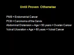
/
Until Proven Otherwise PMB = Endometrial Cancer Until Proven Otherwise PMB = Endometrial Cancer

Until Proven Otherwise PMB = Endometrial Cancer - PowerPoint Presentation

olivia-moreira
olivia-moreira . @olivia-moreira
Follow
368 views
Uploaded On 2018-10-11

Until Proven Otherwise PMB = Endometrial Cancer - PPT Presentation

PCB Carcinoma of the Cervix Abdominal Distension Age gt 50 years Ovarian Cancer Vulval Ulceration Age gt 60 years Vulval Cancer Government Pledges on Waiting Times Maximum Duration of Waiting ID: 687997

woman tumour ovarian cancer tumour woman cancer ovarian answer options item gynaecological cell hpv colposcopy cytology normal repeat uterine

Share:

Link:

Embed:

Download Presentation from below link

Download Presentation The PPT/PDF document "Until Proven Otherwise PMB = Endometrial..." is the property of its rightful owner. Permission is granted to download and print the materials on this web site for personal, non-commercial use only, and to display it on your personal computer provided you do not modify the materials and that you retain all copyright notices contained in the materials. By downloading content from our website, you accept the terms of this agreement.


Presentation Transcript

Slide1

Until Proven Otherwise

PMB = Endometrial CancerPCB = Carcinoma of the Cervix

Abdominal Distension + Age > 50 years = Ovarian Cancer

Vulval

Ulceration + Age > 60 years =

Vulval

CancerSlide2

Government Pledges on Waiting Times

Maximum Duration of Waiting:62 DAYS Urgent Referral for ? Cancer to 1st

Px

31 DAYS Date of Decision to Treat to 1

st

Px

(DTT)

31 DAYS Subsequent Surgery or Radiotherapy or

Anti-Cancer Drug Regimen

14 DAYS Suspected Cancer Symptoms to Seeing a

Specialist Slide3

Gynaecological Malignancy

Growth without controlInvasion

local

distant

Diagnosis

Biopsy

Treatment

remove

destroySlide4

Surgery

RadiotherapyChemotherapySlide5

MDTGCLNSlide6

Referral to the Network(Central) MDT

Ovary/Peritoneum

Uterine

Cervix

Vagina

Vulva

Recurrent cancerSlide7

Role of CNS(Cancer Nurse Specialist)

All woman with a gynaecological cancer should have access to a CNS and is identified as the Key WorkerCNS facilitates the woman and her carer’s needs throughout the cancer pathway from diagnosis

Provision of advice, support and information. Receptive to a full range of social, physical, psychological, cultural, sexual health and spiritual needs.

Link professional who can help patients through the system.

Co-ordinator of care and services

Works closely with surgeons, oncologists, radiotherapists and other professionals

Nurse Led Clinics – Breaking bad news clinics

Cancer Follow up clinics

Holistic Assessment

Transition of care on discharge to GPSlide8

Psychosexual Issues

HRT and menopausal informationLymphoedemaSlide9

Finance

Support agenciesLocal and national support groupsHospice CareSlide10

Classification of Ovarian Malignancy

Epithelial originGerm cell originStromal cell originSlide11

Tumour Markers in Serum

CA125 αFPHCG

Epithelial Ovarian Malignancy

Yolk sac tumour

Trophoblastic tumours choriocarcinoma

Alkaline phosphatase Lactate dehydrogenase

Dysgerminoma

FSH + LH

Granulosa cell tumour

αFP + HCG

Embryonal carcinoma,

PolyembryomaSlide12

Specific HistopathologicalFeatures of Gynaecological Tumour

Call – Exener BodyPsammoma

Body

Schiller Duval Body

Reinké

crystalloids

Granulosa cell tumour

Serous papillary cystadenocarcinoma ovary

Yolk sac tumour

Leydig

cell tumour ovarySlide13

Calculation of the Risk of Malignancy Index (RMI) for a Pelvic Mass

A RMI calculation of greater than 200 indicates a high suspicion of ovarian malignancy.CA125 measurement ··········· AMenopausal statusPremenopausal Score 1

Postmenopausal Score 3

(amenorrhoea >= 1 year, or over 50 years and post hysterectomy)

BSlide14

Ultrasound Score

Multilocular lesionSolid areasBilateral lesionsAscitesIntra abdominal metastasesIf none or one of the above is present score 1, if 2-5 of

above score 3 CSlide15

RMI = A*B*CYoung premenopausal patient with a unilateral, unilocular

cyst on scan with a CA125 of 30. RMI =30*1*1=30c.f. Postmenopausal patient with bilateral solid ovarian tumour and CA125 of 300. RMI=300*3*3=2700.Slide16

SINGLE BEST ANSWER QUESTIONS (SBAs)

Author Mr S J Duthie Theme Gynaecological oncology

Domain

Treatment 1

ITEM 1

Which one of the following is the dose limiting toxicity of cis-

Platin

?

Options

A Central neurotoxicity

B

Myeolotoxicity

C Nephrotoxicity

D Ototoxicity

E Peripheral neurotoxicity

ANSWER

C Slide17

ITEM 2Which one of the following is the dose limiting toxicity of carbo-

Platin? OptionsA Central neurotoxicity B

Myeolotoxicity

C Nephrotoxicity

D Ototoxicity

E Peripheral neurotoxicity

ANSWER

B

ITEM 3

Which one of the following is the dose limiting toxicity of

Oxaliplatin

?

Options

A Aplastic anaemia

B Haemorrhagic cystitis

C Peripheral neuropathy

D Pulmonary fibrosis E Retinopathy

ANSWERC Slide18

SINGLE BEST ANSWER QUESTIONS (SBAs)

Author Mr S J Duthie Theme Gynaecological oncology

Domain

Genetics 1

ITEM 1

Which one of the following is the single most correct description of the genetic transmission of Lynch Syndrome (Hereditary non-polyposis colon cancer syndrome)?

Options

A Autosomal dominant with a high penetrance of 90%

B Autosomal dominant with a low penetrance of 20%

C Autosomal dominant with a moderate penetrance of 50%

D Autosomal recessive with a high penetrance of 90%

E Autosomal recessive with a low penetrance of 20%

ANSWER

A Slide19

SINGLE BEST ANSWER QUESTIONS (SBAs)

Author Mr S J Duthie Theme Gynaecological oncology

Domain

Genetics B

ITEM 1

Women with a mutation in the BRCA1 gene have an increased lifetime risk for fallopian tube/ovarian cancer of approximately which one of the following?

Options

A 30%

B 40%

C 50%

D 60%

E 70%

ANSWER

D Slide20

ITEM 2

Peutz-Jeghers Syndrome is an autosomal dominant condition with a penetration of which one of the following? OptionsA 10%

B 25%

C 75%

D 95%

E Variable

ANSWER

D Slide21

SINGLE BEST ANSWER QUESTIONS (SBAs)Author Mr S J DuthieTheme Gynaecological oncology

Domain Genetics 1ITEM 1In women with sporadic ovarian cancer, a co-existing endometrial tumour occurs in approximately which one of the following? OptionsA 0.1 %B 2 %C 4 %

D 9 %

E 10 %

ANSWER

B Slide22

ITEM 2In women with ovarian cancer associated with Lynch Syndrome (Hereditary non-polyposis colon cancer syndrome) a co-existing endometrial tumour occurs in approximately which one of the following?

OptionsA 5 %B 10 %C 15 %D 20 %E 40 %ANSWERD

Reference

Beirne

J.P., Irwin G.W., McIntosh S.A., Harley I.J.G. and Harkin D. P. The Molecular and Genetic Basis of Inherited Cancer Risk in Gynaecology. TOGSlide23

SINGLE BEST ANSWER QUESTIONS (SBAs)Author Mr S J DuthieTheme Gynaecological oncology

Domain Granulosa cell tumour 1 ITEM 1In a postmenopausal woman with a granulosa cell tumour of the ovary, the serum level of which one of the following is the most likely to be increased?OptionsA Cortisol B Follicle stimulating hormone

C Growth hormone

D Inhibin B

E Luteinising hormone

ANSWER

D Slide24

EXTENDED MATCHING QUESTIONS (EMQs)

Author Mr S J Duthie Theme Gynaecological oncology

Domain

Endometrial Cancer risk factors

Options

A Cigarette consumption

B Diabetes mellitus

C Early menarche

D High body mass index

E History of polycystic ovarian syndrome

F History of unopposed oestrogen therapy

G Hypertension

H Late menopause

I

Nulliparity

J Use of barrier contraception Slide25

ITEM 1A 60 year old nulliparous woman has been “fast tracked” to your clinic as her General Practitioner noted a 5 week history of postmenopausal vaginal bleeding.

You review her history and observe that the woman reached her menarche at the age of 10 years and her menopause at the age of 53 years. The woman had tried to get pregnant in the past, underwent tests which showed that she had polycystic ovarian syndrome and there was a very limited response to ovulation induction. Subsequently, the woman used barrier methods of contraception. Shortly after menopause, the woman was given a prescription for hormone replacement therapy. Inadvertently, the woman received unopposed oestrogen for 2 months, developed irregular vaginal bleeding, the HRT was stopped, assessments at the PMB clinic were reassuring and the woman decided not to use any form of HRT again.

The woman has mild hypertension, adult onset diabetes mellitus which is well controlled by dietary modification and an oral hypoglycaemic agent and she consumes 5 cigarettes daily.

On examination, the woman has a body mass index of 48 kg/ m

2

Pelvic examination shows that the uterus is bulky and histological examination of a

pipelle

biopsy of the endometrium shows adenocarcinoma of the endometrium. Which one of the factors in the list of options is the single most significant risk factor for endometrial cancer in this particular case?

ANSWER

D Slide26

EXTENDED MATCHING QUESTIONS (EMQs)

Author Mr S J Duthie Theme

Gynaecological disorders

Domain

Tumours 1

Options

A Benign cystic

teratoma

B Borderline ovarian tumour

C Borderline serous tumour

D Brenner tumour

E Chocolate cyst

F Cyst of the corpus luteum

G Follicular cyst

H Granulosa cell tumour

I Krukenburg

tumour J Metastatic breast cancer K Mucinous cystadenocarcinoma

L Ovarian fibroma

M Papillary serous cystadenocarcinoma N Theca lutein cyst

Each of the clinical scenarios listed below refers to non pregnant women with a pelvic mass. For each of the items choose the most likely diagnosis from the list of options given above. Each option may be used once, more than once or not at all. Each option may be used once, more than once or not at all.Slide27

ITEM 1

A previously healthy 23 year old woman complains of abdominal pain, abdominal swelling and tiredness. On clinical and radiological assessment the woman is found to have a solid pelvic mass with a diameter of 15 cm which is separate from a normal uterus, abdominal ascites and a left sided pleural effusion. The serum CA 125 is well within the normal range. ANSWER

L

ITEM 2

A 24 year old woman has undergone suction evacuation of the uterus for suspected molar pregnancy. Inspection of the products and histopathological examination confirm the diagnosis. The woman complains of abdominal pain and an ultrasound examination carried out 4 days postoperatively shows;

Empty normal sized uterus, bilateral adnexal masses each of which has a diameter of 8 cm. The lesions are thin walled, cystic and do not appear to contain any solid elements.

ANSWER

N Slide28

EXTENDED MATCHING QUESTIONS (EMQs)

Author Mr S J Duthie Theme Gynaecological Oncology

Domain

Tumour Histopathology 1

Options

A Barr body

B Call-

Exner

body

C Copper sulphate crystals

D Howell- Jolly body

E

Kimmelstiel

-Wilson lesion

F Lines of Zahn

G

Psammoma body H

Reinke’s crystals I Schiller-Duval body

J Sternberg-Reed cells

Select the single most correct term from the list of options for the histopathological findings in the gynaecological tumours described in the items below. Each option may be used once, more than once or not at all. Slide29

ITEM 4A 45 year old woman underwent total abdominal hysterectomy bilateral

salpingo-oophorectomy for suspected ovarian cancer. The woman had completed her family, was known to have a pelvic mass and her case had been discussed at a gynaecological multi-disciplinary team meeting. The operation specimen was examined in the pathology laboratory. The uterus was normal in size, the tubes appeared normal, the right ovary was normal in size and the left ovary contained an infiltrative mass with no obvious capsule. The lesion in the left ovary contained minute concentric

lamellated

calcified bodies visible using light microscopy. Select the term for this feature from the list of options.

ANSWER

G Slide30

EXTENDED MATCHING QUESTIONS (EMQs)Author Mr S J DuthieTheme Gynaecology

Domain Pelvic tumours 1 OptionsA Benign cystic teratoma B Choriocarcinoma C

Endometrioma

D Fallopian tube carcinoma

E Granulosa cell tumour

F

Krukenburg

tumour

G Metastasising melanoma

H Ovarian cancer

I Ovarian fibroma

J Placental site tumour K Theca lutein cyst L Uterine metastasising fibroid

M Uterine sarcoma The list of options contains tumours affecting the female genital tract. Each of the clinical scenarios listed below refers to a woman with a tumour affecting the genital tract. For each of the items choose the single most likely diagnosis from the list of options given above. Each option may be used once, more than once or not at all.Slide31

ANSWERF

ITEM 1You are called to the operating theatre by the surgical team who have just carried out emergency midline laparotomy on a 56 year old woman with a pelvic mass. The specialist trainee in Surgery has discussed the operation with his Consultant (not present) and they request you to attend. You observe that the woman has ascites, bilateral solid ovarian tumours which are not adherent to surrounding structures and peritoneal induration.The uterus is normal and the fallopian tubes are stretched over the ipsilateral ovarian tumours. You telephone your Consultant who is busy with an obstetric emergency. Your Consultant asks you to take multiple representative biopsies, send peritoneal fluid for cytological tests. close the abdomen, to return the woman to the gynaecology ward for postoperative care and

arrange referral to the Gynaecological Multi- Disciplinary team.

Three days later the Pathologist reports that histological diagnosis of the ovarian biopsies show malignant mucin containing signet cell rings.

Options

A Benign cystic

teratoma

B

Choriocarcinoma

C

Endometrioma D Fallopian tube carcinoma E Granulosa cell tumour F

Krukenburg tumour G Metastasising melanoma

H Ovarian cancer

I Ovarian fibroma J Placental site tumour K Theca lutein cyst L Uterine metastasising fibroid

M Uterine sarcoma Slide32

ITEM 2You are called to your Consultant’s office as he is away and the Secretary wants you to look at a histopathology report that has just arrived. The report is on a hysterectomy specimen. Your Consultant had supervised another trainee one week previously to carry out total abdominal hysterectomy on a 40 year old woman of Afro-Caribbean origin who had a uterine fibroid, menorrhagia and who had completed her family.

The pathology report states that there was an intramural fibroid with a diameter of 10 cm. There was a soft friable mass within the uterine fibroid and histological examination of the lesion showed local invasion, spindle cells and 10 mitoses per high- power fields ANSWERM

Options

A Benign cystic

teratoma

B

Choriocarcinoma

C

Endometrioma

D Fallopian tube carcinoma E Granulosa cell tumour F Krukenburg

tumour G Metastasising melanoma H Ovarian cancer

I Ovarian fibroma J Placental site tumour K Theca lutein cyst

L Uterine metastasising fibroid M Uterine sarcoma Slide33

COLPOSCOPY EXAMINATION - ABNORMAL

CIN1 untreatedRepeat in 12/12HR-HPV -

ve

Repeat in 36 months

HR-HPV +

ve

Cytology +

ve

Colposcopy referral

Cytology -

ve

Repeat in 12/12

HR-HPV -

ve

Repeat in 36 months

HR-HPV +

ve

Cytology -ve

Repeat in 36 months

HR-HPV +

veCytology abnormalColposcopy referralSlide34

COLPOSCOPY EXAMINATION - ABNORMAL

>=CIN2TreatmentRepeat in 6/12

HR-HPV -

ve

Repeat in 36 months

HR-HPV +

ve

Cytology normal or abnormal

Colposcopy referralSlide35

COLPOSCOPY EXAMINATION - ABNORMAL

CGIN TreatmentRepeat in 6/12

HR-HPV -

ve

Repeat in 12/12

HR-HPV +

ve

Cytology negative

Refer to colposcopy.

If normal colposcopy,

repeat in 12/12

Cytology abnormal

Refer to colposcopy.

Complete 10-yr

cytology follow-up

12/12 repeat test

Cytology negative

Refer to colposcopy.

If normal colposcopy,

repeat in 12/12

HR-HPV -

ve

Repeat in 36 months

Cytology abnormal

Refer to colposcopy.

Complete 10-yr

cytology follow-up

HR-HPV +

veSlide36

CGIN

Glandular PremalignancyHigh grade / Low gradeMost arise within 1cm of SCJCo-exists with CINNo reliable screening method

No specific

colposcopic

features

Cone biopsy – depth > 25mmSlide37

Precision Medicine

Identify and understand molecular defect Determine presenceCorrect molecular defect Slide38

The End

Thank you very much.

S. J. Duthie