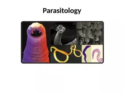
/
Lecture 3 Protozoa/ The Blood Parasites Lecture 3 Protozoa/ The Blood Parasites

Lecture 3 Protozoa/ The Blood Parasites - PowerPoint Presentation

oryan
oryan . @oryan
Follow
28 views
Uploaded On 2024-02-09

Lecture 3 Protozoa/ The Blood Parasites - PPT Presentation

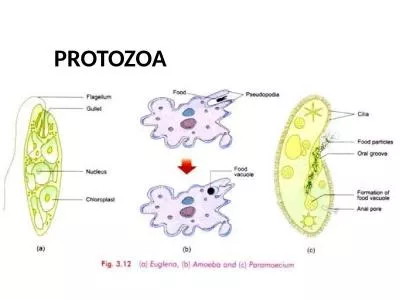
Medical Parasitology Prof Dr Ahmed Ali Mohammed B Blood and tissue flagellates This group of flagellates also known as hemoflagellates They require two hosts in their life cycle man or another susceptible mammal on the one hand and a bloodfeeding insect vector on the other hand They a ID: 1046219

blood host called cells host blood cells called insect parasite stage body form liver life leishmaniasis species infection erythrocytic

Share:

Link:

Embed:

Download Presentation from below link

Download Presentation The PPT/PDF document "Lecture 3 Protozoa/ The Blood Parasites" is the property of its rightful owner. Permission is granted to download and print the materials on this web site for personal, non-commercial use only, and to display it on your personal computer provided you do not modify the materials and that you retain all copyright notices contained in the materials. By downloading content from our website, you accept the terms of this agreement.


Presentation Transcript

1. Lecture 3Protozoa/ The Blood ParasitesMedical ParasitologyProf. Dr. Ahmed Ali Mohammed

2. B- Blood and tissue flagellates:This group of flagellates also known as hemoflagellates. They require two hosts in their life cycle: man, or another susceptible mammal on the one hand, and a blood-feeding insect (vector) on the other hand. They are belonging to two genera, Leishmania and Trypanosoma.During the life cycle of these flagellates, they may appear in four distinct morphological forms distributed between the vertebrate host (the final host) and the arthropod host (the intermediate host). It appears that any of the forms is capable of developing into any other. These forms are different in their morphology, flagellum position (and if it is present or not), the shape of the kinetoplast and its position, and the presence or absence of the undulating membrane. These forms are summarized in the following:

3. Promastigote (leptomonad form)This form occurs only in the insect vector. The body is spindle in shape, the nucleus relatively in the middle of the body. The kinetoplast nearby the anterior end, the flagellum extends from this structure and become free anteriorly (extends outside the body); there is no undulating membrane in this form. Its long flagellum is serving the function of both locomotion through the medium and the attachment to the insect gut wall.

4. 2. Epimastigote (crithidial form)The body is spindle in shape, the kinetoplast found in front of the nucleus which is found nearby the middle of the body. The flagellum grows and extends till the anterior end, attached to the pellicle, producing an undulating membrane then extends freely.3. Trypomastigote (trypanosomal form)The body is spindle in shape, the nucleus in the middle of the body, the kinetoplast in the posterior region of the body; it produces the flagellum which extends on the out margin of the undulating membrane then extends freely.

5. 4. Amastigote (leishmanial form)The body is spherical or ovoid with oval nucleus found nearby the body center; in front of the nucleus, the kinetoplast is laid, which produce a short flagellum extends till the body surface, and there is no undulating membrane in this form. This form usually develops in the vertebrate host cells.

6. Genus LeishmaniaThrough the use of molecular and immunological techniques, a number of species and subspecies of Leishmania have been partially characterized. Of those that infect humans, three clinical manifestations are evident: visceral, cutaneous, and mucocutaneous leishmaniasis. While their life cycles are identical and they are morphologically indistinguishable, they differ in the type and location of the primary lesions they produce in the human host. Leishmaniasis are now endemic in 88 countries on five continents with a total of 350 million people at risk. The vertebrate host of the parasite is the mammals, particularly the human, dogs and many species of rodents. Whereas the invertebrate host (the vector) is the sand fly.

7. Life cycleFor all species of Leishmania, the part of the life cycle spent in mammalian hosts is unbelievable in that the amastigote infects the macrophage, the cells of the mammalian host that constitute its primary defense against invasion by foreign organisms. The parasite, upon entering the macrophage, establishes itself in an endocytotic vacuole called a parasitophorous vacuole. A number of mammals act as natural reservoir hosts for the parasite, the most common being canines, both wild and domestic, and rodents. Leishmaniasis in humans is therefore a zoonosis. In the Vertebrate host, when the sand fly takes a blood meal from man and other mammals, while they are carrying the infective stage of Leishmania (the metacyclic promastigote), it will passively inject these parasites which will be quickly engulfed by the host macrophages in the reticuloendothelial system where the promastigotes revert to the intracellular amastigote form.

8. After sequence longitudinal binary fissions, the cells are filled with the parasites and rupture to release a large number of the amastigotes which are engulfed by other phagocytic cells, thus spreading the infection. At times, many infected phagocytic cells arrive to circulation and to viscera where the parasite will settle down and multiply in the reticuloendothelial cells of the liver, spleen and bone marrow, destroying these cells.

9. The invertebrate host are many species of sand fly from the genus Phlebotomus. When the insect bites the infected vertebrate host, it will suck the blood and the amastigote stage of the parasite, which migrate to the midgut of the insect and transform to promastigote form and start in multiplication by binary fission. After 4 to 5 days from the insect feeding, while attached to the wall of the gut, the promastigotes multiply rapidly by longitudinal binary fission and fill the esophagus. The promastigotes, as they transform to infective metacyclic promastigotes, detach from the gut wall. When they block the esophagus, the insect will push the esophagus contents to the front and back; by this method the insect will inject the infective stages in the victim which are subsequently deposited in the skin of the mammal when the sand fly feeds again.

10. The genus Leishmania includes the following species: 1. Leishmania donovaniThis parasite causes a disease called Kala-azar or Dum-Dum fever or visceral Leishmaniasis or black fever. Its vector is the sand fly (Phlebotomus papatasi).It causes the most severe and often fatal form of leishmaniasis. If left untreated, it has a mortality rate of almost 100%. In the mammalian host, amastigote infected cells are found at numerous sites, e.g. spleen, liver, bone marrow, lymph glands and intestinal mucosa as well as in the endothelial cells of the blood and lymph capillaries of the spleen, liver and bone marrow. Sometimes, the parasite infects the skin again and multiplies there, in this stage the disease called dermal Leishmaniasis or post Kala-azar.

11. Symptoms and Pathology Since leishmaniasis is primarily a disease of the reticulo-endothelial system macrophage, replacement of infected cells produces hyperplasia and consequent enlargement of visceral organs associated with the system, such as the spleen and liver, causes splenomegaly and hepatomegaly. A concomitant decrease in red and white blood cells production results in anemia and leukopenia, facilitating secondary bacterial infections. Without medical treatment, the condition is usually fatal. Surviving individuals, however, commonly acquire long-lasting immunity.The incubation period varies from 10 days to many months, but in the usual case, it is insidious. On the average about 90 days following exposure. The symptoms can be observed are:

12. 1. Greatly enlargement in the spleen and the liver with an increase in size and number of parasitized Kupffer cells. In the advanced cases, enlargement of the liver and spleen produces abdominal distention.2. The bone marrow exhibits markedly increased production of macrophages and decreased erythropoietic function. 3. Thrombocytopenia results in multiple hemorrhages, particularly from the mucous membranes.In the typical acute case:1. Temperature fluctuates daily from 37.5 - 40˚C.2. Bleeding typically occurs from the gum, lips, naris and intestinal mucosa.3. Complications usually observed in kala-azar are principally diarrhea or dysentery and broncho-pneumonia.

13. TreatmentChemotherapy consists of closely monitored intramuscular or intravenous injection of Pentavalent antimonial compounds such as antimony sodium gluconate. The treatment should be given until the aspirate is free of the parasites for at least 2 weeks.2. Leishmania tropicaThis parasite causes a disease called dry or urban cutaneous Leishmaniasis or oriental sore or Baghdad boil or Old world cutaneous Leishmaniasis or tropica sore. The vector is Phlebotomus papatasi and Phlebotomus sergenti.Unlike the amastigote of L. donovani, those of L. tropica are found primarily in the macrophages around the cutaneous sores. Sand flies must feed at these sites in order to acquire the infective amastigotes.

14. The tissue reaction is initiated with the introduction of the promastigotes into the dermis. The macrophages in the vicinity picks up the parasites which rapidly transform into amastigotes and multiply, destroying the host cells. Soon after, there will be a dense concentration of macrophages in the invaded area, all of which are liable to infection and destruction. The lesion then become necrotic at the center, and the margins containing parasitized macrophages which may become infiltrated with giant and plasma cells.Pathology and SymptomsThe lesion appears in the beginning as a macula, then as a papule, with a slightly raised center covered by a thin blister-like layer of epidermis on the skin at the feeding site of the insect. The papule becomes ulcerated after a few weeks, erupts with a discharge of a small amount of clear or purulent exudates, and spreads forming cutaneous lesions most commonly on the hands, feet, legs and face.

15. DiagnosisThe uncomplicated lesion may be mistaken for a variety of infections of the skin; hence, demonstration of the parasite is essential. Examination of Giemsa stained slides of the relevant tissue is still the technique most commonly used to detect the parasite. As well as the serological methods. The most reliable diagnosis is achieved by in vitro culturing of lesion scrapings or aspirates and subsequent identification of the promastigotes in the medium. Antibody detection can prove useful in visceral leishmaniasis but is of limited value where most patients do not develop a significant antibody response.Other diagnostic techniques exist that allow parasite detection and/or species identification using biochemical (isoenzymes), immunologic (immunoassays) and molecular approaches (PCR). Such techniques, however, are not readily available in general diagnostic laboratories.

16. TreatmentTreatment of choice is a daily intramuscular injection of Pentavalent antimony compounds for approximately one week. A second or third course of treatment may be required. Concomitant topical antibiotic treatment is employed in cases of microbial contamination of skin lesions.There is another type of this disease called mucocutaneous Leishmaniasis or Espundia in Brazil or many other tropical parts in America. The infection clinically resembles the oriental sore, but it is caused by Leishmania braziliensis.The infection starts in the skin, but the sores spread to extent regions and appear in a big number on the mucous membranes of the mouth, nasopharynx by extension or metastasis. It is also infecting the ear, nose cartilages and the throat, but not found in the blood and it is seldom in the inner viscera. The life cycle as similar as to that of L. tropica.

17. Malaria parasitePlasmodium vivaxPlasmodium falciparumPlasmodium malariae Plasmodium ovale

18. Malaria is one of the most prevalent and weaken diseases afflicting humans. Regardless of the species responsible for the infection, the certain aspects of the disease such as the life cycle of the infective organism, the chemotherapy and epidemiology are similar, except some medically significant dissimilarities. The four species that causes the disease in the human are:1. Plasmodium vivax.2. Plasmodium falciparum.3. Plasmodium malariae.4. Plasmodium ovale.The entire life span of the four species of Plasmodium that infect humans is spent in two hosts. The insect vector, a female mosquito belonging to the genus Anopheles, and a human host.

19. Malaria parasites as a groupThe significant feature of the life cycle is the alternation of generations phenomenon, which means the life cycle includes an asexual phase (schizogony) alternating with sexual one (gametogony) followed by another asexual phase called (sporogony).In the vertebrate host, the asexual phase develops, and gametocytes are produced. Whereas in the vector host (the invertebrate host), the gametocytes become mature gametes. Following maturation, the microgamete (male gametes) unites with the macrogamete (female gametes) to form the zygote, which then becomes an oocyst and produce the sporozoites. When the numerous sporozoites are introduced into the vertebrate host, they will develop in the asexual stage. Accordingly, there are two separate transfer stages, the gametocytes and the sporozoites.

20.

21. Life cycleSchizogony (in the human):A. Pre-erythrocytic development (Asexual Development out of the R.B.Cs.):The inoculation occurs when an infected female Anopheles mosquito injects saliva containing sporozoites beneath the epidermis of the human victim (in the cutaneous blood vessels) in a preparation to take a blood meal, thus inoculating the sporozoites into the bloodstream. The insect injects a spindle shape bodies (the sporozoites) which circulate in the blood stream.After approximately 1 hour, the sporozoite disappears from the circulation, re-emerging 24 to 48 hours later in the parenchymal cells of the liver where the first colonization takes place and the exoerythrocytic schizogonic phase begins. The specificity of the relationship of the sporozoite with hepatocytes rather than with other cells of the body is due, in part, to the recognition of the surface coat of the sporozoite (circumsporozoite coat) by receptors on the surface of the hepatocytes.Once the sporozoite becomes inside the hepatocyte, the sporozoite develops into a trophozoite, feeding on the host cell cytoplasm.

22. The first evidence of infection would be seen 48 hours to 7 days later in the parenchymal cells of the liver, where young schizonts in active nuclear division would be observed, and these are termed primary exoerythrocytic (EE) schizonts or pre-erythrocytic schizonts. The trophozoite will undergo two schizogony cycles, the first one called (primary exoerythrocytic schizogony) and the second one called (secondary exoerythrocytic schizogony) except in Plasmodium falciparum where the parasite undergoes single schizogony in the liver. The first generation of multiplied trophozoites called cryptozoites and sometimes called merozoites, while the second generation individuals called metacryptozoites. The infected liver cell with one cryptozoite produced (10-40) thousands of cryptozoites (or merozoites).After 7-10 days in the liver, the merozoites rupture from the host cell, enter the blood circulation, and invade the red blood cells, initiating the erythrocytic schizogony.

23.

24. B. Erythrocytic development: (Asexual Development in R.B.Cs.) When merozoites that have developed in pre-erythrocytic foci enters the red blood cells, they grows to the early trophozoite stage. Under the light microscopy, the early trophozoite appears to consist of a ring of cytoplasm and a dotlike nucleus. Due to its resemblance to a finger ring, this stage called the signet ring stage. In reality, the ring stage trophozoite is cup-shaped with a large vacuole filled with host hemoglobin in varying stages of digestion. This early form develops to the mature trophozoite stage. It grows because of its feeding on the R.B.Cs. contents and become rounded then irregular therefore it is called amoeboid shape trophozoite and then undergoes multiple fission and transform into schizonts later, producing a characteristic number of a new generation of merozoites in each infected erythrocyte. In the case of P. falciparum, the R.B.C. become more viscous, so it aggregates in the internal organs and does not appear in circulation. As in the liver, each of these merozoites is capable of infecting a new erythrocyte.

25. One of two fates await this new penetrant; it may become another signet ring trophozoite and begin a new schizogony, or it may become a male microgametocyte (♂) or a female macrogametocyte (♀). These gametocytes continue in the circulation for many weeks, it isn’t growing in the human body. It is important to note that these gametocytes are crescent in case of P. falciparum while they are rounded in the other types. Ring stage. Trophozoite. Schizont.

26.

27.

28. The merozoites spend a regular period from the entrance to the growing and forming new merozoites. This period is 48hr. in P. vivax and P. ovale, 72hr. in P. malariae and 36-48hr. in P. falciparum.2. Gametogony (in the mosquito):Once the ripe gametocytes are ingested by the female Anopheles mosquitoes with the blood meal and reach the midgut, they transform into mature gametes. These stages are unaffected by the digestive juices of the insect. Lysis of the surrounding erythrocytic material releases gametocytes into the lumen of the stomach. There, microgametocytes undergo a maturation process known as exflagellation during which the nucleus undergoes three mitotic divisions, producing 6 to 8 nuclei that migrate to the periphery of the gametocyte. During this period, the macrogametocytes develop into female macrogametes, each of which forms a membrane-derived fertilization cone to be penetrated by the microgamete. One macrogametocyte develops a single macrogamete. The fusion of male and female pronuclei (syngamy) produces a diploid zygote that, after 12 to 24 hours, elongates into a motile, microscopic wormlike ookinete.

29. This ookinete penetrates the gut wall of the mosquito to the area between the epithelium and basal lamina, where it develops into a rounded oocyst just under the outer membrane of the stomach. Following a period of growth during which its diameter increases 4 to 5 times, the oocyst is seen as a bulge on the hemocoel side of the gut. Ookinete.Exflagellation

30. The oocyst grows rapidly and develops internal nuclear centers called sporoblasts within the oocyst. Sporoblast nuclei undergo numerous divisions, producing thousands of delicate, spindle-shaped sporozoites enclosed within the sporoblast membranes. Within 10 to 24 days after the mosquito ingests the gametocytes, the sporozoite-filled oocysts themselves rupture, releasing the sporozoites into the hemocoel. The sporozoites are carried to the salivary gland ducts of the insect and are then ready to be injected into the next victim and initiate a new infection. Oocyst.Sporozoites

31. TRANSMISSIONTO MANTRANSMISSIONTO MANLIVERSporozoitesNucleusHypnozoiteInfected HepatocyteSchizontMerozoitesErythrocyteRingTrophozoiteSchizont43 – 48 hCycle leading to clinical symptomsP. vivax dormant stageGametocytesTRANSMISSIONTO MOSQUITOMacro-gametocyteMacro-gametocyte(Exflagellation)DiploidZygoteOokineteOocystsSporozoites15-30 mins5.4 days9 days15 mins1h12-36h9-12 days

32. SymptomatologyPathology in human malaria is generally manifested in two basic forms: host inflammatory reactions and anemia. Of the four species of Plasmodium responsible for human malaria, P. falciparum is the most virulent and causes, by far, the highest mortality. The initial symptoms of malaria, such as nausea, fatigue, a slight rise in temperature, mild diarrhea and muscular pains, are often mistaken for influenza or gastrointestinal infection. Host inflammatory reactions are triggered by the periodic rupture of infected erythrocytes, which releases malarial pigment such as hemozoin (hemozoin is a disposal product formed from the digestion of blood by some blood-feeding parasites), cellular debris and parasite metabolic wastes into the circulatory system.The incubation period extends from many weeks to months until the symptoms appear, which is sequent paroxysm in regular periods of shivering or chills then fever then sweating; the chills extend for 5-15 min., while the fever from 1-2hrs., and sweating for many hours.

33. Other important symptoms characterized by splenomegaly, hepatomegaly and increase in the bone marrow activity; there is also secondary sings like constipation, diarrhea and anemia (pernicious anemia).A condition known as blackwater fever often accompanies P. falciparum malaria, characterized by massive lysis of erythrocytes. It produces abnormally high levels of hemoglobin in urine and blood, fever, vomiting with blood, and jaundice, and there is a 20 to 50% mortality rate usually due to renal failure. The exact cause of this condition is uncertain; it may be a reaction to quinine, or it may result from an autoimmune phenomenon in which hemolytic antibodies are produced.Host Immune ResponseThe immune response of the human host differs somewhat for each of the two stages in the malarial life cycle i.e., the pre-erythrocytic stage and erythrocytic stage.

34. It is believed that T-cells, notably CD8+ T cells, play an important role in pre-erythrocytic immunity. On the other hand, CD4+ T cell regulation appears to play a critical role in acquired immunity to the erythrocytic stage of malaria infection.DiagnosisBy making (Blood films):1. Thick blood films: are frequently necessary to detect the parasites. This type allows rapid examination of a large volume of blood in a small area on the slide. Staining of thick blood films is carried out according to (Giemsa technique). These films provide concentration of the parasites.2. Thin blood films: it is also essential that thin films be prepared because the malarial species can be more readily identified on these, especially less experienced examiners. It is stain by Giemsa stain or Right stain.

35. Treatment1. All malaria infection except resistant P. falciparum: Chloroquine diphosphate (orally).2. Treatment of attack: If oral dose can’t be given, Quinine dihydrochloride (IV), Chloroquine hydrochloride (IM).