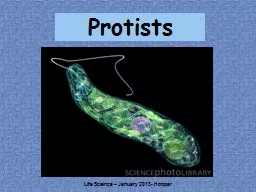
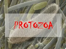
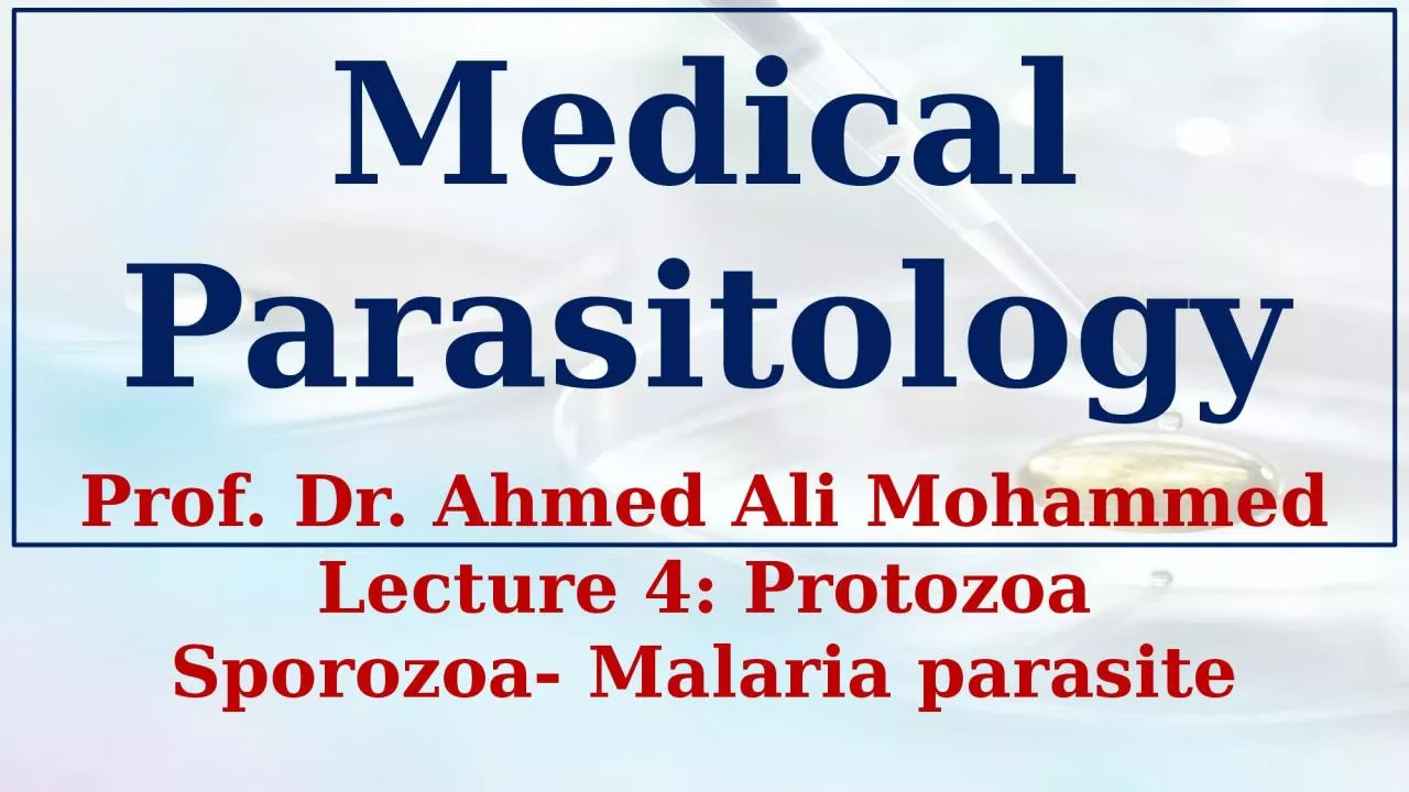
/
Lecture 4: Protozoa Sporozoa- Lecture 4: Protozoa Sporozoa-

Lecture 4: Protozoa Sporozoa- - PowerPoint Presentation

hanah
hanah . @hanah
Follow
0 views
Uploaded On 2024-03-13

Lecture 4: Protozoa Sporozoa- - PPT Presentation

Malaria parasite Medical Parasitology Prof Dr Ahmed Ali Mohammed Malaria parasite Plasmodium vivax Plasmodium falciparum Plasmodium malariae Plasmodium ovale Malaria is one of the most prevalent and weaken diseases afflicting humans Regardless of the species responsible for the inf ID: 1047550

host blood erythrocytic sporozoites blood host sporozoites erythrocytic malaria stage trophozoite called falciparum human gametocytes plasmodium merozoites cells schizogony

Share:

Link:

Embed:

Download Presentation from below link

Download Presentation The PPT/PDF document "Lecture 4: Protozoa Sporozoa-" is the property of its rightful owner. Permission is granted to download and print the materials on this web site for personal, non-commercial use only, and to display it on your personal computer provided you do not modify the materials and that you retain all copyright notices contained in the materials. By downloading content from our website, you accept the terms of this agreement.


Presentation Transcript

1. Lecture 4: ProtozoaSporozoa- Malaria parasiteMedical ParasitologyProf. Dr. Ahmed Ali Mohammed

2. Malaria parasitePlasmodium vivaxPlasmodium falciparumPlasmodium malariae Plasmodium ovale

3. Malaria is one of the most prevalent and weaken diseases afflicting humans. Regardless of the species responsible for the infection, the certain aspects of the disease such as the life cycle of the infective organism, the chemotherapy and epidemiology are similar, except some medically significant dissimilarities. The four species that causes the disease in the human are:1. Plasmodium vivax.2. Plasmodium falciparum.3. Plasmodium malariae.4. Plasmodium ovale.The entire life span of the four species of Plasmodium that infect humans is spent in two hosts. The insect vector, a female mosquito belonging to the genus Anopheles, and a human host.

4. Malaria parasites as a groupThe significant feature of the life cycle is the alternation of generations phenomenon, which means the life cycle includes an asexual phase (schizogony) alternating with sexual one (gametogony) followed by another asexual phase called (sporogony).In the vertebrate host, the asexual phase develops, and gametocytes are produced. Whereas in the vector host (the invertebrate host), the gametocytes become mature gametes. Following maturation, the microgamete (male gametes) unites with the macrogamete (female gametes) to form the zygote, which then becomes an oocyst and produce the sporozoites. When the numerous sporozoites are introduced into the vertebrate host, they will develop in the asexual stage. Accordingly, there are two separate transfer stages, the gametocytes and the sporozoites.

5.

6. Life cycleSchizogony (in the human):A. Pre-erythrocytic development (Asexual Development out of the R.B.Cs.):The inoculation occurs when an infected female Anopheles mosquito injects saliva containing sporozoites beneath the epidermis of the human victim (in the cutaneous blood vessels) in a preparation to take a blood meal, thus inoculating the sporozoites into the bloodstream. The insect injects a spindle shape bodies (the sporozoites) which circulate in the blood stream.After approximately 1 hour, the sporozoite disappears from the circulation, re-emerging 24 to 48 hours later in the parenchymal cells of the liver where the first colonization takes place and the exoerythrocytic schizogonic phase begins. The specificity of the relationship of the sporozoite with hepatocytes rather than with other cells of the body is due, in part, to the recognition of the surface coat of the sporozoite (circumsporozoite coat) by receptors on the surface of the hepatocytes.Once the sporozoite becomes inside the hepatocyte, the sporozoite develops into a trophozoite, feeding on the host cell cytoplasm.

7. The first evidence of infection would be seen 48 hours to 7 days later in the parenchymal cells of the liver, where young schizonts in active nuclear division would be observed, and these are termed primary exoerythrocytic (EE) schizonts or pre-erythrocytic schizonts. The trophozoite will undergo two schizogony cycles, the first one called (primary exoerythrocytic schizogony) and the second one called (secondary exoerythrocytic schizogony) except in Plasmodium falciparum where the parasite undergoes single schizogony in the liver. The first generation of multiplied trophozoites called cryptozoites and sometimes called merozoites, while the second generation individuals called metacryptozoites. The infected liver cell with one cryptozoite produced (10-40) thousands of cryptozoites (or merozoites).After 7-10 days in the liver, the merozoites rupture from the host cell, enter the blood circulation, and invade the red blood cells, initiating the erythrocytic schizogony.

8.

9. B. Erythrocytic development: (Asexual Development in R.B.Cs.) When merozoites that have developed in pre-erythrocytic foci enters the red blood cells, they grows to the early trophozoite stage. Under the light microscopy, the early trophozoite appears to consist of a ring of cytoplasm and a dotlike nucleus. Due to its resemblance to a finger ring, this stage called the signet ring stage. In reality, the ring stage trophozoite is cup-shaped with a large vacuole filled with host hemoglobin in varying stages of digestion. This early form develops to the mature trophozoite stage. It grows because of its feeding on the R.B.Cs. contents and become rounded then irregular therefore it is called amoeboid shape trophozoite and then undergoes multiple fission and transform into schizonts later, producing a characteristic number of a new generation of merozoites in each infected erythrocyte. In the case of P. falciparum, the R.B.C. become more viscous, so it aggregates in the internal organs and does not appear in circulation. As in the liver, each of these merozoites is capable of infecting a new erythrocyte.

10. One of two fates await this new penetrant; it may become another signet ring trophozoite and begin a new schizogony, or it may become a male microgametocyte (♂) or a female macrogametocyte (♀). These gametocytes continue in the circulation for many weeks, it isn’t growing in the human body. It is important to note that these gametocytes are crescent in case of P. falciparum while they are rounded in the other types. Ring stage. Trophozoite. Schizont.

11.

12.

13. The merozoites spend a regular period from the entrance to the growing and forming new merozoites. This period is 48hr. in P. vivax and P. ovale, 72hr. in P. malariae and 36-48hr. in P. falciparum.2. Gametogony (in the mosquito):Once the ripe gametocytes are ingested by the female Anopheles mosquitoes with the blood meal and reach the midgut, they transform into mature gametes. These stages are unaffected by the digestive juices of the insect. Lysis of the surrounding erythrocytic material releases gametocytes into the lumen of the stomach. There, microgametocytes undergo a maturation process known as exflagellation during which the nucleus undergoes three mitotic divisions, producing 6 to 8 nuclei that migrate to the periphery of the gametocyte. During this period, the macrogametocytes develop into female macrogametes, each of which forms a membrane-derived fertilization cone to be penetrated by the microgamete. One macrogametocyte develops a single macrogamete. The fusion of male and female pronuclei (syngamy) produces a diploid zygote that, after 12 to 24 hours, elongates into a motile, microscopic wormlike ookinete.

14. This ookinete penetrates the gut wall of the mosquito to the area between the epithelium and basal lamina, where it develops into a rounded oocyst just under the outer membrane of the stomach. Following a period of growth during which its diameter increases 4 to 5 times, the oocyst is seen as a bulge on the hemocoel side of the gut. Ookinete.Exflagellation

15. The oocyst grows rapidly and develops internal nuclear centers called sporoblasts within the oocyst. Sporoblast nuclei undergo numerous divisions, producing thousands of delicate, spindle-shaped sporozoites enclosed within the sporoblast membranes. Within 10 to 24 days after the mosquito ingests the gametocytes, the sporozoite-filled oocysts themselves rupture, releasing the sporozoites into the hemocoel. The sporozoites are carried to the salivary gland ducts of the insect and are then ready to be injected into the next victim and initiate a new infection. Oocyst.Sporozoites

16. TRANSMISSIONTO MANTRANSMISSIONTO MANLIVERSporozoitesNucleusHypnozoiteInfected HepatocyteSchizontMerozoitesErythrocyteRingTrophozoiteSchizont43 – 48 hCycle leading to clinical symptomsP. vivax dormant stageGametocytesTRANSMISSIONTO MOSQUITOMacro-gametocyteMacro-gametocyte(Exflagellation)DiploidZygoteOokineteOocystsSporozoites15-30 mins5.4 days9 days15 mins1h12-36h9-12 days

17. SymptomatologyPathology in human malaria is generally manifested in two basic forms: host inflammatory reactions and anemia. Of the four species of Plasmodium responsible for human malaria, P. falciparum is the most virulent and causes, by far, the highest mortality. The initial symptoms of malaria, such as nausea, fatigue, a slight rise in temperature, mild diarrhea and muscular pains, are often mistaken for influenza or gastrointestinal infection. Host inflammatory reactions are triggered by the periodic rupture of infected erythrocytes, which releases malarial pigment such as hemozoin (hemozoin is a disposal product formed from the digestion of blood by some blood-feeding parasites), cellular debris and parasite metabolic wastes into the circulatory system.The incubation period extends from many weeks to months until the symptoms appear, which is sequent paroxysm in regular periods of shivering or chills then fever then sweating; the chills extend for 5-15 min., while the fever from 1-2hrs., and sweating for many hours.

18. Other important symptoms characterized by splenomegaly, hepatomegaly and increase in the bone marrow activity; there is also secondary sings like constipation, diarrhea and anemia (pernicious anemia).A condition known as blackwater fever often accompanies P. falciparum malaria, characterized by massive lysis of erythrocytes. It produces abnormally high levels of hemoglobin in urine and blood, fever, vomiting with blood, and jaundice, and there is a 20 to 50% mortality rate usually due to renal failure. The exact cause of this condition is uncertain; it may be a reaction to quinine, or it may result from an autoimmune phenomenon in which hemolytic antibodies are produced.Host Immune ResponseThe immune response of the human host differs somewhat for each of the two stages in the malarial life cycle i.e., the pre-erythrocytic stage and erythrocytic stage.

19. It is believed that T-cells, notably CD8+ T cells, play an important role in pre-erythrocytic immunity. On the other hand, CD4+ T cell regulation appears to play a critical role in acquired immunity to the erythrocytic stage of malaria infection.DiagnosisBy making (Blood films):1. Thick blood films: are frequently necessary to detect the parasites. This type allows rapid examination of a large volume of blood in a small area on the slide. Staining of thick blood films is carried out according to (Giemsa technique). These films provide concentration of the parasites.2. Thin blood films: it is also essential that thin films be prepared because the malarial species can be more readily identified on these, especially less experienced examiners. It is stain by Giemsa stain or Right stain.

20. Treatment1. All malaria infection except resistant P. falciparum: Chloroquine diphosphate (orally).2. Treatment of attack: If oral dose can’t be given, Quinine dihydrochloride (IV), Chloroquine hydrochloride (IM).