/
HIFU Dr Prashant Bansal Two basic forms of thermal therapy for treatment of prostate cancer HIFU Dr Prashant Bansal Two basic forms of thermal therapy for treatment of prostate cancer

HIFU Dr Prashant Bansal Two basic forms of thermal therapy for treatment of prostate cancer - PowerPoint Presentation

pamella-moone
pamella-moone . @pamella-moone
Follow
357 views
Uploaded On 2019-06-23

HIFU Dr Prashant Bansal Two basic forms of thermal therapy for treatment of prostate cancer - PPT Presentation

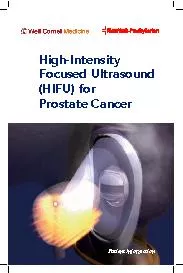
HighIntensity Focused Ultrasound which uses focused sound energy to heat tissue to destroy it HIFU is different from cryotherapy in that the focal zone is extremely discrete with little or no tissue effects in areas immediately adjacent to the treatment zone ID: 760022

Share:

Link:

Embed:

Download Presentation from below link

Download Presentation The PPT/PDF document "HIFU Dr Prashant Bansal Two basic forms ..." is the property of its rightful owner. Permission is granted to download and print the materials on this web site for personal, non-commercial use only, and to display it on your personal computer provided you do not modify the materials and that you retain all copyright notices contained in the materials. By downloading content from our website, you accept the terms of this agreement.


Presentation Transcript

Slide1

HIFU

Dr Prashant Bansal

Slide2

Two basic forms of thermal therapy for treatment of prostate cancer have been developed: cryotherapy, which freezes tissue to destroy it, and HIFU (

High-Intensity Focused Ultrasound), which uses focused sound energy to heat

tissue to destroy it.

HIFU is different from cryotherapy in that the focal zone is extremely discrete with little or no tissue effects in areas immediately adjacent to the treatment zone.

Slide3

HIFU Technology

HIFU uses high-energy ultrasound waves to destroy tissue at focal point of a transducer without injuring the intervening tissue.

Strong ultrasound waves in the inaudible sound range and approximately 10,000 times stronger than diagnostic ultrasound are generated by a transducer with a parabolic configuration.

This parabolic configuration focuses these sound waves into a discrete focal point measuring approximately 3 mm × 3 mm × 11 mm.

This focal point is located 3 to 4 cm distant from the transducer, and its size and shape is dependent on the energy emitted by the transducer, the geometric configuration of the transducer, and the characteristics of the tissue.

At the focal point of the transducer, ultrasound energy is concentrated, is absorbed by the tissue, and generates temperatures that can exceed 80° C, resulting in coagulative necrosis and the destruction of tissue.

Slide4

HIFU is generally performed under general or regional anaesthesia.

A transducer probe placed in a balloon filled with liquid (cooled or room temperature) is inserted into rectum (serves as an acoustical interface and to cool the rectal wall)

There are two transducers on these devices

Low-energy transducers (3 to 4

Mhz

) for imaging

High-energy transducers for treatment.

The prostate is imaged in both the sagittal and coronal planes, and the target treatment zone is outlined.

There is a treatment cycle in which the treatment zone is heated and then a cooling period during which the computer-controlled device moves to the next treatment zone distant from the first.

Slide5

The procedure is performed by imaging the prostate in the coronal and sagittal planes with a transrectally placed stationary probe.

This probe allows both imaging and treatment of the prostate. The treating physician outlines the areas to be treated in both planes

This focal point is then sequentially moved through the tissue to be destroyed by a computer-driven system that moves the parabolic transducer sagittally and coronally until the entire selected area has been coagulated.

The primary advantage of HIFU technology is precision.

In order to prevent the conductive spread of thermal energy through the prostate, the computer-controlled transducer is moved throughout the treatment to allow treated areas to cool down.

Because of the lack of adjacent tissue damage, HIFU is a repeatable precise technology.

Slide6

HIFU results in coagulative necrosis of the treated tissue.

In the case of complete prostate ablation with HIFU, the prostate gland retains its general anatomic configuration but shrinks to a small size (≈15 mL) over 3 to 6 months post therapy.

Post-HIFU biopsies show fibrosis only.

GENERAL CLINICAL USE of HIFU

Spleen, liver, kidney, breast, uterus, and bone

HIFU has been most well studied in treatment of prostatic conditions

HIFU has not been widely used for the treatment of symptomatic BPH because of its relatively invasive nature in comparison to other thermal-based modalities such as microwave and radiofrequency ablation.

Currently, HIFU does not have USFDA approval.

HIFU currently has FDA approval for the treatment of uterine fibroids

2 HIFU devices are available for the treatment of prostate:- Ablatherm and Sonoblate -500

Slide7

Slide8

Indications:

Ideal indications for HIFU as primary therapy are:

Older patients (≥70 years) with clinical stage T

1

-T

2

N

0

M

0

prostate cancer

Gleason score < 7

PSA < 15 ng /ml

Prostate vol < 40 ml

Particularly if patient refuses/is unsuitable for radical therapy

In this situation a lymph node evaluation is unnecessary and best chance for local control of cancer seems to be present.

Slide9

Safety

MC complications after primary therapy with HIFU are

U

TI

S

UI

E

D

R

ectal burn and rectourethral fistula/

R

etention

S

tenosis or strictures (Urethral/bladder neck)

With the technical improvements of the Ablatherm device c/c have decreased considerably

The use of TURP before HIFU also significantly decreases incidence of SUI, UTI, stenosis / strictures and BOO requiring TURP.

TURP is therefore indicated before HIFU. But this does not have any affect on disease control

Slide10

HIFU in Localized Prostate Cancer

Madersbacher and colleagues (1995) were the first to examine the feasibility of HIFU for treatment of localized prostate cancer.

They demonstrated that HIFU resulted in a sharply demarcated lesion with no treatment effects in areas immediately adjacent to the treatment zone no heat damage was noted to rectum/neurovascular bundle even though HIFU was extended to prostatic capsule

Slide11

HIFU after Radiation Failure

HIFU devices have also been tested for use as salvage therapy after radiation failure

The original

ASTRO

consensus panel recommendations were that treatment failure

after external beam radiotherapy should be defined

as two consecutive rises in the PSA after the occurrence of the PSA nadir. It specifically stated that this criterion for failure was not appropriate for other forms of radiation, for radiation combined with hormonal therapy, or for cryotherapy (and therefore other thermal-based approaches).

biochemical failure after radiation be defined as a rise of 2 ng/mL after nadir occurred, the so-called Phoenix criteria

Slide12

Treatment of HIFU Failures

HIFU remains unique compared with other modalities for localized prostate cancer in that it results in much less adjacent tissue damage.

This makes it a repeatable technology and thus potentially more salvageable by other techniques when it fails.

Slide13

Endorectal HIFU devices

There are currently two commercially available clinical HIFU instruments:

Ablatherm

, by

EDAP TMS

, and

Sonablate

manufactured by

Focus Surgery Inc.

Slide14

Ablatherm

Has a 4 cm focal length driven at 2.25-3 MHz and has an intensity of 1000 W/ sq. cm.

Slide15

Sonablate system

Consists of

chiller

, probe, and console. The

chiller

circulates and cools water flowing around the HIFU transducer in order to cool the probe. The probe itself is also motorized, allowing for computer-driven movement and also includes both ultrasound imaging capability and HIFU treatment.

Slide16

The

Sonablate system probe uses a 4 MHz PZT transducer that produces a beam of 3, 3.5, or 4 cm focal length with an intensity at the focus of 1680-2000 W/ sq. cm.

Slide17

Treatment Monitoring

In current practice, this is achieved in one of two ways–either by using real-time ultrasound, or MRI.

A t contrast enhanced MRI is the gold standard method.

Slide18

Why HIFU?

Achieves the necessary temperature rise in the timescale required to ablate tumours.

Margin of ablation is accurate.

Ablated tissue is re-absorbed by the body

Non-invasive (extracorporeal transducers)

No blood loss

No therapeutic interference with other modalities

Repeatability.

Slide19

Therapeutic Applications

Liver

tumours

.

Prostatic lesions.

Bladder.

Kidney.

Pancreatic malignancy.

Breast lesions.

Slide20

Therapeutic applications.

Neuro

surgical.

Obstetrics and Gynecology.

Role in

Thrombolysis

.

Role in Gene therapy.

Body sculpting.

Haemostasis

.

Slide21

Role of HIFU in liver.

Mainly are two indication HCC and Metastatic lesions.

Even though the surgery is mainstay of treatment, even after surgical resection survival rate of after 5 yrs only 25-30% so this makes HIFU as attractive non invasive treatment option.

Slide22

Prostatic Lesions

BHP

Role of HIFU in treatment of BHP is not rewarding. Even though the symptoms are reduced in immediate post procedure period, but these results were temporary.

Symptoms

reccured

by 1 yr. HIFU is not recommended as alternative to Minimally invasive TURP

Slide23

Prostatic Carcinoma

The treatment of carcinoma of the prostate (

CaP

) is more complex than that of BPH. Different treatment modalities have a place in the different disease stages.

The therapeutic choice for organ confined

CaP

lies essentially between radical prostatectomy and external beam radiotherapy, but the ability of HIFU to destroy the to destroy entire

tumours

successfully, makes HIFU as additive tool in the treatment of

CaP

Slide24

Pre-HIFU TURP is indeed now standard for treatment in Europe.

Complications

Stress incontinence

Significant

hematuria

.

Immediate post procedure retention.

Erectile dysfunction

UTI.

Slide25

Bladder

Superficial bladder cancer has also been identified as a potential target for treatment with extracorporeal HIFU.

Diagnostic ultrasound has been proposed as an alternative to

cystoscopy

.

If this is combined with HIFU this will obviate the need of invasive

cystoscopy

.

Slide26

Kidney.

Although studies are not numerous, accuracy and reliability of HIFU techniques seems provide a useful non-invasive alternative to surgery for the treatment of advanced renal

tumours

.

Slide27

Pancreatic Malignancy

HIFU is mainly used as palliative procedure in patient with advanced pancreatic malignancy.

The mean survival of the patients treated with HIFU increased from 6 to 12 months.

High-intensity focused ultrasound might be an effective treatment option for pain control, particularly in patients with tumors infiltrating the celiac plexus and in whom conventional pain treatments are not considered an effective option.

Slide28

Breast

Main indications are

fibroadenoma

and

localised

malignancy.

Promising results are seen in treatment of

localised

malignancy.

Main advantage is repeatability after

tumour

reccurence

.

Slide29

Neurosurgical

Earliest clinical research HIFU was in the field of neurosurgery, Parkinson’s disease were treated at this time these early studies were all limited in part by the lack of accurate imaging facilities, and in part by the need for

craniotomy to provide an acoustic window in the skull.

Slide30

Obstetrics and Gynecology

Uterine fibroids

Infertility treatment.

Umbilical vessel ablation

Slide31

Role in Thrombolysis

Pulsed HIFU enhanced the

tPA

mediated

thrombolysis

of clot by 50%.

But does not show any significant increase in

thrombolysis

with the clot treated with only Pulsed HIFU.

There was corresponding increase in

thrombolysis

with increasing number of pulses and total acoustic power.

Slide32

Gene therapy

“small, reparable tears or holes, which permeate a cell membrane and allow the insertion” of a substance into a cell without compromising its viability.

So HIFU has got tremendous role and great hope in transforming knowledge of Genetics into Medical treatments.

Slide33

Body Sculpting

Recently, the medical use of HIFU has been extended to include the ablation of unwanted subcutaneous adipose tissue as a non-invasive method of body sculpting.

Slide34

Haemostasis

Effective in arresting

haemorrhage

from either organs or vessels.

Slide35

Limitations

As ultrasound waves cannot pass through the air filled structures, so HIFU cannot be used in

tumours

of lung.

Ablation of

tumours

lying in close proximity to bowel or indeed of the bowel wall itself, would run the risk of visceral perforation.

Treatment times- longer usually it will take few hours to complete the procedure

Slide36

Requires

anaesthesia

.

Movement of organs at the time of exposure of HIFU limit the efficacy of treatment.