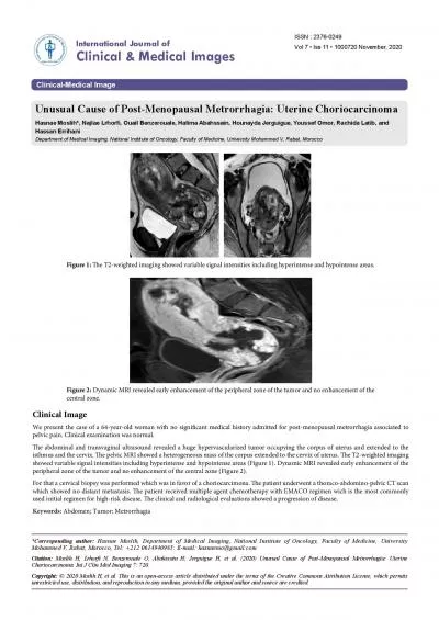
/
Introduction to medical imaging Introduction to medical imaging

Introduction to medical imaging - PowerPoint Presentation

pamella-moone
pamella-moone . @pamella-moone
Follow
401 views
Uploaded On 2016-05-09

Introduction to medical imaging - PPT Presentation

Dr Fadhl Alakwaa Biomedical Engineering program fadlworkgmailcom The thing you must have when you graduate Things you must have when you graduate Self confident Critical thinking Problem solving ID: 312346

image imaging ultrasound ray imaging image ray ultrasound resolution spatial contrast medical information tissue mri emission clinical spinal tomography

Share:

Link:

Embed:

Download Presentation from below link

Download Presentation The PPT/PDF document "Introduction to medical imaging" is the property of its rightful owner. Permission is granted to download and print the materials on this web site for personal, non-commercial use only, and to display it on your personal computer provided you do not modify the materials and that you retain all copyright notices contained in the materials. By downloading content from our website, you accept the terms of this agreement.


Presentation Transcript

Slide1

Introduction to medical imaging

Dr

Fadhl

Alakwaa

Biomedical Engineering program

fadlwork@gmail.comSlide2

The thing you must have when you graduate?Slide3

Things you must have when you graduate?

Self confident

Critical thinking

Problem solving

Team work

Communication skills

Fast learningSlide4

COURSE INFORMATION Slide5

GRADING SYSTEMSlide6
Slide7

Medical Imaging

The overall objective of medical imaging is to acquire useful information about physiological processes or organs of the body by using external or internal sources of energy.Slide8

Radiography

Fluoroscopy

Mammography

Computed Tomography (CT)

Nuclear Medicine Imaging

Single Photon Emission Computed Tomography (SPECT)

Positron Emission Tomography (PET)

Magnetic Resonance Imaging (MRI)

Ultrasound Imaging

Doppler Ultrasound Imaging

X-RAYSlide9

Image properties

Contrast

Spatial resolutionSlide10

ContrastSlide11

Contrast

X-ray contrast is produced by differences in tissue composition, which affect the local x-ray absorption coefficient.

Contrast in MRI is related primarily to the proton density and to relaxation phenomena (i.e., how fast a group of protons gives up its absorbed energy).

Contrast in ultrasound imaging is largely determined by the acoustic properties of the tissues being imaged.Slide12

Spatial resolution

resolve fine detail in the patient.

RESOVE= separate into constituent parts

the ability to see small detail, and an imaging system has

higher spatial resolution

if it can demonstrate the presence of

smaller objects in the image.

The limiting spatial resolution is the size of the smallest object that an imaging system can

resolve.

In ultrasound imaging, the wavelength of sound is the fundamental limit of spatial resolution. At 3.5 MHz, the wavelength of sound in soft tissue is about 0.50 mm. At 10 MHz, the wavelength is 0.15 mm.Slide13

Spatial resolutionSlide14

MEDICAL IMAGING: FROM PHYSIOLOGY TO INFORMATION

1.

Understanding Image medium:

tissue density is a static property that causes attenuation of an external radiation beam in X-ray imaging modality. Blood flow, perfusion and cardiac motion are examples of dynamic physiological properties that may alter the image of a biological entity.Slide15

MEDICAL IMAGING: FROM PHYSIOLOGY TO INFORMATION

2 Physics of Imaging:

The next important consideration is the principle of imaging to be used for obtaining the data. For example, X-ray imaging modality uses transmission of X-rays through the body as the basis of imaging. On the other hand, in the nuclear medicine modality, Single Photon Emission Computed Tomography (SPECT) uses emission of gamma rays resulting from the interaction of radiopharmaceutical substance with the target tissue.Slide16

MEDICAL IMAGING: FROM PHYSIOLOGY TO INFORMATION

3.

Imaging instrumentation:

The instrumentation used in collecting the data is one of the most important factors defining the image quality in terms of signal-to

ratio,resolution

and ability to show diagnostic information.

Source specifications of the instrumentation directly affect imaging capabilities. In addition, detector responses such as non-linearity, low efficiency and long decay time may cause artifacts in the image.Slide17

MEDICAL IMAGING: FROM PHYSIOLOGY TO INFORMATION

4.

Data Acquisition Methods for Image formation:

The data acquisition methods used in imaging play an important role in image formation. Optimized with the imaging instrumentation, the data collection methods become a decisive factor in determining the best temporal and spatial resolution.Slide18

MEDICAL IMAGING: FROM PHYSIOLOGY TO INFORMATION

5.

Image Processing and Analysis:

Image processing and analysis methods are aimed at the enhancement of diagnostic information to improve manual or computer-assisted interpretation of medical images.Slide19
Slide20
Slide21

What you want to know about each modalities?

(1) a short history of the imaging modality,

(2) the theory of the physics of the signal and its interaction with tissue,

(3) the image formation or reconstruction process,

(4) a discussion of the image quality,

(5) the different types of equipment in use today {block diagram + implementation},

(6) examples of the clinical use of the modality,

(7) a brief description of the biologic effects and safety issues, and

(8) some future expectations.Slide22

Safety

MR and ultrasound, which do not produce any

ionising

radiation, could perform diagnostic roles that were traditionally the preserve of X-ray radiology.Slide23

How does the referring doctor decide to request an MRI rather than an X-ray, CT or ultrasound image?

In general, the investigation chosen is the simplest, cheapest and safest able to answer the specific question posed.Slide24

X-ray

Because of the high contrast between bone and soft tissue, the X-ray is particularly useful in the investigation of the skeletal system.

An X-ray image of the chest, for example, reveals a remarkable amount of information about the state of health of the lungs, heart and the soft tissues in the

mediastinum

(the area behind the breast bone).Slide25

X-ray

In contrast, soft tissue organs such as the spinal cord, kidneys, bladder, gut and blood vessels are very poorly resolved by X-ray. Imaging of these areas necessitates the administration of an artificial contrast medium to help delineate the organ in question.Slide26

CT

In general, CT images are only obtained after a problem has been identified with a single projection X-ray or ultrasound image; however, there are clinical situations (a head injury, for example) in which the clinician will request a CT image as the first investigation.

CT is particularly useful when imaging soft tissue organs such as the brain, lungs,

mediastinum

, abdomen and, with newer ultra-fast acquisitions, the heart.Slide27

Gamma imaging: SPECT

Single Photon Emission Computed Tomography

Like X-ray images, gamma investigations are limited by the dose-related effects of

ionising

radiation and their spatial resolution, even with

tomographic

enhancement, means that they are poorly suited for the imaging of anatomical structure. However, the technique has found an important niche in the imaging of

function

, that is to say, how well a particular organ is working.Slide28

Gamma imaging

In practice, function equates to the amount of

labelled

tracer taken up by a particular organ or the amount of

labelled

blood-flow to a particular region. The radionuclide is usually injected into a vein and activity measured after a variable delay depending on the investigation being performed. A quantitative difference in ‘function’ provides the contrast between

neighbouring

tissues, allowing a crude image to be obtained.Slide29

Gamma imaging

In kidney scans, an intravenous injection of 99mTc

labelled

diethylenetriaminepentaacetic

acid (DTPA) helps quantify the ability of each kidney to extract and excrete the tracer.Slide30

An Introduction to the Principles of Medical Imaging, Chris Guy, 2005.Slide31

PET

Positron Emission Tomography

In contrast, PET, first proposed in the 1950’s, has taken much longer to be accepted as a clinical tool. The problem is related in part to the cost of the scanner and its ancillary services the cyclotron and

radiopharmacy

— and in part to the absence of a defined clinical niche. Thus, while PET has a number of theoretical advantages over SPECT such as its higher spatial resolution and its use of a number of biologically interesting

radionuclides

, in practice, it remains a research tool, found in a handful of national specialist

centres

, used

in the investigation of

tumours

or heart and brain function.Slide32

MRI

it has already found a particular place in the imaging of the brain and spinal cord.

One reason is its ability to detect subtle changes in

cerebral and spinal cord anatomy

that were not resolvable with CT (a slipped disc pressing on a spinal nerve or a small brain

tumour

, for example).Slide33

MRI

This advantage of MRI over CT is due in part to the superior spatial resolution of the technique and in part to the fact that MR images are insensitive to bone — in CT, the proximity of bony vertebrae to the spinal cord make this region difficult to image as a result of partial volume effects.

Furthermore, patients with pacemakers, artificial joints or surgical clips cannot be scanned and there are technical problems in scanning unconscious patients that require monitoring or artificial ventilation.Slide34

Ultrasound

Ultrasound is an effective and safe investigative tool. It offers only limited spatial resolution but can answer a number of clinical questions without the use of

ionising

radiation and, unlike MRI, the equipment required is portable, compact and relatively inexpensive.

It has found a particular place in the imaging of pregnancy, but it is also used to image the liver, spleen,

kidneys, pancreas, thyroid and prostate glands, and is also used as a screening tool in interventional radiology .

Ultrasound plays an important role in the investigation of the heart and blood vesselsSlide35

Ultrasound

However, there are a number of specific clinical situations in which ultrasound cannot be used. Structures surrounded by bone, such as the brain and spinal cord, do not give clinically useful images, and the attenuation of the ultrasound signal at air/tissue boundaries means that the technique is not suitable for imaging structures in the lung or abdominal organs obscured by gas in the overlying bowel.